Polymorphonuclear cells (PMNs) contribute to the initiation and progression of the immune response by mediating cytotoxicity, phagocytosis, and cytokine secretion. Because CD44 serves as a cytotoxic-triggering molecule on PMNs, it was hypothesized that it could also trigger cytokine production. In this study, the effect of anti-CD44 antibodies on interleukin-6 (IL-6) production in human PMNs was assessed. By using a reverse transcriptase–polymerase chain reaction, it was shown that PMNs stimulated with a mouse monoclonal or a rabbit polyclonal F(ab)2 anti-CD44 transcribe IL-6 messenger RNA. A similar effect was obtained when an anti-CD44 antibody was replaced with hyaluronic acid (HA). Kinetic studies showed that anti-CD44 and HA induced IL-6 gene transcription, initiated 3 hours after stimulation, peaked between 12 and 24 hours, and disappeared after 48 hours. Analogous results were achieved when secreted IL-6 protein was measured by enzyme-linked immunosorbent assay in the PMN culture supernatants. To characterize which metabolic pathways regulated CD44-dependent IL-6 production in PMNs, an RNA polymerase inhibitor, actinomycin D, and 2 protein kinase inhibitors, such as genistein and staurosporine, were tested. Actinomycin D and genistein blocked IL-6 production, whereas staurosporine did not, suggesting that CD44-dependent IL-6 production requires gene transcription and tyrosine kinase activity. Furthermore, the relationship between CD44 and cytokines that affect PMN function, including interferon γ (IFNγ) and IL-2, was investigated. Without CD44 cross-linking, IFNγ did not trigger IL-6 production. However, on CD44 cross-linking, IFNγ produced a strong synergistic effect on IL-6 syntheses in human PMNs.
Introduction
Polymorphonuclear cells (PMNs) have long been considered as cells only endowed with effector functions, including phagocytosis and killing of target cells. PMN-specific cell surface receptors, such as the Fc fragment of the constant portion of immunoglobulin G (IgG),1 regulate PMN cellular mechanisms by releasing reactive oxygen intermediates and cytoplasmic lytic enzymes. Therefore, they were not considered to produce significant protein levels.
A large body of new findings has highlighted that resting or stimulated human purified PMNs make a variety of messenger RNA (mRNA) and translate their relative proteins. PMNs stimulated with lipopolysaccharide (LPS) synthesize interleukin-1β (IL-1β),2 IL-1 receptor antagonist (IL-1ra),3 interleukin-8 (IL-8),4 tumor necrosis factor α,5 transforming growth factor β6, and interleukin-12 (IL-12).7 PMNs stimulated with granulocyte-colony stimulating factor (G-CSF) produce interferon α (IFN-α),8 whereas granulocyte-macrophage colony-stimulating factor (GM-CSF) induces IL-89production. Ionomycine plus phorbol 12-myristate 13-acetate stimulates the production of GM-CSF and interleukin-3 (IL-3).10 Finally, it has been reported that the monoclonal antibody (mAb) stimulation of myeloid cytotoxic triggering molecules, such as FcγRI or FcγRII, induces IL-6 gene transcription and translation.11
CD44 surface receptors are heavily expressed on progenitors and on terminally differentiated myeloid cells12 and on a variety of cell types, including lymphocytes, macrophages, erythrocytes, fibroblasts, and epithelial cells. Alternative splicing of CD44 gene provides at least 12 isoforms, which are reported to be involved in cancer metastasis and cellular activation.13 The major ligand for CD44 receptor is hyaluronic acid (HA), a main glycosaminoglycan component of the extracellular matrix. HA is a polysaccharide found in all tissue and is synthesized in the cellular plasma membrane. It is expressed on the cell surface and is bound to the extracellular matrix. HA binds a group of molecules termed hyaladherins that include cartilage link protein, aggrecan, and P-32.14 Osteopontin has been found to bind CD44 as well.15 Ligation of CD44 with mAb or HA results in the stimulation of several myeloid and lymphoid functions, including normal and leukemic cellular differentiation,16 PMNs, and natural killer (NK) cellular cytotoxicity.17,18 In addition, CD44 plays a major role in the regulation of cell migration through the vascular wall and inflammation.19 20 Thus, PMNs could be a potential target for studying the CD44-immunoregulatory role in myeloid cells.
In this study we assessed the ability of anti-CD44 mAbs or polyclonal antibodies or CD44 natural ligand HA to induce IL-6 production in freshly isolated PMNs. We found that, on CD44 cross-linking, PMNs efficiently promoted IL-6–RNA transcription and translation. Because IL-6 affects the processes of inflammation and cell-mediated immune response, these findings provide new information about the role of CD44 in the management of innate and adaptive immune response.
Materials and methods
Antibodies and reagents
Rabbit antihuman CD44 was produced by immunizing 2 rabbits with a genetically engineered extracellular-soluble CD44 fragment produced in bacterial inclusion bodies and Freund Adjuvant. Polyclonal anti-CD44 was purified from rabbit serum by using a protein A affinity-purified column. NIH44.1 and polyclonal anti-CD44 F(ab)2 fragments were produced by using a commercially available kit (Immunopure F(ab′)2 Preparation Kit, Pierce, Rockford, IL). The polyclonal anti-CD44 specificity was tested in vitro by enzyme-linked immunosorbent assay (ELISA) and by Western blot analysis of a refolded recombinant extracellular CD44 against which it was raised. NIH44.1 (anti-CD44; IgG1) and 3G8 (anti-CD16; IgG1) were provided by Dr David Segal (Experimental Immunology Branch, National Cancer Institute, Bethesda, MD). Whole mouse monoclonal 32.2 (anti-CD64; IgG1) and IV-3 (anti-CD32; IgG2b) were kind gifts from Dr Yashwant M. Deo (Medarex, Annandale, NJ). Fluorescein isothiocyanate (FITC)-conjugated anti-FcγRI, FITC–anti-FcγRII, and phycoerythrin (PE)-conjugated anti-FcγRIII were purchased from Pharmingen (San Diego, CA). Unlabeled anti-CD56 was purchased from Becton Dickinson (San Jose, CA); FITC-conjugated HA was a kind gift from Karen Hatchock (Experimental Immunology Branch, National Cancer Institute). Genistein, actinomycin D, staurosporine, propidium iodide, and endotoxin-free HA were purchased from Sigma (St Louis, MO).
Cell isolation
PMNs were isolated from buffy coats from normal, voluntary S Eugenio Blood Bank donors by Ficoll-Hypaque density gradient separation. PMNs were collected from the top layer over the red cell surface. With the use of distilled water, contaminating red cells were lysed by hypotonic shock and washed 3 times. PMN purity was demonstrated by Wright-Giemsa staining and flow cytometry analysis by using a FITC-conjugated anti-CD16 and FITC–anti-CD32. Throughout the study, PMNs were cultured in RPMI 1640 (Life Technologies, Gaithersburg, MD) supplemented with heat-inactivated fetal bovine serum (Mascia Brunelli S.p.A., Milano, Italy), glutamine (2 mM), streptomycin (100 U/mL), penicillin (100 U/mL), thereafter referred to as complete medium. Peripheral blood mononuclear cells (PBMCs) were separated by Ficoll-Hypaque density separation. Monocytes and NK cells were purified from PBMCs, using a Vario MACS system with an NK or monocytes isolation kit (Miltenyi Biotec, Auburn, CA).
CD44, FcγRI, and FcγRII stimulation on PMNs
Wells of 24-well plates (Becton Dickinson) were incubated with 0.25 μg/mL NIH44.1 F(ab)2, 0.25 μg/mL rabbit polyclonal anti-CD44 F(ab)2, various doses of soluble HA (range 500 to 1 μg/mL), or whole anti-FcγRs (anti-FcγRI or anti-FcγRII) in complete medium at 37°C, whereas, for HA immobilization, 96-well plates (Becton Dickinson) were incubated overnight at room temperature with 200 μL distilled water containing HA (range, 5 mg to 50 μg/mL). Unbound HA was removed and replaced with complete media at 37°C. Three hours later, freshly purified PMNs (2.5 × 10⁶/mL) were added to each well, and the plates were incubated at 37°C in 5% CO2. At the desired time point (range between 3 and 48 hours), PMN culture supernatants were collected and stored at −80°C until a human IL-6 ELISA assay was performed, whereas PMNs were harvested and total RNA was isolated by using Trizol reagent (Life Technologies).
Reverse transcriptase–polymerase chain reaction
Total RNA was extracted from 4 × 10⁶ PMNs using Trizol reagent; complementary DNA (cDNA) first strand was produced using Moloney murine leukemia virus reverse transcriptase (Life Technologies) with an oligo(dt)12-18 antisense primer (Life Technologies). IL-6 cDNA was amplified for 30 cycles using ATGAACTCCTTCTCCACAAGCGC sense primer and GAAGAGCCCTCAGGCTGGACTG antisense primer, amplifying a transcript of 628 bases. FcγRI cDNA was amplified for 28 cycles using AGATCTATGTGGTTCTTGACAACTCTG sense primer and CCATGGCCAGTGGAAAAACTTAAAGGC antisense primer. Finally, FcγRII was amplified for 30 cycles using CATTCAGTGGTTCCACAATGGGAA sense primer and GAAATCCGCTTTTTCCTGCAGTAG antisense primer. Amplified fragments were analyzed in 1% agarose gel electrophoresis in the presence of ethidium bromide (Sigma).
IL-6 protein measurement
Peripheral blood PMNs (2 × 10⁶/mL) were incubated in complete media for 24 to 48 hours (the percentage of nonapoptotic cells was 70% at 24 hours and 30% at 48 hours) in the presence or absence of rabbit or mouse anti-CD44 F(ab)2 or HA. Cell supernatants were collected at the desired times and stored at −80°C until the assays were performed. IL-6 content in supernatants was measured by a standard quantitative commercially available ELISA kit (Endogen, Woburn, MA). The minimum detectable IL-6 level using this assay was less than 1 pg/mL.
Flow-cytometry analysis of HA-binding and PMN cell cycle
Freshly isolated PMNs (1 × 10⁶) were incubated in ice for 30 minutes with FITC-conjugated HA and washed twice. The cells were then analyzed by using a Becton Dickinson FACScan flow cytometer. To assess the specificity of HA-binding on PMNs, in some experiments, unlabeled HA was successfully used to saturate the FITC-labeled HA. For the assessment of apoptosis, 0.5 × 10⁶ PMNs were harvested in FACS tubes and fixed for 15 minutes in 1% formaldehyde and washed once. The cell pellet was resuspended in 1 mL permeabilizing buffer (0.1% sodium citrate, 0.1% triton-X) and incubated overnight at 4°C in the presence of 25 μL of 1 mg/mL propidium iodide stock and 10 μg/mL RNAse. PMN red fluorescence was analyzed by flow cytometry. Apoptotic PMNs were easily distinguished from the normal PMNs on the basis of the lower binding of propidium iodide to apoptotic cells (these cells show as a distinct peak at the left of the G0/G1 peak).
Results
FcγR expression on peripheral blood PMNs
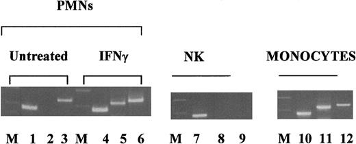
Because the PMN population could enclose contaminating monocytes, reverse transcriptase–polymerase chain reaction (RT-PCR) for FcγRI and FcγRII mRNA analyzed the purity of human peripheral PMNs used in this study. Figure 1 (left panel) shows that unstimulated PMNs were FcγRI mRNA− (lane 2) and FcγRII mRNA+ (lane3). In contrast, the addition of IFNγ to PMN cultures induced the expression of FcγRI (lane 5) without affecting FcγRII gene expression (lane 6). Figure 1 (middle and right panels) shows that NK cells and monocytes had a different profile of FcγR expression. In fact, NK cells did not express FcγRI and FcγRII genes (lanes 8 and 9), whereas monocytes expressed both genes (lanes 11 and 12). The presence of β-actin gene transcripts confirmed the quality and the purity of DNA on the agarose gels (lanes 1, 4, 7, and 10). Flow cytometry analysis of unstimulated PMNs showed that 97% of the cells expressed FcγRII and FcγRIII but not FcγRI or CD56, suggesting that we were working with highly purified PMNs.
Analysis of freshly purified PMN purity by FcγRI gene expression.
PMNs were isolated from a buffy coat and incubated overnight in the presence or absence of IFNγ as indicated. NK and monocytes were purified from freshly isolated PBMCs by magnetic cell sorting. With the use of specific primers, we analyzed FcγRI, FcγRII, and β-actin mRNA expression by RT-PCR as described in “Materials and methods.” The present data are given in the following order: Markers (M), β-actin (lanes 1, 4, 7, and 10), FcγRI (lanes 2, 5, 8, and 11), and FcγRII (lanes 3, 6, 9, and 12).
Ligation of CD44 on freshly purified PMNs induced IL-6 gene expression
Because PMNs constitutively express high levels of the hyaluronate receptor, CD44,17 we first assessed IL-6 gene expression in PMNs stimulated with an anti-CD44 F(ab)2. Resting PMNs do not express IL-6 (Figure 2, lane 1). By contrast, PMNs treated with NIH44.1 F(ab)2 produced detectable amounts of IL-6 gene transcript (Figure 2, lane 4). Resting PMNs are FcγRII+ and FcγRI−. As expected, the stimulation of PMNs with an anti-FcγRII mAb, IV-3, resulted in IL-6 gene transcription (Figure 2, lane 3), whereas an anti-FcγRI, 32.2, was fully ineffective (Figure 2, lane 2). In these experiments the amplification of the β-actin gene transcript confirmed the integrity of the experimental procedures analyzed. These data suggest that CD44 can control IL-6 transcription in resting PMNs of normal donors. Figure 2 shows a representative experiment of 10 performed with similar results.
CD44 ligation with mAbs induces IL-6 gene expression in peripheral blood PMNs.
PMNs were isolated from a buffy coat from a normal donor and incubated for 18 hours in complete medium in the absence or presence of different anti-FcγRs mAbs. RT-PCR, using specific primers, assessed IL-6 and β-actin mRNA expression, as described in “Materials and methods.” The present data are expressed in the following order: unstimulated PMNs (lane 1), anti FcγRI, 32.2 (lane 2), anti FcγRII, IV-3 (lanes 3), and NIH44.1 F(ab)2 (lane 4). This figure shows a representative experiment of 10 performed with similar results.
Ligation of CD44 with HA induced IL-6 gene transcription in resting PMNs
Although some mouse and human tumor cell lines bind HA, resting human T and NK cells do not. To investigate whether a soluble HA binds CD44 on human PMNs, we first analyzed the binding of a FITC-conjugated HA to PMNs by flow cytometry analysis. Figure3A shows that freshly isolated human PMNs bound a perceptible amount of FITC-conjugated HA. Subsequently, we did competition experiments in which unlabeled HA and a PE-conjugated anti-CD44 mAb competed for CD44 binding on human PMNs. Figure 3B shows that unlabeled HA inhibited the anti-CD44 binding. However, the inhibition was not complete, suggesting that HA other than CD44 may recognize other molecules on PMNs. We evaluated also the effects of HA on IL-6 gene expression by the addition of a logarithmic dose concentration of soluble HA (0 to 100 μg/mL) or immobilized HA (0 to 1000 μg/mL) to resting peripheral blood PMNs. Unstimulated PMNs did not show evidence of IL-6 gene expression (Figure3C, lane 1). In contrast, PMNs stimulated overnight with 10 (Figure 3C, lane 3) to 100 μg/mL (Figure 3C, lane 4) of soluble HA transcribed IL-6 gene, but a lower dose concentration did not (Figure 3C, lane 2). Similar results were obtained when we used immobilized HA (Figure 3C, lanes 5, 6, and 7). These data suggest that CD44 on PMNs could be physiologically involved in the regulation of IL-6 gene transcription.
IL-6 gene expression on peripheral blood PMNs on in vitro stimulation with HA.
(A) Normal freshly isolated PMNs were stained without (dash line) or with FITC-conjugated HA (solid line). (B) PMNs were stained with a PE-conjugated anti-CD44 in the absence (thick, solid, line) or in the presence of 25 μg (dotted line) and 50 μg (thin, solid line) unlabeled HA. The dashed line represents the red fluorescence of PMNs stained with a PE-conjugated, matching, isotype mAb. (C) PMNs were isolated from a buffy coat from a normal donor and stimulated for 18 hours in the absence or presence of logarithmic dose concentrations of soluble HA: 0 μg/mL (lane 1), 1 μg/mL (lane 2), 10 μg/mL (lane 3), and 100 μg/mL (lane 4), as well as linear dose concentrations of immobilized HA: 0 μg/mL (lane 5), 250 μg/mL (lane 6), and 1000 μg/mL (lane 7). Using specific sense and antisense primers, RT-PCR analyzed IL-6 or β-actin gene expression. The figure shows 1 of 3 significant experiments performed with similar results. FL1-H and FL2-H indicate the logarithmic numbers of green and red fluorescence, respectively.
Kinetics of IL-6 gene expression and protein production in PMNs stimulated with an anti-CD44 or HA
We were interested in the kinetics of IL-6 gene expression during anti-CD44 or HA stimulation. Thus, we stimulated PMNs obtained from buffy coats of normal donors with NIH44.1 F(ab)2 or HA. Aging PMNs easily undergo spontaneous apoptosis associated with the impairment of PMN functions. Therefore, we have also assessed the percentage of apoptotic PMNs used in these studies. Table1 shows that most of the PMNs with or without stimulation are apoptotic 48 hours after in vitro incubation. Figure 4A shows that IL-6 transcripts were undetectable in unstimulated PMNs (lane 1). However, they started to be visible 3 hours after CD44 stimulation with NIH44.1 F(ab)2 (lane 2) or HA (lane 3), remained detectable at 6 hours (lanes 6 and 7), at 12 hours (lanes 10 and 11), and at 24 hours (lanes 14 and 15), and disappeared completely at 48 hours of stimulation (lanes 18 and 19). Notably, as shown in Table 1, the absence of IL-6 transcripts 48 hours after PMN incubations could be due to the reduction of vital PMNs. However, the presence of β-actin transcripts suggested that the surviving PMNs could resolve IL-6 gene transcription. The next question was whether IL-6 protein production accompanied the IL-6 gene transcription. IL-6 was detectable in the supernatant of PMNs stimulated with NIH44.1 or HA 10 hours after stimulation. Around 24 hours after stimulation, we observed the highest concentration of IL-6, rapidly decreasing thereafter (Figure 4B, left side). On CD44 ligation, PMNs released an amount of IL-6 similar to that induced on LPS stimulation. In addition, PMNs stimulated overnight with immobilized HA induced a significant secretion of IL-6 in a dose-dependent manner (Figure 4B, right side). These data suggest that CD44 could play a major role in the regulation of PMNs proinflammatory mechanisms.
Kinetics of in vitro apoptosis of human peripheral blood polymorphonuclear cells
| . | Apoptotic PMNs (%) . | ||
|---|---|---|---|
| After 3 h in culture . | After 24 h in culture . | After 48 h in culture . | |
| Unstimulated | 1 | 38 | 70 |
| F(ab)2NIH44.1 | 1 | 26 | 74 |
| HA | 1 | 29 | 80 |
| LPS | 1 | 23 | 76 |
| . | Apoptotic PMNs (%) . | ||
|---|---|---|---|
| After 3 h in culture . | After 24 h in culture . | After 48 h in culture . | |
| Unstimulated | 1 | 38 | 70 |
| F(ab)2NIH44.1 | 1 | 26 | 74 |
| HA | 1 | 29 | 80 |
| LPS | 1 | 23 | 76 |
Polymorphonuclear cells (PMNs) from the same donor as in Figure 4were incubated in complete medium (unstimulated) or in complete medium containing NIH44.1 F(ab)2 0.25 μg/mL, hyaluronic acid (HA) 100 μg/mL, and lipopolysaccharide (LPS) 12.5 μg/mL and were harvested at the indicated times. The assessment of apoptosis in PMNs was obtained by propidium iodide staining of permeabilized cells by flow cytometry. The percentages of apoptotic PMNs with subdiploid DNA are indicated.
Kinetics of IL-6 gene expression and protein production after CD44 ligation on normal peripheral blood PMNs.
Freshly purified PMNs were isolated from a buffy coat from a normal donor and stimulated in complete medium. At the indicated times after stimulation, PMNs were harvested as source of total RNA, and the supernatants were collected for ELISA IL-6 protein assays. (A) This panel shows that, using sense and antisense specific primers, RT-PCR analyzed IL-6 and β-actin gene expression: lanes 1, 5, 9, 13, and 17, unstimulated PMNs; lanes 2, 6, 10, 14, and 18, NIH44.1 F(ab)2; lanes 3, 7, 11, 15, and 19, HA 100 μg/mL; and lanes 4, 8, 12, 16, and 20, LPS 12.5 ng/mL. (B) Left side shows IL-6 production measured in the supernatants collected at the indicated times: unstimulated PMNs (open squares), PMNs stimulated with NIH44.1 F(ab)2 (closed circles), HA 100 μg/mL (open circles), and LPS 12.5 ng/mL (closed triangles). Right side shows the IL-6 production measured in the supernatants of human PMNs stimulated overnight with immobilized or soluble HA at the indicated doses.
Gene transcription and tyrosine kinase activity are required for CD44-dependent IL-6 production
To determine whether CD44-dependent IL-6 production required protein synthesis, PMNs were incubated for 18 hours either in the absence or in the presence of the transcription inhibitor, actinomycin D (Figure 5A). Without actinomycin D, PMN stimulation with NIH44.1 F(ab)2 or HA promptly induced IL-6 gene transcription and protein secretion (Figure 5A, white bars), whereas, in the presence of actinomycin D, such expression was completely suppressed (Figure 5A, black bars), suggesting that IL-6 production requires de novo protein synthesis. To test which early signaling pathway is required for the regulation of IL-6 production, after CD44 ligation, we incubated PMNs in the absence or in the presence of genistein,21 a potent tyrosine kinase inhibitor, or staurosporine,22 a broad serine-threonine kinase inhibitor. PMNs stimulation with anti-CD44, HA, and anti-FcγRII induced a significant level of IL-6 release (Figure 5B-C, white bars). In the presence of genistein (Figure 5B, black bars) at the concentration equal to or higher than its reported 50% inhibitory concentration, IL-6 production was completely inhibited. Surprisingly, staurosporine only partially blocked IL-6 secretion (Figure 5C, black bars), suggesting that protein tyrosine kinase activity was the major requirement for CD44-dependent IL-6 production in human peripheral blood PMNs.
CD44-dependent IL-6 production requires protein transcription and tyrosine kinase activity.
Freshly purified PMNs isolated from a buffy coat from a normal donor were stimulated as indicated in the presence (closed bars) or absence (open bars) of metabolic inhibitors: actinomycin D (A), genistein (B), and staurosporine (C). The figure shows 1 of 3 experiments performed with similar results.
Synergistic effect of IFNγ on IL-6 production induced by CD44 ligation
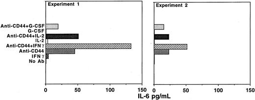
We then evaluated the relationship between anti-CD44 and cytokine activation of PMNs on IL-6 production. Because PMNs express the β and γ chain of IL-2 receptors and their IL-2 stimulation results in cytokine production and apoptosis inhibition, we included IL-223 among the cytokines used in these types of experiments. Figure 6 shows that IFNγ, IL-2, and G-CSF did not trigger IL-6 secretion. However, the ligation of CD44 by a polyclonal anti-CD44 F(ab)2 induced PMNs to release a significant amount of IL-6 in the supernatants (similar results were obtained using NIH44.1 F(ab)2). Notably, the addition of IFNγ enhanced IL-6 production induced by anti-CD44, but the addition of IL-2 or G-CSF did not. To characterize the mechanism by which IFNγ potentiated CD44-dependent IL-6 production, we analyzed CD44 expression in untreated or IFNγ-treated human peripheral PMNs by a flow-cytometry analysis of a direct immunofluorescence staining. We did not observe any changes in CD44 expression (data not shown), suggesting that IFNγ may rather potentiate intracellular signaling pathways arising from CD44 stimulation that lead to IL-6 transcription and translation.
Synergistic effect between IFNγ and CD44 stimulation on IL-6 production in human peripheral blood PMNs.
Freshly purified PMNs isolated from buffy coats from normal donors were stimulated for 18 hours in the absence or presence of the indicated stimuli. In experiment 1 and 2, PMNs were treated as follows: rabbit anti-CD44 (Fab)2, 0.25 μg/mL; IFNγ, 200 IU/mL; IL-2, 300 IU/mL; and G-CSF, 100 ng/mL. The figure shows 2 of 4 experiments performed with similar results.
Discussion
In the past decade, several studies have shown that, following cross-linking of specific signaling molecules expressed on the cell surface of freshly purified PMNs, a number of cytokines are induced and secreted. The major molecule that regulates IL-6 production in resting PMNs is CD14.24,25 However, Ericson et al11showed that cross-linking of FcγRI or FcγRII or the exposure to G-CSF results in IL-6 synthesis in freshly isolated PMNs. Although CD44 is heavily expressed and mediates PMN cytotoxic functions,17 18 it is not known whether it also affects cytokine production in resting PMNs. In this study we show that the ligation of CD44 with an anti-CD44 F(ab)2 or HA specifically induces the IL-6 gene expression. Furthermore, the synthesis and release of IL-6 following CD44 cross-linking required RNA transcription and tyrosine kinase activity.
Although it is definitively accepted that activated PMNs release several cytokines, there are conflicting reports about IL-6 production.26,27 Lloyd and Oppenheim26reported that PMNs treated with different stimuli secrete IL-6. However, Bazzoni et al28 could not confirm these data, suggesting that the presence of IL-6 mRNA could be a marker for monocytes contaminating PMN population. Because of this controversy, we have carefully evaluated PMN purity used in this study.
Resting human PMNs constitutively express FcγRII and FcγRIII but not FcγRI.29 However, the stimulation with IFNγ or G-CSF or some infectious diseases induce FcγRI expression on PMNs.30-34 As demonstrated by RT-PCR analysis, in the experiments conducted in this study, freshly purified PMNs were FcγRI mRNA− and FcγRII+ (Figure 1). Flow cytometry analysis confirmed these data. Furthermore, most of the cells analyzed by flow cytometry were FcγRIII+ and CD56−(data not shown). In addition, the stimulation of resting PMNs with IV-3 did induce IL-6 gene transcription, but the stimulation with 32.2 did not (Figure 2), suggesting that we were working with highly purified PMNs. However, in individual experiments, we noted that unstimulated PMNs showed a weak FcγRI expression but never sufficient to cause IL-6 production. Our results contrast with those obtained by Ericson et al11 who also stimulated PMNs with 32.2, but interestingly they observed IL-6 production. Perhaps PMNs may have a low-level expression of FcγRI that is difficult to detect but sufficient to trigger IL-6 production after a very efficient FcγRI cross-linking. In fact, both studies failed to show IL-6 gene expression in PMNs stimulated with 32.2. In contrast, a further FcγRI cross-linking with a goat anti-mouse F(ab)2 led to IL-6 gene expression and protein production.
In comparison to our results, Ericson et al11 found that unstimulated PMNs constitutively expressed FcγRI. This diversity may be due to different intervals in which we have studied FcγRI gene expression. In fact, it may be possible that FcγRI expression on PMNs could only be detected following 18 hours of culture.
We did not observe constitutive expression of IL-6 at gene and protein level in PMNs, but we noted that it was constitutively expressed in unstimulated PBMCs isolated by Ficoll-Hypaque gradient separation from each buffy coat assessed (data not shown). These data are different from those obtained by Melani et al35 and Palma et al36 who found constitutive expression of IL-6 mRNA in human venous blood PMNs. The data suggest that differences in PMN manipulation may affect IL-6 expression.
Because FcγRII ligation on PMNs triggers IL-6 release, our observation could be attributed to a nonspecific binding of contaminating Fc fragments in the NIH44.1 F(ab)2. However, we propose 2 lines of evidence indicating that NIH44.1 F(ab)2 specifically triggered IL-6 production in human PMNs: (1) The usage of a matching isotype whole mAb anti-FcγRI, 32.2, failed to stimulate IL-6 production in resting PMNs (Figure 1). (2) A different F(ab)2 such as a polyclonal rabbit anti-CD44 induced IL-6 production in resting PMNs.
During the local tissue phase of tissue inflammation, monocytes and resident macrophages are the major contributors to the immune response. They release proinflammatory cytokines and chemokines, which can induce leukocyte adhesion to the endothelial cells through sialyl Lewis X (SleX)/E-selectin and LFA/intercellular adhesion molecule (ICAM) interaction with subsequent migration to the extralymphoid tissues.37,38 A recent study has shown that a second pathway for lymphocyte adhesion and migration may exist. This pathway is CD44/HA dependent and is controlled by proinflammatory cytokines released from mononuclear cells during the local phase of infection. These cytokines induce HA expression on endothelial cells, exposing a docking site to CD44+ circulating leukocytes.19,39 Therefore, we propose that CD44 on PMNs interacts with HA on activated endothelium, inducing IL-6 secretion locally or into the blood flow. Local release of IL-6 may influence cell–cell and cell–endothelium interactions possibly by enhancing PMN adhesiveness through ICAM and CD18 upregulation.40Increasing levels of circulating IL-6 in the blood may result in systemic inflammatory effects mainly by the stimulation of acute-phase protein production.
After having crossed the endothelial wall and migrated to the source of the infection, PMNs meet an intercellular microenvironment rich in the extracellular matrix (ECM). ECM is composed of collagen proteins and glycosaminoglycans that bind with different affinities to CD44. Therefore, CD44 on PMNs could bind such molecules and secrete IL-6 locally at the site of inflammation. Increasing levels of local IL-6 could stimulate the proliferation of nonlymphoid cells involved in tissue damage and repair.
Ericson et al11 showed that IL-6 secretion resulting from FcγRI cross-linking is secondary to IL-6 gene transcription and translation. However, Terebuh et al41 reported that circulating murine IL-6 mRNA− PMNs contain preformed IL-6 in the intracellular storage. To obtain an insight into the mechanism involved in CD44-dependent IL-6 production, a metabolic inhibitor, actinomycin D, was added to PMN cultures undergoing CD44 stimulation. Actinomycin D completely blocked IL-6 secretion, showing that IL-6 production requires de novo protein expression. Although the CD44 intracellular tail lacks tyrosine residues and is phosphorylated on Ser323 and Ser325,42 the mechanism of signal transduction via CD44 is not well clarified yet. CD44 was found to be associated with Src family protein tyrosine kinases in T lymphocytes and in plasma membrane of human peripheral blood lymphocytes.43 44 On the basis of these data, it could be possible that either serine threonine or tyrosine kinase pathways may be involved in the regulation of CD44-dependent IL-6 production. Surprisingly, genistein completely blocked IL-6 secretion, whereas staurosporine did not, suggesting that CD44-dependent IL-6 production is under control of the Src family protein tyrosine kinases.
The relationship between cytokines affecting PMN functions and CD44 ligation was studied by stimulating PMN cultures in the presence of an anti-CD44 with IFNγ, IL-2,23 or G-CSF. We found a marked synergy between IFNγ and CD44 stimulation on IL-6 production in human peripheral PMNs. In contrast, IL-2 and G-CSF did not. To characterize the mechanism by which IFNγ enhanced CD44-dependent IL-6 production, we analyzed the level of CD44 expression on PMNs undergoing IFNγ stimulation. We did not observe up-regulation of CD44 during IFNγ treatment, suggesting that IFNγ has no role in the regulation of CD44 expression. Alternatively, IFNγ may increase IL-6 production by improving the efficiency of IL-6 gene transcription.
In conclusion, we have identified a novel mechanism by which PMNs may, in part, regulate the inflammatory response and other leukocyte functions by releasing IL-6 proinflammatory cytokine.
We thank Giulio C Spagnoli (Research Division, Department of Surgery, University of Basel) and Daruka Mahadevan (Hematology/Oncology, University of Arizona) for their critical review of the manuscript.
Supported by a grant from the Italian National Council Research.
The publication costs of this article were defrayed in part by page charge payment. Therefore, and solely to indicate this fact, this article is hereby marked “advertisement” in accordance with 18 U.S.C. section 1734.
References
Author notes
Giuseppe Sconocchia, Institute of Tissue Typing and Dialysis, S. Eugenio Hospital-Clinical Surgery, Ple dell'Umanesimo, 10, 00144, Roma, Italy; e-mail:g.sconocchia@rm.cnr.it.


This feature is available to Subscribers Only
Sign In or Create an Account Close Modal