Abstract
Interstitial deletion or loss of chromosome 5 is frequent in malignant myeloid disorders, including myelodysplasia (MDS) and acute myeloid leukemia (AML), suggesting the presence of a tumor suppressor gene. Loss of heterozygosity (LOH) analysis was used to define a minimal deletion interval for this gene. Polymorphic markers on 5q31 were identified using a high-resolution physical and radiation hybrid breakpoint map and applied to a patient with AML with a subcytogenetic deletion of 5q. By comparing the DNA from leukemic cells to buccal mucosa cells, LOH was detected with markers D5S476 and D5S1372 with retention of flanking markers D5S500 to D5S594. The D5S500–D5S594 interval, which covers approximately 700 kb, thus represents a minimal localization for the tumor suppressor gene. Further refinement of the physical map enabled the specification of 9 transcription units within the encompassing radiation hybrid bins and 7 in flanking bins. The 9 candidates include genes CDC25, HSPA9, EGR1, CTNNA1, and 5 unknown ESTs. Reverse-transcription polymerase chain reaction confirms that all of them are expressed in normal human bone marrow CD34+ cells and in AML cell lines and thus represent likely candidates for the MDS–AML tumor suppressor gene at 5q31.
Interstitial loss of the long arm of chromosome 5, del(5q), or complete loss of the entire chromosome −5 is a frequent finding in malignant myeloid disorders, including acute monocytic leukemia (AML) and myelodysplastic syndrome (MDS).1-4 These chromosomal losses are especially prevalent in AML arising after prodromal MDS or in MDS and AML arising after previous cancer treatment with alkylating agents or radiotherapy. In these therapy-related myeloid disorders, chromosome 5 deletions occur in approximately 40% of patients.5,6 Chromosome 5 deletions are less prevalent in de novo AML and occur in an estimated 5% to 7% of patients, but they are increased in the elderly.1 AML with del(5q) usually shows trilineage involvement, pronounced dysplasia, and characteristic megakaryocytic abnormalities. Additional cytogenetic abnormalities are frequent; the most common is del(7q) or −7.6,7 Regardless of the etiology, del(5q) is among the worst prognostic indicators in AML because it is characterized by poor response to chemotherapy, low complete remission rate (less than 30%), and a median survival time of 4 months.7 8
The prognostic significance of chromosome 5 deletions in MDS, however, is variable. In therapy-related MDS, or in RAEB and RAEB-t arising de novo, del(5q) or −5 generally implies a rapid progression to leukemia and poor outcome.5,6 On the other hand, a relatively indolent form of MDS is also associated with the loss of 5q. This disorder, called 5q− syndrome, is characterized by transfusion-dependent anemia with little or no cytopenia, usually shows an FAB subtype of RA or RARS, and rarely progresses to leukemia.3,9 10 It is clear that the 5q− syndrome is a distinct clinical entity with a different natural history than the more aggressive forms of MDS–AML. Thus, the impact of the 5q deletion in myeloid disorders must be interpreted in light of other clinical information.
The recurrent nature of these chromosomal deletions suggests that 5q contains tumor suppressor genes important to hematologic transformation. To localize these genes, others and we11-14have attempted to define a consistently deleted region using cytogenetic and molecular methods to map the extent of deletions in clinical samples. These studies suggest that the critical tumor suppressor gene for AML and some forms of MDS is located at band 5q31. This is in contrast to the common deletion interval for the 5q− syndrome, which has been localized to 5q32.15 The specification of 2 genomic intervals implies that different genes are responsible for each of these 2 myeloid disorders, as clinical findings suggest. Identification of each gene will provide valuable insight into the process of malignant myeloid transformation and useful markers for diagnosis, prognosis, and targets of therapy.
Previously our group proposed an interval of approximately 1.5 Mb within 5q31 as a tumor suppressor gene site based on overlapping deletions in clinical samples of MDS and AML, which included a patient with a submicroscopic chromosomal deletion.13 In the current study, we reexamined this case to further narrow the deletion interval, using additional polymorphic markers and a corrected marker order based on a high-resolution physical, genetic, and transcript map of 5q31.16 Here we report the result of this analysis, showing that the minimal deletion interval spans approximately 700 kb of 5q31 and thus would be expected to be deleted in all patients with AML with del (5q). Further refinement of the existing physical map by radiation hybrid (RH) analysis led to the identification of 9 transcription units that lie within this interval and that represent candidates for the MDS–AML tumor suppressor gene.
Materials and methods
Polymerase chain reaction analysis with microsatellite markers
Bone marrow and buccal smear samples were collected as sources of tumor DNA and normal DNA, respectively. Polymerase chain reaction (PCR) was performed as described,13 using 0.1 μg sample DNA or 5 μL buccal smear extract under the following conditions: 10 mmol/L Tris–HCl, pH 8, 50 mmol/L KCl, 1.5 mmol/L MgCl2, 200 μmol/L dATP, dGTP, and dTTP, 50 μmol/L dCTP, 1 μCi32P dCTP (3000 Ci/mmol), 5% glycerol, 0.1% Triton X-100, 0.5 U Taq polymerase (Perkin–Elmer/Cetus, Norwalk, CT), and 100 ng each primer in a 20 μL reaction. The reactions were run in a Perkin–Elmer model 480 thermocycler using the following parameters: initial denaturing at 94°C for 3 minutes; 35 cycles at 94°C (40 seconds), 55°C (30 seconds), and 72°C (30 seconds); final extension at 72°C for 5 minutes. Then 1 to 5 μL each PCR reaction was separated on a 6% polyacrylamide gel with 8 mol/L urea and exposed to x-ray film (Eastman Kodak, Rochester, NY). Loss of heterozygosity (LOH) was scored visually. The criteria for scoring LOH was as follows: retention of 2 alleles in normal tissue was considered informative, whereas the presence of only 1 allele in normal tissue was not informative. Therefore, LOH was considered when there was a loss of 1 allele in the tumor DNA when compared with the informative control DNA of 2 alleles.
Radiation hybrid analysis
Expression studies in myeloid tissues
Expression of candidate genes was examined in normal human bone marrow CD34+ cells and 3 human myeloid leukemia cell lines, KG-1, HL-60, and AML-193 obtained from ATCC (Rockville, MD). The CD34+ cells were isolated and purified, as previously described,19 from bone marrow obtained from normal donors with informed consent under the guidelines provided by the University of Illinois. Briefly, low-density marrow cells were prepared using Ficoll-Paque (Pharmacia Biotech, Piscataway, NJ) centrifugation and were further purified using a CD34+ Progenitor Cell Isolation Kit (Miltenyi Biotec, Auburn, CA). Purity of CD34+ cells was confirmed by FACS analysis using anti-CD34 antibodies. Total RNA was extracted from approximately 107cells using TRIZol Reagent (Gibco Technologies, Gaithersburg, MD) according to the recommended protocol.
Reverse transcription (RT)–PCR was performed using either the Titan 1 Tube RT-PCR System (Boehringer Mannheim, Indianapolis, IN) or the Enhanced Avian RT-PCR kit (Sigma, St. Louis, MO) according to the manufacturer's protocol. Primers were designed based on published or publicly available database sequences. Optimal Mg2+concentrations and annealing temperatures were determined for each primer pair. All reactions were performed in duplicate, including control reactions that did not include RT or RNA. RT-PCR products were visualized on 1.5% agarose gels stained with EtBr.
The following β2-microglobulin primers were used as positive controls for RT-PCR: forward 5′TGCCTGTACACTGCTCTTTATG 3′; reverse 5′GAATGTTTGGTGAACCTTCCT 3′. Because primers span an intron, the expected size of the RNA-derived PCR product was 245 bp whereas that from genomic DNA was 860 bp.
Results
Microsatellite analysis of AML with a minimal deletion
Our previously published high-resolution physical map spanning 6 Mb of 5q3116 was used to identify polymorphic markers for this study. This map includes all PCR-formatted polymorphisms that could be identified in existing databases; marker order was independently confirmed by typing on a CEPH meiotic breakpoint panel.20 Selected microsatellite markers from 5q31 were used to continue the analysis of a patient with AML whom we previously reported to have a submicroscopic deletion of chromosome 5.13 This clinical sample, designated case 24 in that study, is a diagnostic bone marrow sample from a 48-year-old man who had a relapse of AML of FAB M5a morphology and a white blood cell count of 104 000/μL. The karyotype was t,6;11del7(q31q36),+18, showing no visible abnormalities of chromosome 5, and allelotype analysis revealed LOH limited to a small interval on 5q31.
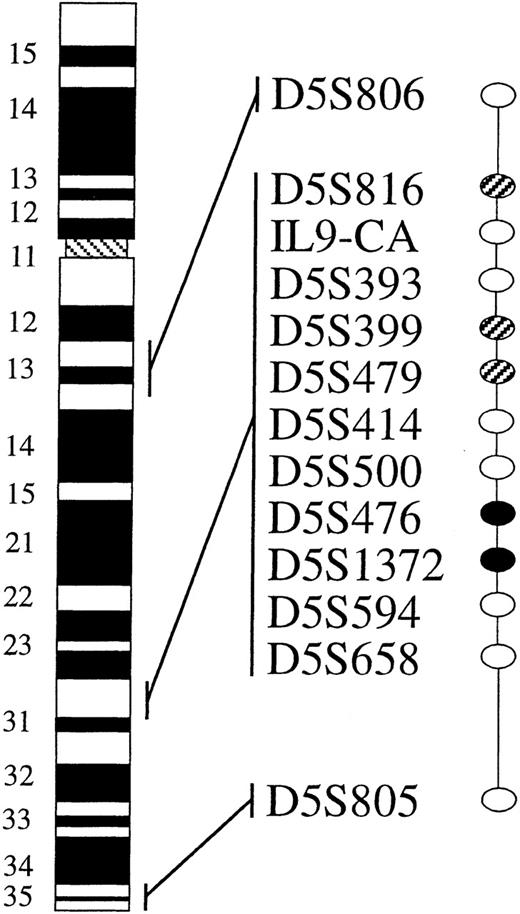
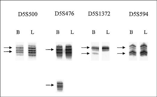
In the current study, LOH was similarly assessed by comparing the amplification patterns of the selected polymorphic markers in the leukemia sample to that of the buccal smear. Figure1 depicts a diagrammatic summary of these results relative to marker order. The study revealed loss of single alleles in 2 contiguous markers, D5S476 and D5S1372, with retention of both alleles in flanking markers, D5S500 and D5S594 (Figure2). Note that LOH with D5S476 and D5S1372 was reported in the previous study, but the placement of the markers within the map was in error; they were thought to be proximal to D5S500.13 The corrected marker order presented here is based on a high-resolution integrated map13,20 and is consistent with that reported elsewhere.14 Note also that marker AFMB350YB1, which had previously been interpreted as LOH in case 24, was found to be noninformative on further testing. LOH results thus led us to conclude that the minimal deletion in case 24 results in the loss of internal markers D5S476 and D5S1372 and is completely contained within the flanking markers D5S500 and D5S594. The size of this minimal interval is estimated to be approximately 700 kb, based a radiation hybrid distance between flanking markers of 29 cR and using the approximation of 1 cR10,000 = 29 kb for the Stanford G3 radiation hybrid panel.18
Allelotype results for AML case 24.
Markers are shown in order from centromere (top) to telomere (bottom), with their approximate cytogenetic band locations indicated on the chromosome 5 histogram. Filled oval, marker is informative with loss of heterozygosity; open oval, marker is informative with no loss; hatched oval, marker is not informative.
PCR amplification patterns for critical markers in case 24.
Marker names indicated on the top of the figure are shown in order from telomere (left) to centromere (right). PCR analysis is shown for DNA isolated from normal buccal smear (B) compared with DNA from leukemia cells (L) isolated from the patient's bone marrow sample. For each marker, the 2 alleles in the buccal DNA are indicated by arrows. Markers D5S500 and D5S594 show retention of both alleles in the leukemia sample, whereas loss of 1 allele is apparent for markers D5S476 and D5S1372.
Refinement of the physical map and identification of expressed sequences within the deletion interval
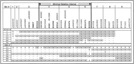
Expressed sequences within the minimal deletion interval were identified with reference to our previously published RH breakpoint map. The construction of this map resulted in the placement of 122 expressed sequences within 5q31, including both mapped expressed sequence tags (ESTs) from the Human Gene Map and unmapped ESTs from the Radiation Hybrid Database (RHdb). To localize more accurately the expressed sequences within 5q31 and to order them relative to the candidate gene interval, it was necessary to improve the resolution of the RH breakpoint map. This was done by additional PCR typing of ESTs on the Stanford G3 RH panel. The G3 panel has a higher resolution (more radiation hybrid breaks per given genomic distance) than the GB4 panel, where most of the ESTs were typed. Our RH breakpoint panel of 5q31 as published consisted of 13 hybrids from both the G318 and the GB417 panels; from this panel of 13 hybrids, 8 of them had breakpoints within the interval and 4 contained the interval within its entirety. Selected PCR markers were typed or confirmed on this panel as necessary (Figure3). Radiation hybrid “bins” were then defined by the breakpoints on G3 and GB4 hybrids, and markers were placed in different bins based on visual inspection of their PCR typing results relative to the breakpoints (Figure 3). The list of ESTs and their localizations has been updated from the previous publication.16
Radiation hybrid breakpoint map of 5q31 interval.
Vertical bars indicate the boundaries of the RH bins based on the presence of a breakpoint in any hybrid in the panel. The extent of the minimal deletion interval is labeled and indicated by bold vertical bars. Markers used in the study are listed in the row above the panel, from centromere (left) to telomere (right). Expressed sequences are italicized; known genes are italicized in bold and appear next to an identifying EST, if known. Polymorphic markers are shown in nonitalicized bold script. Identifying numbers for radiation hybrids are listed in the left column for the Stanford G3 panel (upper half) and the Genebridge 4 panel (lower half). Shaded squares (+) indicate that the marker scored positive; (−) indicates that the marker scored negative; (2) indicates an ambiguous signal; blanks indicate the marker has not been tested.
The relative order of the RH bins is fixed, but the order of markers within each bin cannot be further resolved by the RH panel. The breakpoint map was compared to the deletion interval, and all ESTs within bins 4 to 6, encompassing the flanking markers and intervening sequences, were considered to be primary candidate genes (Figure 3). ESTs from adjacent bins 3 and 7, which lie outside the deletion interval, were considered secondary candidates because their corresponding coding sequences could potentially overlap into the deletion interval. A summary of candidate genes or ESTs and secondary candidates in flanking bins is provided in Table1.
Candidate genes and ESTs in 5q31 deletion interval
| EST . | Bin No.† . | RHdb No. . | Genbank ID . | Unigene Cluster No. . | Description . |
|---|---|---|---|---|---|
| CDC25C* | 4 | RH69126 | M34065 | Hs.656 | CDC25C |
| SGC32445 | 4 | RH60414 | H11651 | Hs.28088 | Unknown EST |
| EGR1* | 4 | RH59849 | T61077 | Hs.738 | Early growth response gene |
| HSPA9* | 4 | RH9797 | Z19246 | Hs.3069 | Mortalin |
| D5S1856 | 5 | RH25090 | G05741 | Hs.114169 | KIAA0416 |
| CTNNA1* | 5 | RH70257 | T28827 | Hs.178452 | α-Catenin |
| stSG8723 | 5 | RH16367 | H82946 | Hs.174323 | Unknown EST |
| WI-17966 | 5 | RH60368 | R96021 | Hs.35495 | Unknown EST |
| WI-15469 | 6 | RH60079 | H48434 | Hs.198992 | Unknown EST assigned to Unigene for cDNA sequence of Matrin 3; see detailed information |
Known genes are listed in italics.
Refers to RH breakpoint map (Figure 3).
Within the deletion interval, 9 expressed sequences were found, including 4 known genes, CTNNA1, CDC25C, early growth response gene-1 (EGR1), and HSPA9. CTNNA1 (αE-catenin)encodes a cadherin-associated protein that plays an important role in cell–cell association and cell differentiation.21,22CTNNA1 is a promising candidate for a tumor suppressor gene; it has recently been shown to contain mutations in colon cancer cell lines, and it can function to suppress invasion.23CDC25C encodes a cell cycle regulatory protein required for cell entry into mitosis.24EGR1 encodes a zinc-finger protein essential for the differentiation of monocytic cells.25 Both CDC25C and EGR1 have been evaluated as tumor suppressor genes but have not been found to contain mutations in clinical AML samples.14HSPA9(Mortalin) encodes a cellular immortalization protein that is implicated in the control of cell proliferation and cellular senescence.26 It is also a good candidate for the AML because since its loss may promote cellular immortalization. Among the EST candidates, WI-15469 is linked to the Unigene cluster that represents Matrin 3, Unigene cluster Hs.198992. The assignment of WI-15469 to Hs.198992 was made since our previous map was completed,20 and we believe that this association is a database error because WI-15469 shares no sequence homology with the complete cDNA sequence of Matrin 3(Genbank M63483). Additionally, WI-15469 does not enter the TIGR cluster for the Matrin 3 gene but is instead assigned to the cluster THC252032, an unidentified partial cDNA. We are trying to obtain additional sequence and resolve this inconsistency, which probably resulted from an incorrect or chimeric cDNA sequence in Unigene. The other 4 ESTs have no known homologies.
RH bins 3 and 7 contain the secondary candidates, including thyroid hormone receptor coactivator protein (THRCP). This protein is a tyrosine kinase and nuclear receptor coactivator, which regulates the expression of target genes resulting from thyroid hormone and other cellular proliferation signals.27 Gene CDC23(located in the flanking bin 3) was recently proposed as a candidate tumor suppressor gene, but it has been excluded because of the lack of mutations in leukemia cells with the loss of5q.28 The EST stSG30881 encodes a protein that shows some homology to murine kinesin-like protein RabKinesin. Kinesins are microtubule-dependent molecular motors involved in intracellular transport and mitosis.29
Expression of candidate genes and ESTs in myeloid tissue
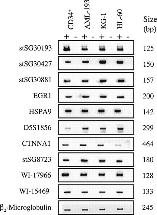
As a first step in the evaluation of these 9 genes as AML tumor suppressor gene candidates, their expression in myeloid cells was investigated. RNA was isolated from 3 human myeloid leukemia cell lines and CD34+ cells obtained from normal human bone marrow. The HL-60 cell line was established from the peripheral blood cells of a patient with acute promyelocytic leukemia,30 KG-1 from the bone marrow of a patient with acute erythroblastic leukemia,31 and AML-193 from a patient with acute monocytic leukemia.32 Both KG-133 and HL-6030are reported to have chromosome 5 deletion or loss and consistently show single alleles on allelotyping (data not shown). AML-193, on the other hand, has retained both chromosome 5 alleles by karyotype32 and allelotype assessment (data not shown). Primers were designed using available cDNA and EST sequences (Table2) and were tested on the RNA by RT-PCR. The analysis showed that all sequences were expressed in the cell lines studied (Figure 4), confirming that these ESTs originated from expressed sequences and were appropriately expressed in myeloid tissues, as would be expected of a leukemia tumor suppressor gene.
Polymerase chain reaction primers for candidate genes in 5q31 deletion interval
| Gene/EST . | Primer Sequence (5′-3′) . | Product Size (bp) . | Annealing Temp (°C) . | Mg2+ (mmol/L) . |
|---|---|---|---|---|
| stSG30193* | f: AGCCAGCCAGATCACAGAGT | 125 | 56 | 1.5 |
| r: CACCCACCTCAACCAGAACT | ||||
| stSG30427† | f: TCTCCAAGAAACAGCCCG | 150 | 53 | 2.5 |
| r: TTCACTCAGCACCTGGGAG | ||||
| stSG30881* | f: TTGCATCCTGGATATAATTCCC | 157 | 56 | 1.5 |
| r: TCCCCTTTACTCAAATCTGGG | ||||
| CDC25C* | f: AACGGTCTTGCATAGCC | 127 | 60 | 1.5 |
| r: CAGCCTTGAGTTGCATAGAG | ||||
| SGC32445* | f: AGTTTGGTTTATTGGCTCATCC | 125 | 60 | 1.5 |
| r: TTCAGTCAGGGCCAGGAG | ||||
| EGR1* | f: GGAATCATGCCTTATGTAGTCAC | 200 | 56 | 1.5 |
| r: GGACACATGACGTTTGCCTAGA | ||||
| HSPA9* | f: TCAGCAAGAGTGACATAGGAGAAGTGA | 142 | 56 | 1.5 |
| r: CACAGCCTCATCAGGATTGACAGCTT | ||||
| D5S1856* | f: GGCTCAAGGGCTTTAGTCAA | 299 | 57 | 3 |
| r: AGACTCCATCTTTCCAATAAAAATG | ||||
| CTNNA1* | f: CTTGGCCGCACCATTGCAGACCAT | 464 | 56 | 1.5 |
| r: GGCCGGCCTGGGCAGACTTAGATG | ||||
| stSG8723† | f: CACCATCACCTATGCCCTCT | 180 | 56 | 1.5 |
| r: ATAGGCAAAGCCACCCTTTT | ||||
| WI-17966* | f: ACAAAACTTGCCTGTACACTGC | 128 | 54 | 1.5 |
| r: CAAGAGCCATTTTTCTTTTTGG | ||||
| WI-15469* | f: GCACATAATGCTTTATGTACCTGC | 133 | 57 | 3 |
| r: GGTGATGATTTTAATGTGACATGC |
RT-PCR performed using Boehringer Mannheim Titan One RT-PCR System.
RT-PCR performed using Sigma Enhanced Aviann RT-PCR Kit.
Expression analysis of candidate genes/ESTs.
RT-PCR was performed on human bone marrow CD34+ cells and myeloid cell lines, AML-193, KG-1, and HL-60, as indicated, and was visualized by agarose gel electrophoresis and ethidium bromide staining. The left column lists the gene or EST that was tested, using the PCR primers indicated in Table 2. The right column indicates the product size in base pairs. The PCR results are displayed for each primer set, with or without the addition of RT, indicated by (+) or (−), respectively. Negative controls for template pairs (no RNA added) is not shown. β2-Microglobulin primers were used as the positive control for RT-PCR.
Discussion
In this study we present results that define a minimal deletion interval for the proposed tumor suppressor gene in 5q31, which is implicated in AML and some forms of MDS. In previous work, others and we11 specified a region on 5q31 that is consistently deleted in patients with AML and MDS with del(5q). The smaller interval, which we define here, is contained within this larger region; thus it would be expected to be deleted in all patients with AML with del(5q). We have identified 9 expressed sequences within the interval and 7 in adjacent regions as primary and secondary candidates, respectively, for this tumor suppressor gene.
The minimal deletion interval of approximately 700 kb in size was specified based on the application of a high-density panel of polymorphic markers that enabled the identification of more closely spaced flanking markers, D5S500 and D5S594. It should be noted that the assignment of this genomic location for the tumor suppressor gene is based on a marker order that differed from that in our previous article.13 The resultant minimal deletion interval is not only smaller, it has been transposed telomerically from IL9-D5S414 to D5S500-D5S594. This transposition is the result of an error in our previous marker order, which was based on an unpublished YAC contig. This error has since been corrected, such that the map used in the current study16 is now in agreement with other physical maps, including that published by Zhao et al.14
The minimal interval defined in the current study overlaps partially with that specified by Zhao et al14 and Fairman et al.12 Zhao et al14 used fluorescence in situ hybridization analysis to define the minimal overlap of cytogenetically visible deletions in myeloid malignancies; this overlap included D5S479 to D5S500. Fairman et al12 placed the minimal interval between IL-9 and EGR1, using LOH analysis of a deletion accompanying a translocation involving 5q. Given the different methods and clinical samples used and the slight discrepancies in map positions for some markers, there is fairly good concordance in the resultant deletion intervals. It is therefore reasonable to assume that all these investigators are localizing the same gene. Combining the data from all 3 reports, it seems most probable that the gene of interest is located within the centromeric portion of our interval near the EGR1gene. Continued LOH studies in AML and MDS are underway in our group to try to identify additional incidences of LOH that further narrow the deletion interval.
Defining a minimal interval for a disease permits the identification of candidate genes within the interval. Although there are a number of positional cloning strategies by which this can be done, the approach presented here is rapid and does not require additional cloning steps but instead uses publicly available database information. RHdb is such a database, supported by the Human Genome Program, that aims to generate a transcript map of the human genome by typing ESTs on the RH panels34; the resultant transcript map, Genemap, has been estimated to have achieved at least 50% coverage of all human genes. The current version of Genemap has a resolution that is too low for our purposes, but additional data exist because there are many ESTs in RHdb that have been typed but have not been positioned on Genemap. Thus, additional analysis is still required to complete a transcript map of a small genomic interval such as ours. The analysis presented here enabled the placement of 9 confirmed ESTs or 1 per 80 kb. It is reasonable to estimate that we have identified at least half the transcription units that are present based on the estimated coverage of RHdb. Additional genes will undoubtedly be discovered by complementary methods such as exon trapping and with continued updating of the various Human Genome Project databases. Of note, a major sequencing effort for 5q31 is already underway at 2 centers (Lawrence Berkeley Laboratory, http://www-hgc.lbl.gov/seq/, and Washington University,http://www.genome.wustl.edu/gsc/) that will help to validate these results and to identify additional genes in the interval.
The candidate genes reported here include 6 known genes (THRCP, HSPA9, EGR1, CTNNA1, CDC23, and CDC25C) and 1EST homologous to kinesin. The remaining are ESTs of unknown function that have not yet been characterized or sequenced. All candidate genes identified in this study were confirmed to be expressed in myeloid tissues and cell lines; however, it was not possible to evaluate their expression in case 24, or in the del(5q) cases reported previously by us13 because only DNA was then available.
Although there is no prior information about the function of this myeloid malignancy tumor suppressor gene, the minimal criteria possibly required are that the gene be expressed in normal myeloid tissue, that 1 allele be lost by the deletion of 5q, and that the remaining allele be mutationally inactivated in AML or MDS cells with del(5q). Thus we expect that the coding sequences of the gene will harbor mutations in clinical samples. Such criteria have been successfully applied to confirm tumor suppressor genes in other diseases and to exclude candidates at 5q31 (eg, the gene CDC23).28Alternatively, expression might be decreased sufficiently to inactivate the gene. Efforts are now underway to further characterize these genes by obtaining full-length cDNA sequences and evaluating them for expression and mutations in AML and MDS tumor samples.
Acknowledgments
We thank Dr Ronald Hoffman for kindly providing CD34+cells. We also thank Dr Ignatius Gomez and Dr Zhenbo Hu for helpful discussion and critical reading of the manuscript, and Seby Edassery and Brent Chyna for invaluable help in the preparation of the manuscript.
Supported by the W. M. Keck Foundation (C.A.W.) and by Public Health Service grants R01CA72593 and P01CA75606 (C.A.W.).
Reprints:Carol A. Westbrook, Section of Hematology/Oncology, Department of Medicine, University of Illinois at Chicago, 900 S Ashland Ave, M/C 734, Chicago, IL 60607; e-mail: cwcw@uic.edu.
The publication costs of this article were defrayed in part by page charge payment. Therefore, and solely to indicate this fact, this article is hereby marked “advertisement” in accordance with 18 U.S.C. section 1734.


This feature is available to Subscribers Only
Sign In or Create an Account Close Modal