Abstract
The tumor-associated antigen MUC1 is overexpressed on various hematological and epithelial malignancies and is therefore a suitable candidate for broadly applicable vaccine therapies. It was demonstrated that major histocompatibility complex (MHC)-unrestricted cytotoxic T cells can recognize epitopes of the MUC1 protein core localized in the tandem repeat domain. There is increasing evidence now that MHC-restricted T cells can also be induced after immunization with the MUC1 protein or segments of the core tandem repeat. Using a computer analysis of the MUC1 amino acid sequence, we identified two novel peptides with a high binding probability to the HLA-A2 molecule. One of the peptides is derived from the tandem repeat region and the other is derived from the leader sequence of the MUC1 protein, suggesting that, in contrast to previous reports, the MUC1-directed immune responses are not limited to the extracellular tandem repeat domain. Cytotoxic T cells (CTL) were generated from several healthy donors by primary in vitro immunization using peptide-pulsed dendritic cells. The addition of a Pan-HLA-DR binding peptide PADRE as a T-helper epitope during the in vitro priming resulted in an increased cytotoxic activity of the MUC1-specific CTL and a higher production of cytokines such as interleukin-12 and interferon-γ in the cell cultures, demonstrating the importance of CD4 cells for an efficient CTL priming. The peptide induced CTL lysed tumors endogenously expressing MUC1 in an antigen-specific and HLA-A2–restricted fashion, including breast and pancreatic tumor cells as well as renal cell carcinoma cells, showing that these peptides are shared among many tumors. The use of MUC1-derived peptides could provide a broadly applicable approach for the development of dendritic cell-based vaccination therapies.
MUC1 IS A HIGHLY glycosylated type I transmembrane glycoprotein with a unique extracellular domain consisting of a variable number of tandem repeats (VNTR) of 20 amino acids (PDTRPAPGSTAPPAHGVTSA).1,2 It is abundantly overexpressed on the cell surface of many human adenocarcinomas such as breast and ovarian cancers and hematological malignancies, including multiple myeloma and B-cell lymphoma, making MUC1 an attractive and broadly applicable target for immunotherapeutic strategies.3-9 It was demonstrated that major histocompatibility complex (MHC)-unrestricted T cells from ovarian, breast, pancreatic, and multiple myeloma tumors can recognize epitopes of the MUC1 protein core localized in the tandem repeat.10-14 However, there is increasing evidence from murine and human studies that MHC-restricted T cells can be induced in mice and humans after immunization with the MUC1 protein or MUC1 antigenic epitopes.15-19
Immunization methods using self-antigens have often resulted in the induction of low-affinity CTL responses and, consequently, lack of a sufficient recognition of naturally processed antigens by these CTL.20,21 Presentation of antigens by professional antigen-presenting cells (APC) may be critical for the effectiveness of an induced immune response, and the nature of the APC can determine the outcome, ranging from immunity to tolerance.22 Dendritic cells (DC) are recognized to be very potent APC, with the unique capacity to activate naive resting T cells and initiate primary T-cell responses when pulsed with antigenic peptides or proteins, making them an interesting tool for cancer vaccine therapies.23-30
CTL recognize antigenic peptides when presented in the groove of the MHC molecule. The MHC class I peptides are usually derived from cytosolic antigens by the action of the proteasoms and transported into the endoplasmatic reticulum (ER) lumen by an adenosine triphosphate-dependent transporter associated with antigen presentation (TAP). In the ER, a chaperone-mediated assembly generates a stable complex containing MHC class I heavy chain, β2-microglobulin, and an antigenic peptide. This complex traffics to the cell surface, where it can be recognized by CD8+ T cells.31-33 The definition of MHC class I allele-specific motifs allowed investigators to define epitopes contained within a given antigen and opened new opportunities for developing vaccine therapies.33 To identify HLA-A2–binding peptides derived from the MUC1 protein, we performed a computer analysis of the amino acid sequence of the MUC1 protein to screen for epitopes with HLA-A2-binding motifs.34,35 Two peptides with a high binding probability were identified, synthesized, and analyzed for CTL responses in vitro. For CTL induction, we used autologous DC generated from peripheral blood monocytes as APC.36 37 One of the peptides is derived from the tandem repeat region of the MUC1 protein, referred to as M1.1. The second peptide (referred to as M1.2) is localized within the signal sequence of MUC1, indicating that the MUC1-directed immune response is not limited to the tandem repeat and that this peptide can probably be presented by tumor cells independent of TAP. We show here that the CTL generated from several healthy donors by primary in vitro immunization using peptide-pulsed DC elicited an antigen-specific, HLA-A2–restricted cytotoxic activity against targets pulsed with the cognate synthetic peptide or tumor cells endogenously expressing MUC1.
MATERIALS AND METHODS
Tumor cell lines.
Tumor cell lines used in the experiments were grown in RP10 medium (RPMI 1640 supplemented with 10% heat-inactivated fetal calf serum [FCS], 2 mmol/L L-glutamine, 50 μmol/L 2-mercaptoethanol, and antibiotics). The following HLA-A2–expressing tumors were used: MCF-7 (breast cancer), A 498 (renal cell carcinoma), HPAF (pancreatic cell line), MZ 1774-RCC, NWI-RCC (renal cell carcinoma lines; kindly provided by Alexander Knuth, Department of Medicine, Northwest Hospital, Frankfurt, Germany), HCT 116 (colon carcinoma), 3D7 (Epstein-Barr virus [EBV]-immortalized B-cell line), and T2 cells (174xCEM.T2 hybridoma, TAP1 and TAP2 deficient). Croft (HLA-A2) is an EBV-immortalized B-cell line and was kindly donated by O.J. Finn (Pittsburgh, PA). SK-OV-3 (HLA-A3) is an ovarian cell line, Caki-2 (HLA-A1, HLA-A11) and ACHN (HLA-A26) are derived from renal cell carcinoma (RCC). The proerythroblastic HLA-antigen–negative cell line K562 was used as a target cell in cytotoxicity assays to test for natural killer (NK) activity.
Cell isolation and generation of DC from adherent peripheral blood mononuclear cells (PBMNC).
Generation of DC from peripheral blood monocytes was performed as described previously.36-38 In brief, PBMNC were isolated by Ficoll/Paque (GIBCO-BRL, Grand Island, NY) density gradient centrifugation of heparinized blood obtained from buffy coat preparations of healthy volunteers (n = 3) from the blood bank of the University of Tübingen (Tübingen, Germany). Cells were seeded (1 × 10⁷ cells/3 mL per well) into 6-well plates (Costar, Cambridge, MA) in RP10 media. After 2 hours of incubation at 37°C, nonadherent cells were removed and the adherent blood monocytes (purity >95%) were cultured in RP10 medium supplemented with the following cytokines: human recombinant granulocyte-macrophage colony-stimulating factor (GM-CSF; Leukomax; 100 ng/mL; Novartis, Nürnberg, Germany), interleukin-4 (IL-4; 1,000 IU/mL; Genzyme, Cambridge, MA), and tumor necrosis factor-α (TNF-α; 10 ng/mL; Genzyme). The phenotype of DC was analyzed by flow cytometry after 7 days of culture.
Immunostaining.
Cell staining was performed using fluorescein isothiocyanate (FITC)- or phycoerythrin (PE)-conjugated mouse monoclonal antibodies (MoAbs) against CD86 and CD40 (Pharmingen, Hamburg, Germany); CD80, HLA-DR, CD54, and CD14 (Becton Dickinson, Heidelberg, Germany); HLA-A, HLA-B, and HLA-C (W6/32; Dako, Glostrup, Denmark); CD83 (Coulter-Immunotech, Hamburg, Germany); and CD1a (OKT6; Ortho Diagnostic Systems, Seattle, WA). Appropriate mouse IgG isotypes were used as controls (Becton Dickinson). The level of HLA-A2 expression was analyzed using a purified MoAb specific for HLA-A2 (BB7.2). The MUC1 expression was determined using unlabeled antibodies MAM-6 (IgG2b; BioGenex, San Ramon, CA) and HMFG-1 (IgG1; Novocastra Laboratories, Newcastle, UK), followed by FITC-conjugated goat antimouse antibody (Becton Dickinson). The samples were analyzed on a FACScan Calibur (Becton Dickinson).
Induction of antigen-specific CTL response using HLA-A2–restricted synthetic peptides.
MUC1 peptides with a high probability of being presented by HLA-A2 were predicted using the PAP program and the HLA-A*0201 peptide motif.34,35 Among the high-scoring peptides were signal sequence or transmembrane region-derived sequences and only one peptide from the tandem repeat domain. There are 3 of 44 VNTRs in the MUC1 sequence that vary in their amino acid sequence. The M1.1 peptide (amino acids 950-958: STAPPVHNV) is derived from the last VNTR and varies in two positions (V in position 6 and N in position 8) from the previously described STAPPAHGV peptide.15,18 The presence of the V substitution in position 6 increases the binding of the M1.1 peptide to the HLA-A2 molecule. The MUC1-derived peptide M1.2 (amino acids 12-20: LLLLTVLTV) is from the leader sequence. Both MUC1 peptides as well as the Her-2/neu–derived peptide E75 (amino acids 369-377: KIFGSLAFL), the IMP peptide (influenza matrix protein; amino acids 58-66: GILGFVFTL), and the Pan-HLA-DR binding PADRE peptide [a(X)VAAWTLKAAa]39 were synthesized using standard Fmoc chemistry on a peptide synthesizer (432A; Applied Biosystems, Weiterstadt, Germany) and analyzed by reversed-phase high-performance liquid chromatography (HPLC) and mass spectrometry. The HLA-A2 binding of the synthetic peptides was confirmed by the T2 stabilization assay.
For CTL induction, 5 × 10⁵ DC were pulsed with 50 μg/mL synthetic peptide for 2 hours, washed, and incubated with 2.5 × 10⁶ autologous PBMNC in RP10 medium. To analyze the effect of a helper epitope on antigen-specific CTL induction, DC were incubated with a Pan-HLA-DR binding peptide PADRE (50 μg/mL) in addition to the HLA-A2 binding antigenic peptides.38 39
To analyze the influence of MUC1 protein on CTL induction, soluble MUC1 protein (kindly provided by Dr S. Kaul, Heidelberg, Germany) purified from T47D cells by 12H12 affinity chromotography and gelfiltration40 was added to the cell cultures at the concentration of 50 U/mL.
After 7 days of culture, cells were restimulated with autologous peptide-pulsed PBMNC and 1 ng/mL human recombinant IL-2 (Genzyme) was added on days 1, 3, and 5. The cytolytic activity of induced CTL was analyzed on day 5 after the last restimulation in a standard51Cr-release assay.
CTL assay.
The standard 51Cr-release assay was performed as described.20 38 Target cells were pulsed with 50 μg/mL peptide for 2 hours and labeled with [51Cr]-sodium chromate in RP10 for 1 hour at 37°C. Cells (104) were transferred to a well of a round-bottomed 96-well plate. Varying numbers of CTL were added to give a final volume of 200 μL and incubated for 4 hours at 37°C. At the end of the assay, supernatants (50 μL/well) were harvested and counted in a beta-plate counter. The percentage of specific lysis was calculated as follows: 100 × (experimental release − spontaneous release/maximal release − spontaneous release). Spontaneous and maximal release were determined in the presence of either medium or 1% Triton X-100, respectively.
Antigen specificity of tumor cell lysis was further determined in a cold target inhibition assay by analyzing the capacity of peptide pulsed unlabeled T2 cells to block lysis of tumor cells at a ratio of 20:1 (inhibitor to target ratio).
For antibody blocking experiments, cells were incubated for 30 minutes with 10 μg/mL of MoAb BB7.2 (IgG2b) recognizing HLA-A2, MoAb T8 (IgG1) recognizing CD8 and BMA 031, anti–Pan-TCR-αβ (IgG2b), and isotype antibodies before seeding in 96-well plates. Antibodies were purchased from Coulter-Immunotech.
Cytokine determination.
Cytokine concentrations in cell cultures during the primary in vitro immunizations using peptide-pulsed DC were measured by commercially available two-site sandwich enzyme-linked immunosorbent assays (ELISAs) from Genzyme (IL-12, IL-6, interferon-γ [IFN-γ], soluble IL-2 receptor [sIL2-R], and TNF-α) and R&D Systems (Minneapolis, MN; IL-10) according to the manufacturers’ instructions. Experiments were performed in triplicates and supernatants were collected on day 5 of the CTL induction.
Statistical analysis.
Each experiment was performed at least three times. Representative experiments are shown. The Student’s t-test was performed to evaluate the significance of the results.
RESULTS
Induction of antigen-specific CTL using peptide-pulsed DC.
Adherent PBMNC were grown in RP10 medium supplemented with GM-CSF, IL-4, and TNF-α. Analysis of surface markers after 7 days of culture showed high levels of expression of MHC class I and II molecules, CD83, CD80, CD86, CD40, and CD54 corresponding to phenotypic characteristics of mature DC (data not shown).
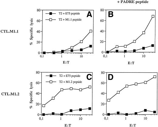
To identify peptides with a high probability of HLA-A2 binding, we performed a computer analysis of the amino acid sequence of the MUC1 protein to screen for peptides with HLA-A2–binding motifs.34,35 Two predicted MUC1-derived peptides M1.1 and M1.2 were synthesized and used for CTL induction in vitro using DC as APC. As shown in Fig 1, CTL lines obtained after 3 weekly restimulations demonstrated peptide specific killing. T cells only exhibited a cytotoxic response against T2 cells coated with the cognate peptide, whereas they did not lyse T2 cells coated with an irrelevant HER-2/neu–derived peptide E75. CTL induced in presence of the Pan-DR binding peptide PADRE39 elicited a higher cytotoxic activity (Fig 1), demonstrating the importance of CD4 cells for the induction of a efficient CTL response. In line with these results, higher concentrations of IL-12, TNF-α, IFN-γ, IL-6, and sIL-2R were found in supernatants from cultures containing DC pulsed with the M1.1 and the PADRE peptide (Table1). Similar results were obtained when the M1.2 peptide was used for CTL induction (data not shown).
Induction of CTL responses by peptide-pulsed DC. Adherent PBMNC were grown for 7 days in RP10 medium supplemented with GM-CSF, IL-4, and TNF-. DC pulsed with the synthetic peptides derived from the MUC1 protein (M1.1 and M1.2) were used to induce a CTL response in vitro. In addition to the MUC1 peptide DC were incubated with the Pan-DR binding peptide PADRE as a T-helper epitope. Cytotoxic activity of induced CTL was determined in a standard 51Cr-release assay using T2 cells as targets pulsed for 2 hours with 50 μg of the cognate (open symbols) or irrelevant Her-2/neu protein-derived E75 peptide (solid symbols).
Induction of CTL responses by peptide-pulsed DC. Adherent PBMNC were grown for 7 days in RP10 medium supplemented with GM-CSF, IL-4, and TNF-. DC pulsed with the synthetic peptides derived from the MUC1 protein (M1.1 and M1.2) were used to induce a CTL response in vitro. In addition to the MUC1 peptide DC were incubated with the Pan-DR binding peptide PADRE as a T-helper epitope. Cytotoxic activity of induced CTL was determined in a standard 51Cr-release assay using T2 cells as targets pulsed for 2 hours with 50 μg of the cognate (open symbols) or irrelevant Her-2/neu protein-derived E75 peptide (solid symbols).
Production of Cytokines During Induction of MUC1-Specific CTL Using Peptide-Pulsed DC
| . | No Peptide . | M1.1 Peptide . | +PADRE* . |
|---|---|---|---|
| IL-12 (pg/mL) | 43.3 ± 17 | 349 ± 140 | 786 ± 62 |
| IFN-γ (pg/mL) | 0.8 ± 1.2 | 16.8 ± 1.6 | 33 ± 9.5 |
| TNF-α (pg/mL) | 36.8 ± 5.5 | 205 ± 17 | 306 ± 21 |
| IL-10 (pg/mL) | 17.2 ± 2.2 | 24 ± 4.2 | 32 ± 1.9 |
| IL-6 (pg/mL) | 2.4 ± 1.7 | 6.5 ± 3.5 | 16 ± 4.6 |
| sIL-2R (U/mL) | 56.8 ± 8.1 | 201 ± 26 | 304 ± 39 |
For CTL induction, DC were pulsed with the synthetic MUC1 peptide M1.1 and incubated with autologous PBMNC in RP10 medium. To analyze the effect of a helper epitope on antigen-specific CTL induction, DC were further incubated with a Pan-HLA-DR binding peptide PADRE in addition to the HLA-A2–binding antigenic peptides. After 5 days of culture, cytokine concentrations in the supernatants were determined using commercially available ELISAs. Experiments were performed in triplicates and the results are expressed as the mean ± SD.
Recently, it was demonstrated that the MUC1 protein expression and secretion in cancer patients is associated with high metastatic potential and poor prognosis.41,42 In vitro studies have shown that the MUC1 protein can induce apoptosis or inhibit T-cell proliferation.43,44 The presence of purified MUC1 protein40 in the cell cultures during the primary in vitro immunization had no inhibitory effects on the induction of MUC1 peptide-specific CTL and their cytotoxic activity (data not shown).
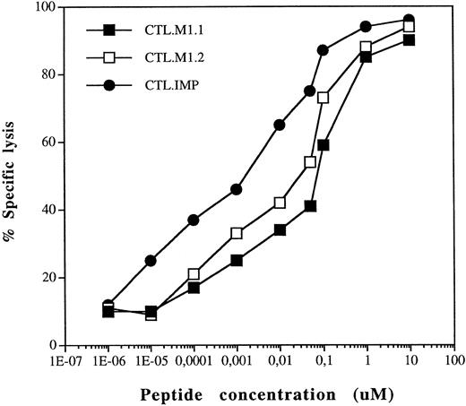
To test the sensitivity of the induced MUC1-specific CTL, T2 cells were incubated with titrated amounts of the synthetic peptides and effector cells were added after a preincubation time of 30 minutes at an E:T ratio of 20:1. As shown in Fig 2, the MUC1-specific CTL CTL.M1.1 and CTL.M1.2 lysed the target cells in an antigen concentration-depending fashion. However, the sensitivity of an in vitro-induced CTL line CTL.IMP38 specific for an influenza matrix protein-derived viral peptide IMP is about 10 times higher as compared with the CTL specific for the MUC1 self-peptides.
Peptide sensitivity of in vitro-induced MUC1-specific CTL. T2 cells were incubated with titrated amounts of the MUC1 peptides M1.1 and M1.2 as well as the influenza matrix protein peptide IMP. Corresponding specific CTL were added to the target cells incubated with the cognate peptide at a ratio of 20:1.
Peptide sensitivity of in vitro-induced MUC1-specific CTL. T2 cells were incubated with titrated amounts of the MUC1 peptides M1.1 and M1.2 as well as the influenza matrix protein peptide IMP. Corresponding specific CTL were added to the target cells incubated with the cognate peptide at a ratio of 20:1.
The lysis of allogeneic breast cancer cells by CTL specific for MUC1 is antigen-specific and HLA-A2–restricted.
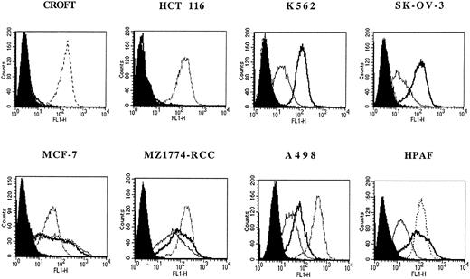
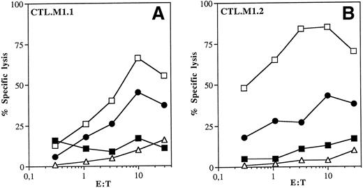
The expression of MUC1 and HLA-A2 molecules on tumor cells was determined by flow cytometry, and results are presented in Fig 3. To analyze the ability of CTL.M1.1 and CTL.M1.2 to lyse endogenously MUC1-expressing tumor cells, the MUC1-positive, HLA-A2–expressing breast cancer cell line MCF-7 was used as target cell in a standard 51Cr-release assay. As shown in Fig 4A and B, both CTL lines were able to efficiently lyse MCF-7 (HLA-A2+/MUC1+) tumor cells and peptide-pulsed Croft cells (HLA-A2+/MUC1−). There was no lysis of the ovarian cancer cells SK-OV-3 (MUC1+/HLA-A3+) or Croft cells pulsed with an irrelevant peptide E75 derived from the Her-2/neu protein. These results indicate the necessity of both the presence of MUC1 epitopes on the target cells and HLA-A2 restriction of the induced CTL. Furthermore, these data show that the two novel MUC1 peptides can be efficiently processed and presented in an HLA-restricted manner by tumor cells endogenously expressing MUC1 on the cell surface.
Flow cytometric analysis of HLA-A2 and MUC1 expression on the human tumor cell lines. The level of HLA-A2 expression (dotted line) was analyzed using a purified MoAb specific for HLA-A2, BB7.2. The MUC1 expression was determined using unlabeled antibodies HMFG-1 (bold solid line) and MAM-6 (thin solid line), followed by staining with FITC-conjugated goat antimouse antibody. Solid histograms represent isotype-matched controls.
Flow cytometric analysis of HLA-A2 and MUC1 expression on the human tumor cell lines. The level of HLA-A2 expression (dotted line) was analyzed using a purified MoAb specific for HLA-A2, BB7.2. The MUC1 expression was determined using unlabeled antibodies HMFG-1 (bold solid line) and MAM-6 (thin solid line), followed by staining with FITC-conjugated goat antimouse antibody. Solid histograms represent isotype-matched controls.
Lysis of cancer cells endogenously expressing MUC1 by CTL.M1.1 (A) and CTL.M1.2 (B). Human breast cancer cell line MCF-7 (HLA-A2+/MUC1+), ovarian cancer cell line SK-OV-3 (HLA-A2−/MUC1+), and the immortalized B-cell line Croft (HLA-A2+/MUC1−) were used as targets in a stardard 51Cr-release assay. Croft cells were pulsed with the MUC1 peptides or an irrelevant Her-2/neu–derived peptide E75. (▪) Croft + E75 peptide; (□) Croft + M1.1 peptide; (•) MCF-7; (▵) SK-OV-3.
Lysis of cancer cells endogenously expressing MUC1 by CTL.M1.1 (A) and CTL.M1.2 (B). Human breast cancer cell line MCF-7 (HLA-A2+/MUC1+), ovarian cancer cell line SK-OV-3 (HLA-A2−/MUC1+), and the immortalized B-cell line Croft (HLA-A2+/MUC1−) were used as targets in a stardard 51Cr-release assay. Croft cells were pulsed with the MUC1 peptides or an irrelevant Her-2/neu–derived peptide E75. (▪) Croft + E75 peptide; (□) Croft + M1.1 peptide; (•) MCF-7; (▵) SK-OV-3.
Lysis of RCC cells by MUC1-specific CTL.
Flow cytometric analysis of MUC1 expression of different tumor cell types using two specific MoAbs showed MUC1 expression on several tumor cell lines, including the proerythroblastic K562 cells and several RCC cell lines. We therefore analyzed the presentation of MUC1-derived peptides by these cell lines and used them as targets in a standard Cr-release assay. As shown in Fig 5A and B and Table 2, CTL.M1.1 and CTL.M1.2 did lyse tumor cell lines expressing MUC1 and HLA-A2, suggesting that the identified MUC1 peptides are presented by these tumors. In contrast, there was no lysis of MUC1-positive but HLA-negative K562 cells, demonstrating that the observed cytotoxicity was not mediated by NK cells and confirming the MHC necessity and restriction.
Lysis of renal carcinoma cells by MUC1-reactive CTL.M1.1 (A and C) and CTL.M1.2 (B and D). Human RCC cell lines A-498 (HLA-A2+/MUC1+), MZ1774-RCC (HLA-A2+/MUC1+), Caki-2 (HLA-A2−/MUC1+), ovarian cancer cell line SK-OV-3 (HLA-A2−/MUC1+), and K562 (HLA-A2−/MUC1+) were used as targets in a stardard 51Cr-release assay. The antigen specificity of the CTL lines was tested in the presence of unlabeled cold targets, T2 cells coated with the cognate, or an irrelevant peptide at an inhibitor:target ratio of 20:1 (C and D). For (A) and (B), (◊) K562; (▴) CAKI-2; (▹) SK-OV-3; (⧫) A498; (└) MZ1774-RCC. For (C) and (D), (▪) MZ1774-RCC + M1.1 peptide; (□) MZ1774-RCC; (○) MZ1774-RCC + T2-M1.1 peptide; (•) MZ1774-RCC + T2-E75 peptide.
Lysis of renal carcinoma cells by MUC1-reactive CTL.M1.1 (A and C) and CTL.M1.2 (B and D). Human RCC cell lines A-498 (HLA-A2+/MUC1+), MZ1774-RCC (HLA-A2+/MUC1+), Caki-2 (HLA-A2−/MUC1+), ovarian cancer cell line SK-OV-3 (HLA-A2−/MUC1+), and K562 (HLA-A2−/MUC1+) were used as targets in a stardard 51Cr-release assay. The antigen specificity of the CTL lines was tested in the presence of unlabeled cold targets, T2 cells coated with the cognate, or an irrelevant peptide at an inhibitor:target ratio of 20:1 (C and D). For (A) and (B), (◊) K562; (▴) CAKI-2; (▹) SK-OV-3; (⧫) A498; (└) MZ1774-RCC. For (C) and (D), (▪) MZ1774-RCC + M1.1 peptide; (□) MZ1774-RCC; (○) MZ1774-RCC + T2-M1.1 peptide; (•) MZ1774-RCC + T2-E75 peptide.
Lysis of Human Tumor Cell Lines by MUC1-Specific CTL
| Tumor Cell Lines* . | HLA-A2 Expression . | MUC1 Expression . | CTL.M1.1 % Specific Lysis at E:T . | CTL.M1.2 % Specific Lysis at E:T . | ||||
|---|---|---|---|---|---|---|---|---|
| 30 . | 10 . | 3 . | 30 . | 10 . | 3 . | |||
| MCF-7 | + | + | 39 | 30 | 16 | 58 | 41 | 23 |
| MZ1774-RCC | + | + | 38 | 33 | 18 | 43 | 18 | 12 |
| A-498 | + | + | 39 | 22 | 14 | 29 | 19 | 14 |
| NWI-RCC | + | + | 51 | 27 | 22 | 36 | 25 | 10 |
| HPAF | + | + | 36 | 24 | 16 | 41 | 28 | 11 |
| ACHN | − | + | 9 | 3 | 3 | 7 | 3 | 1 |
| Caki-2 | − | + | 5 | 5 | 2 | 3 | 1 | 0 |
| SK-OV-3 | − | + | 4 | 3 | 3 | 4 | 1 | 1 |
| HCT 116 | + | − | 9 | 5 | 0 | 7 | 3 | 4 |
| Croft | + | − | 9 | 3 | 3 | 13 | 10 | 5 |
| 3D7 | + | − | 8 | 4 | 0 | 7 | 6 | 1 |
| K562 | − | + | 9 | 3 | 2 | 6 | 3 | 3 |
The cytotoxic activity of CTL.M1.1 and CTL.M1.2 against human tumor cell lines was analyzed in a standard 51Cr-release assay.
The antigen specificity and MHC restriction mediated by the in vitro-induced CTL was confirmed in a cold target inhibition assay. The lysis of MZ1774-RCC cells could be blocked by the addition of T2 cells pulsed with the cognate peptide, whereas T2 cells pulsed with an irrelevant peptide showed no effect on cytotoxic lysis (Fig 5C and D).
Blocking of target cell lysis by MoAbs against HLA-A2, CD8, and TCR.
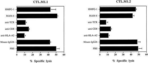
To further analyze the association of the MUC1 peptide presentation with HLA-A2, blocking experiments using a monoclonal anti–HLA-A2 antibody BB7.2 were performed. As shown in Fig 6, incubation of MZ 1774-RCC cells with the BB7.2 antibody resulted in the inhibition of the target cell lysis, whereas anti-MUC1 MoAb MAM-6, used as an isotype control (mouse IgG2b), had no effect. Incubation of the CTL effector cells with anti-CD8 or anti-TCR Abs before adding to the assay abolished their lytic activity, suggesting that the target cell lysis is HLA-A2–restricted and mediated by CD8+ T cells. Isotype-matched control Abs (MAM-6 and HMFG-1) did not block the lysis of tumor cells.
Inhibition of the cytotoxic activity of MUC1-reactive CTL.M1.1 (A) and CTL.M1.2 (B) by MoAbs. For HLA-A2 blocking experiments, target cells (MZ1774-RCC) were incubated for 30 minutes with 10 μg/mL of MoAbs (BB7.2 (IgG2b) recognizing HLA-A2. For blocking of CD8 molecules or the TCR on the effector cells, CTL were incubated with 15 μg/mL of T8 (IgG1) MoAb recognizing CD8 and anti–Pan-TCR-β BMA 031 MoAb (IgG2b) before adding to the assay. Isotype-matched antibodies (MAM-6, HMFG-1) were used as controls. The assay was performed at E:T ratio of 20:1. The data are shown as the mean and standard deviation from 6 replicate wells.
Inhibition of the cytotoxic activity of MUC1-reactive CTL.M1.1 (A) and CTL.M1.2 (B) by MoAbs. For HLA-A2 blocking experiments, target cells (MZ1774-RCC) were incubated for 30 minutes with 10 μg/mL of MoAbs (BB7.2 (IgG2b) recognizing HLA-A2. For blocking of CD8 molecules or the TCR on the effector cells, CTL were incubated with 15 μg/mL of T8 (IgG1) MoAb recognizing CD8 and anti–Pan-TCR-β BMA 031 MoAb (IgG2b) before adding to the assay. Isotype-matched antibodies (MAM-6, HMFG-1) were used as controls. The assay was performed at E:T ratio of 20:1. The data are shown as the mean and standard deviation from 6 replicate wells.
DISCUSSION
Mucins are transmembrane type I glycoproteins with a unique extracellular domain consisting mostly of 20 to 60 tandem repeats.1,2 The MUC1 protein is overexpressed on many epithelial and nonepithelial malignant cells and is therefore a suitable candidate for broadly applicable vaccine therapies.3-9 Several previous reports have demonstrated that the tandem repeat of the MUC1 protein is highly immunogenic and that both antibody-mediated and T-cell–mediated responses recognizing epitopes derived from this domain can be induced in mice and humans. It was further reported that MUC1 reactive T cells found in patients with breast cancer, pancreatic cancer, and multiple myeloma recognize directly the MUC1 molecule in an MHC-unrestricted manner.8-14,44-46 It was proposed that normally cryptic T-cell epitopes from the MUC1 core domain were recognized by MUC1-specific CTL as a result of underglycosylation in tumor cells and cross-linking of the T-cell receptor by the highly multivalent epitopes of tandemly repeated 20 amino acid peptides on a single MUC1 molecule. However, there is now increasing evidence from mouse and human studies indicating that T cells induced against the MUC1 protein can be MHC-restricted. In a recently published study using HLA-A0201/Kb transgenic mice, two HLA-A2–binding peptides were identified. Interestingly, one of these peptides was also shown to induce HLA-A11–restricted CTL in humans.15-19
Using a computer-assisted analysis, we screened the MUC1 protein for HLA-A2–binding peptides.34,35 Using this approach, we were able to identify two novel 9-mer peptides, M1.1 and M1.2, with a high binding probability to HLA-A2. The M1.1 peptide is derived from the VNTR domain of the MUC1 protein. There are 3 of 44 tandem repeats that differ in their amino acid sequence from the PDTRPAPGSTAPPAHGVTSA sequence. The M1.1 peptide is localized in the last VNTR (amino acids 950-958: STAPPVHNV) and varies in two positions from the previously described STAPPAHGV peptide.15,18 The presence of the V substitution in position 6 increases the binding of the M1.1 peptide to the HLA-A2 molecule. To analyze whether these epitopes are presented by tumor cells endogenously expressing MUC1, we induced MUC1 peptide-specific CTL responses by primary in vitro immunization and used these CTL to determine the presentation of MUC1 epitopes on human tumor lines. Autologous DC generated from peripheral blood monocytes in the presence of GM-CSF, IL-4, and TNF-α were pulsed with the identified peptides M1.1 and M1.2 and used as APC for CTL priming. DC used here demonstrated a high ability to initiate an MUC1-specific CTL response. The antigen-specific cytotoxic activity of the induced CTL could be further increased by the addition of a T-helper epitope, the Pan-HLA-DR binding peptide PADRE.39This finding could be of potential importance for the developing of cancer vaccination protocols.
In a previous report,47 it was demonstrated that a strong antigen-specific response could be induced in vitro when human peripheral blood lymphocytes (PBL) pulsed with a liposome-encapsulated MUC1 peptide consisting of 25 amino acids from the VNTR were used for CTL induction. Proliferative and cytotoxic T-cell responses as well as the secretion of IFN-γ were already detected after 2 weekly restimulations, comparable to the results obtained in our study and demonstrating that this approach could be effective and feasible for the development of cancer vaccines.
In our study, we show that MUC1 peptide-specific CTL were able to recognize tumor cells endogenously expressing the MUC1 protein in an HLA-A2–restricted manner. Furthermore, the MUC1-specific CTL lysed not only breast cancer cells, but also pancreatic and RCC cell lines expressing MUC1 and HLA-A2. The cytotoxicity against tumor cells was blocked by cold HLA-A2+ targets pulsed with the cognate peptide in a cold-target inhibition assay and by anti–HLA-A2 MoAb. Our results extend the list of epithelial tumors that present MUC1-derived T-cell epitopes that increases the possible clinical use of peptide-pulsed DC as a cancer vaccine.
Of particular interest is the finding that the M1.2 peptide is derived from the signal sequence of MUC1, showing that the T-cell–mediated immune responses to the MUC1 protein are not restricted to the tandem repeat domain. Furthermore, the M1.2 peptide could be presented in absence of a functional TAP by malignant cells. It was recently shown that proteolysis of peptides derived from the signal sequence in the endoplasmatic reticulum represent a second pathway for the generation of class I MHC-associated peptides and that this peptides are presented independent of TAP.48 49
Recently, it was demonstrated that the MUC1 protein expression and secretion in cancer patients is associated with high metastatic potential and poor prognosis.41,42 In vitro studies have shown that the MUC1 protein can induce apoptosis or inhibit T-cell proliferation.43,44 The growth inhibition was mediated by the whole MUC1 protein or large synthetic tandem repeats of the MUC1 core peptide and was reversible by addition of IL-2, anti-CD28 MoAb, or short 16-amino acid MUC1 peptide,44 indicating that active specific immunotherapy using short synthetic peptides in combination with professional APC and/or IL-2 may overcome the observed immunosuppression in cancer patients. In MUC1 transgenic mice, the tolerance to human MUC1 antigen could be reversed by immunizing the animals with fusions of DC- and MUC1-expressing tumors.50Interestingly, the presence of purified MUC1 protein in the cell cultures during primary in vitro immunization with peptide-pulsed DC did not inhibited the induction of MUC1 peptides-specific CTL responses or the cytotoxic activity elicited by these CTL (data not shown).
Because protocols for maintenance and expansion of human DC generated from bone marrow-derived progenitors or peripheral blood monocytes have recently been established,38,39,51-55 it is now possible to generate sufficient numbers of DC from patients and apply them in vaccination therapies. Vaccination with DC pulsed with antigenic peptides was shown to be effective in patients with malignant lymphoma and melanoma.56 57 The use of DC- and MUC1-derived peptides could reverse the observed immunosuppression and MUC1 tolerance in cancer patients and provide an additional broadly applicable approach to established therapies of hematologic and epithelial malignancies, such as multiple myeloma and renal cell, breast, and pancreatic carcinoma.
ACKNOWLEDGMENT
We thank S. Kurtz and Y. Hoffmann for their excellent technical assistance.
Supported in part by grants from Deutsche Forschungsgemeinschaft (SFB 510) and Deutsche Krebshilfe.
The publication costs of this article were defrayed in part by page charge payment. This article must therefore be hereby marked “advertisement” in accordance with 18 U.S.C. section 1734 solely to indicate this fact.
REFERENCES
Author notes
Address reprint requests to Wolfram Brugger, MD, Department of Hematology, Oncology, Rheumatology and Immunology, University of Tübingen, Otfried-Müller-Strasse-10, D-72076 Tübingen, Germany.


This feature is available to Subscribers Only
Sign In or Create an Account Close Modal