Abstract
Early intensification of chemotherapy with high-dose cytarabine either in the postremission or remission induction phase has recently been shown to improve long-term relapse-free survival (RFS) in patients with acute myeloid leukemia (AML). Comparable results have been produced with the double induction strategy. The present trial evaluated the contribution of high-dose versus standard-dose cytarabine to this strategy. Between March 1985 and November 1992, 725 eligible patients 16 to 60 years of age with newly diagnosed primary AML entered the trial. Before treatment started, patients were randomized between two versions of double induction: 2 courses of standard-dose cytarabine (ara-C) with daunorubicin and 6-thioguanine (TAD) were compared with 1 course of TAD followed by high-dose cytarabine (3 g/m2every 12 hours for 6 times) with mitoxantrone (HAM). Second courses started on day 21 before remission criteria were reached, regardless of the presence or absence of blast cells in the bone marrow. Patients in remission received consolidation by TAD and monthly maintenance with reduced TAD courses for 3 years. The complete remission (CR) rate in the TAD-TAD compared with the TAD-HAM arm was 65% versus 71% (not significant [NS]), and the early and hypoplastic death rate was 18% versus 14% (NS). The corresponding RFS after 5 years was 29% versus 35% (NS). An explorative analysis identified a subgroup of 286 patients with a poor prognosis representing 39% of the entire population; they included patients with more than 40% residual blasts in the day-16 bone marrow, patients with unfavorable karyotype, and those with high levels of serum lactate dehydrogenase. Their CR rate was 65% versus 49% (p = .004) in favor of TAD-HAM and was associated with a superior event-free survival (median, 7 v 3 months; 5 years, 17% v 12%; P = .012) and overall survival (median, 13 v 8 months; 5 years, 24% v 18%;P = .009). This suggests that the incorporation of high-dose cytarabine with mitoxantrone may contribute a specific benefit to poor-risk patients that, however, requires further substantiation. Double induction, followed by consolidation and maintenance, proved a safe and effective strategy and a new way of delivering early intensification treatment for AML.
RESULTS OF LARGE scale therapeutic trials in acute myeloid leukemia (AML) reported during the 1980s and 1990s have shown a gradual improvement in the results of chemotherapy. During this period of time, an increasing majority of patients have achieved complete remission (CR)1-6 and the proportion of patients remaining in permanent remission (still a minority) has also improved.1-3,5,7,8 When remission induction had reached some standard dose levels,1 further increase in the remission rate appeared associated with progress in supportive care rather than intensification in antileukemic treatment.
However, in contrast with remission rates, chemotherapy dose effects have been found in the duration of remissions and relapse-free survival (RFS). This was first shown for the effect of prolonged myelosuppressive maintenance treatment after 1 course of consolidation at standard induction doses. Patients assigned to maintenance had a markedly improved RFS compared with patients assigned to consolidation alone.2 Similarly, dose effects could be demonstrated for the immediate postremission phase, in that 4 courses of cytarabine at either 3 g/m2 or 400 mg/m2 or 100 mg/m2 resulted in a dose-dependent RFS.5Furthermore, it has been demonstrated that, after 4 courses of induction/consolidation chemotherapy, the addition of myeloablative chemotherapy plus total body irradiation and autologous bone marrow transplantation substantially improved RFS when compared with no further treatment.9 More recently, it has been shown that the dosage of cytarabine, even when included from the beginning of the induction treatment, clearly affected RFS,8,10 with a probability of 41% at 5 years when cytarabine, at a dose of 3 g/m2, was incorporated in the protocol.8 The present trial now addressed the question of the dose effect of induction treatment by comparing a regimen containing high-dose cytarabine with a regimen containing the drug at a standard dose, both included in the second courses of double induction. The response nonadapted double induction strategy provides an ideal basis for the comparison of dose effects, because the amount of induction treatment depends only on the randomization and not on an individualized number of induction courses. Although little is known of which subtypes of AML may benefit from intensification strategies, the curative impact of postremission high-dose cytarabine appeared to be restricted to favorable and intermediate karyotype abnormalities in one study.11 The effect of intensification of induction therapy on the outcome in special prognostic groups has been analyzed exploratively in this trial.
MATERIALS AND METHODS
Patients.
Patients 16 to 60 years of age with AML according to the French-American-British (FAB) classification12,13 who had never received antileukemic therapy were eligible. In common with other comparable trials5,6 8 and to optimize homogeneity, patients with a history of myelodysplasia or other antecedent hematologic disorder or who had previous exposure to cytotoxic drugs or radiotherapy were excluded, as were patients with pre-existing non–leukemia-related liver disease or renal or heart failure. Written informed consent was obtained before a patient entered the study.
Study design.
Before treatment started, all patients were randomized, by a phone call to the statistical center, between one of the two induction therapy arms. All patients then received the first induction course, consisting of 100 mg/m2 cytarabine by continuous intravenous infusion daily on days 1 and 2 and subsequently by infusion over 30 minutes every 12 hours on days 3 through 8; 60 mg/m2 daunorubicin by 30 minutes of intravenous infusion on days 3, 4, and 5; and 6-thioguanine 100 mg/m2 orally every 12 hours on days 3 through 9 (TAD).14 On day 16 of therapy, the bone marrow was examined for the percentage of blast cells. On day 21, all patients received a second induction course (double induction). According to the randomization, the second course was either 3 g/m2cytarabine by 3 hours of intravenous infusion every 12 hours on days 1 through 3 with 10 mg/m2 mitoxantrone by 30 minutes of intravenous infusion on days 3, 4, and 5 (HAM)15 or a repetition of the first TAD induction course. If after the second course the bone marrow contained ≥5% blasts or similar features reappeared in weekly bone marrow sampling, the patient was treated off study. Patients who went into CR received a consolidation course of TAD. After consolidation, maintenance treatment was administered to all patients and consisted of monthly courses of 100 mg/m2cytarabine by subcutaneous injection every 12 hours for 5 days, with a second drug being administered in rotation, including 45 mg/m2 daunorubicin by 30 minutes of intravenous infusion on days 3 and 4 (course 1), 100 mg/m2 6-thioguanine orally every 12 hours on days 1 through 5 (course 2), 1 g/m2cyclophosphamide intravenous injection on day 3 (course 3), or again 6-thioguanine (course 4) and restarting with daunorubicin (course 5). If absolute neutrophil counts decreased to less than 500/μL and/or platelets to less than 20,000/μL after 2 sequential courses, the doses of all antileukemic drugs were reduced to 50%, permanently. Using this policy it was found that from the third maintenance course, the vast majority of patients required adjustment and continued at 50% of full dosage. Maintenance treatment continued until the patient was 3 years in remission.2 As an alternative to maintenance chemotherapy, allogeneic bone marrow transplantation in first remission was offered to all patients up to 50 years of age who had a histocompatible sibling.
Evaluation.
Patients underwent full physical examinations and assessment of blood counts and liver and renal function tests before each maintenance course. Bone marrow examinations were performed before alternating maintenance courses and every 3 months after the end of maintenance, unless earlier bone marrow examinations were indicated by peripheral blood changes inconsistent with CR.
Criteria for response.
A CR was defined by a bone marrow with normal hematopoieses of all cell lines, less than 5% blast cells, and a peripheral blood with at least 1,500 neutrophils and 100,000 platelets/μL. Therapeutic failures were classified as persistent leukemia, death less than 7 days after completion of the first induction therapy course (early death) and death during treatment-induced bone marrow hypoplasia, irrespective of the time after chemotherapy (hypoplastic death). Relapse was defined as reinfiltration of the bone marrow by 25% or more leukemic blasts5 or a proven leukemic infiltration at any other site. Relapse-free interval was measured from the achievement of CR until relapse and RFS from CR until relapse or death in remission. Survival was recorded from randomization until death. Event-free survival was recorded from randomization until nonachievement of remission, relapse, or death. Analyses considering day-16 bone marrow blasts began from the date when this marrow was collected. Patients receiving bone marrow transplantation were censored at the time of transplantation.
Cytogenetics.
Cytogenetic examination was performed on pretreatment bone marrow specimens. Chromosome analysis was performed after short-term cultures using standard protocols for G- or R-banding techniques. Karyotype changes were interpreted according to the 1995 ISCN nomenclature.16 All cytogenetic results were centrally reviewed by the study reference laboratory.
Statistical analysis.
The primary objective of the study was the randomized comparison of the two versions of double induction TAD-TAD and TAD-HAM with respect to event-free survival. The size of the study was based on power calculations. The type I error was fixed at α = .05 and the median event-free survival was expected to be at least 7 months. Patient accrual should last at least 3 years. The participating centers expected about 100 randomizations per year. The follow up period was set at a minimum of 2 years. Thus, the study had a power of about 0.8 to detect a minimum difference of 3 months in median event-free survival.
After the target number of 300 patients had been exceeded, it was decided to extend this number substantially. The main reason for this decision was the increasing availability of cytogenetics for the study and the new evidence on the impact of the karyotype on patients’ outcome. Thus, further substantiation of the role of cytogenetic changes was incorporated as an objective of the trial.
Comparison of the rates of CR and failures was evaluated by Pearson’s χ2 test. Distributions of time to event variables were estimated by the Kaplan-Meier method,17 and comparisons were based on the log-rank-test.18 All P values reported are two-sided. Potential prognostic factors were tested by multiple regression analysis using logistic regression for response to induction treatment and Cox proportional hazard model19 for RFS and overall survival. Randomization was stratified by center, which was ignored in the statistical analyses. There was no further stratification.
RESULTS
Patient population.
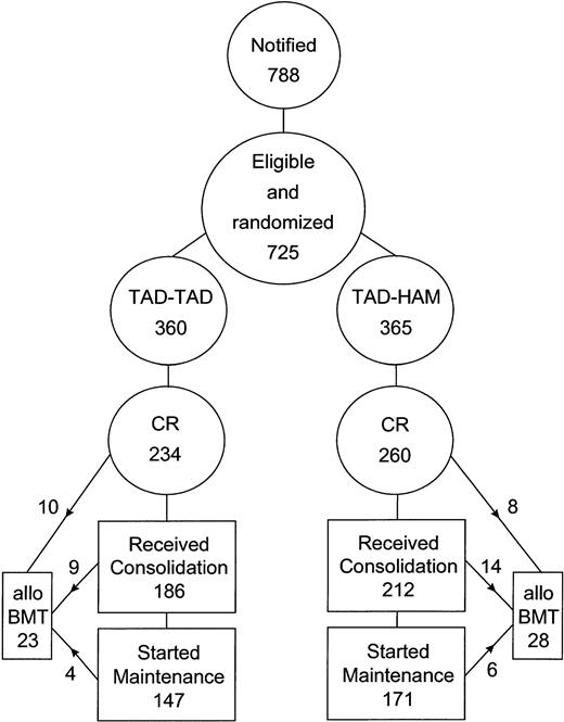
Between March 1985 and November 1992, a total of 788 patients from 45 participating institutions entered the trial. Sixty-three patients were excluded according to protocol criteria, including medical contraindications to intensive chemotherapy in 46 patients, missing consent of 10 patients, and protocol violation, mainly through nonrandomized treatment, in 7 patients. A total of 725 patients were eligible and randomized. Patient numbers evaluable according to the treatment groups are shown in Fig 1.
Flow diagram showing evaluable patient numbers according to treatment arms and numbers of patients receiving the assigned treatment.
Flow diagram showing evaluable patient numbers according to treatment arms and numbers of patients receiving the assigned treatment.
Patient characteristics.
Table 1 shows the pretreatment characteristics of patients in the two randomized arms. Cytogenetics of the bone marrow cells were obtained from 47% of all patients. Karyotypes classified as favorable included translocations t(15;17), t(8;21), and inversion 16, whereas deletions and losses of chromosomes 5 and 7, abnormalities involving 11q23, and complex karyotype with three or more numerical or structural abnormalities were considered unfavorable. This karyotype classification was similar to that used in other large multicenter series.11 20
Patient Characteristics by Randomization
| . | TAD-TAD . | TAD-HAM . |
|---|---|---|
| No. of patients | 360 | 365 |
| Median age (range) | 44 (16-60) | 44 (16-60) |
| Female/Male | 193/167 | 196/169 |
| Median WBC/μL ×103 (range) | 17.2 (0.1-405) | 21.0 (0.5-331) |
| Median LDH (U/L; range) | 400 (90-3,600) | 443 (102-2,868) |
| FAB subtypes (% of patients) | ||
| M0 | 0 | 1 |
| M1 | 16 | 17 |
| M2 | 26 | 33 |
| M3 | 7 | 5 |
| M4 total (M4Eo) | 34 (7) | 29 (6) |
| M5 | 12 | 11 |
| M6 | 4 | 3 |
| M7 | 1 | 1 |
| Cytogenetics (no. available) | 172 | 171 |
| Different karyotypes in percentages | ||
| t (15; 17) | 6 | 5 |
| t (8;21) | 6 | 10 |
| inv (16) | 8 | 9 |
| −5/5q− | 3 | 1 |
| −7/7q− | 4 | 3 |
| Abnormal 11q23 | 1 | 4 |
| Complex karyotype | 8 | 4 |
| Other abnormal karyotype | 11 | 18 |
| Normal karyotype | 53 | 46 |
| Cytogenetics (no. not available) | 188 | 194 |
Drug delivery.
Double induction with both courses was administered to 665 of the entire 725 patients (91%), with 322 patients (89%) in the TAD-TAD arm and 340 patients (93%) in the TAD-HAM arm. On 37 and 51 occasions, in the TAD-TAD and TAD-HAM sequence, respectively, second courses were postponed to the postremission period as additional consolidation courses, following protocol guidelines to avoid excessive toxicity. Thus, 79% of the patients in both arms received double induction as was planned to be administered before CR criteria were achieved. The remaining 63 (9%) patients only received 1 induction course due to early death (8%) or contraindications. Among the 494 patients going into CR (234 in the TAD-TAD arm and 260 in the TAD-HAM arm), 186 and 212 patients, respectively, went on to consolidation. The reasons for not receiving consolidation in the TAD-TAD arm and in the TAD-HAM arm were early relapse in 10 and 4 patients, respectively; early death in remission in 1 patient in each group; toxicity in induction treatment in 17 and 28 patients, respectively; refusal of consolidation by 10 and 7 patients, respectively; and planned allogeneic bone marrow transplantation in 10 and 8 patients, respectively. Maintenance treatment was started in 147 patients in the TAD-TAD arm and in 171 patients in the TAD-HAM arm. The reasons for not administering maintenance were death in remission in 3 and 8 patients, respectively; toxicity in consolidation in 7 and 8 patients, respectively; refusal of maintenance by 11 and 4 patients, respectively; relapse in 9 and 7 patients, respectively; and planned allogeneic bone marrow transplantation in 9 and 14 patients, respectively. Twenty-three and 28 patients went to allogeneic bone marrow transplantation, respectively: 10 and 8 of them without and 9 and 14 after having received consolidation. Another 4 and 6 patients went to transplantation after having received maintenance courses. For patient assignment and flow, see Fig 1.
Therapeutic outcome by treatment.
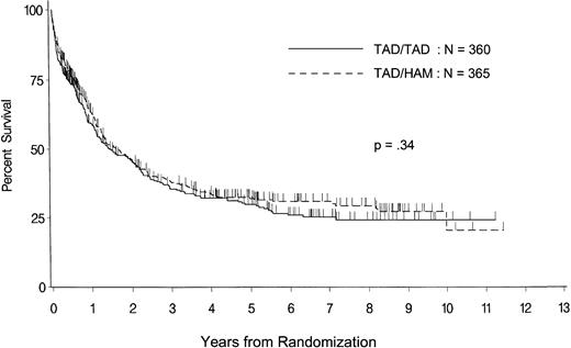
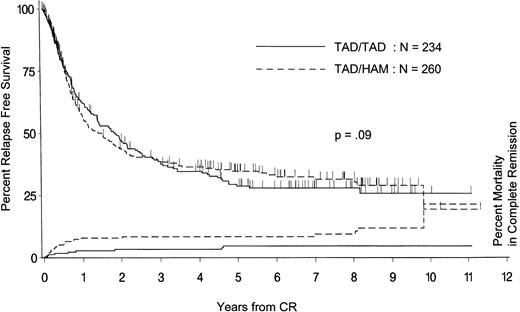
The essential data on patients’ outcome are listed in Table 2 for the total population and for the two randomized treatment arms. The median observation time for survival and remaining in remission is 6 years. Kaplan Meier life table plots for all randomized patients are shown for overall survival in Fig 2 and for RFS and mortality in remission in Fig 3. Among the 360 and 365 patients assigned to TAD-TAD and TAD-HAM double induction, respectively, 228 and 218 have died. Among the 234 and 260 patients going into remission, respectively, 131 and 127 relapsed and another 9 and 24 patients died in remission.
Treatment Outcome by Randomization
| . | Total . | TAD-TAD . | TAD-HAM . | P . |
|---|---|---|---|---|
| Patients randomized | 725 | 360 | 365 | |
| CR (%) | 68 (64-72) | 65 (59-70) | 71 (66-76) | .072 |
| Persistent leukemia (%) | 16 (13-19) | 17 (13-22) | 15 (11-20) | .491 |
| Early and hypoplastic death (%) | 16 (13-19) | 18 (14-23) | 14 (10-18) | .108 |
| Event-free survival | ||||
| Median (mo) | 9 (7.5-11.5) | 9 (6-12) | 10 (8-12) | .208 |
| 5 yrs (%) | 22 (18-26) | 19 (14-24) | 25 (19-30) | |
| Overall survival | ||||
| Median (mo) | 19 (15.5-24) | 18 (13.5-25) | 20 (14.5-25) | .338 |
| 5 yrs (%) | 31 (27-35) | 30 (24-36) | 32 (26-38) | |
| Patients with CR | 494 | 234 | 260 | |
| RFS | ||||
| Median (mo) | 20 (15-24) | 23 (16.5-30) | 18 (12-24) | .897 |
| 5 yrs (%) | 32 (27-37) | 29 (22-36) | 35 (28-42) | |
| Responders’ survival | ||||
| Median (mo) | 36 (29-47.5) | 38 (27.5-58.5) | 35 (25-49) | .640 |
| 5 yrs (%) | 42 (36-47) | 42 (34-50) | 41 (34-49) |
In parentheses are 95% CI.
Overall survival from randomization for all patients entering the trial in the two randomized treatment arms. Tick marks indicate patients alive and patients censored at the time of allogeneic bone marrow transplantation.
Overall survival from randomization for all patients entering the trial in the two randomized treatment arms. Tick marks indicate patients alive and patients censored at the time of allogeneic bone marrow transplantation.
RFS from achievement of remission and mortality in remission for the two randomized treatment arms. Tick marks indicate patients alive and in remission and patients censored at the time of allogeneic bone marrow transplantation.
RFS from achievement of remission and mortality in remission for the two randomized treatment arms. Tick marks indicate patients alive and in remission and patients censored at the time of allogeneic bone marrow transplantation.
Toxicity.
Toxicity and adverse events during the period of induction and consolidation treatment were classified in the two arms according to the World Health Organization criteria and are listed for grades 3 and 4 in Table 3. No significant difference was found for any kind of adverse events. Myelotoxicity was measured by the recovery time of blood neutrophils and platelets from the end of the second induction course until 500/μL absolute neutrophils and 100,000/μL platelets. Median time to recovery for those who recovered was 16 days (range, 15 to 17 days) in the TAD-TAD arm and 20 days (range, 19 to 21 days) in the TAD-HAM arm (P = .0001). Nineteen percent of patients in the TAD-TAD arm and 16.5% in the TAD-HAM arm did not fulfill criteria of recovery.
Adverse Events WHO Grades 3 and 4 During Induction and Consolidation
| Events . | Induction . | Consolidation . | ||
|---|---|---|---|---|
| TAD-TAD . | TAD-HAM . | TAD-TAD . | TAD-HAM . | |
| Nausea/vomiting | 17 (13-21) | 22 (18-27) | 16 (11-22) | 9 (5-13) |
| Stomatitis | 10 (7-14) | 9 (6-12) | 1 (0-4) | 3 (1-6) |
| Diarrhea | 12 (9-16) | 10 (7-14) | 3 (1-7) | 4 (2-8) |
| Hemorrhage | 11 (8-15) | 10 (7-14) | 2 (1-5) | 4 (2-8) |
| Infection | 33 (28-38) | 40 (35-45) | 11 (7-16) | 11 (6-15) |
| Cardiac events | 8 (5-11) | 6 (4-9) | 2 (1-5) | 1 (0-3) |
| CNS toxicity | 1 (0-3) | 2 (1-4) | 0 (0-2) | 1 (0-3) |
Values are percentages. In parentheses are 95% CI.
Prognostic factors.
The multiple regression analysis of potential prognostic factors predictive for achieving CR included initial white blood cell count (WBC), lactate dehydrogenase (LDH) in serum, karyotype, and FAB subtype. Independent prognostic factors were FAB-M4Eo (odds ratio, 2.62; 95% confidence interval [CI], 1.21 to 5.67) and unfavorable karyotype (odds ratio, 0.47; 95% CI, 0.26 to 0.87). In addition to these potential prognostic factors, the percentage of residual bone marrow blasts on day 16 of treatment was also analyzed for its impact on RFS and overall survival. Factors found to be independently predictive are listed in Table4. LDH values have been available in the great majority of patients in this trial. Homogeneity testing did not detect any disparity at the 5% level between the centers. To define a poor prognostic group according to LDH, we selected patients with greater than 700 U/L, representing the upper quartile of the population, with the cut-off point being approximately 3 times the upper limit of normal. Although karyotype was available from only 47% of the patients, availability did not cause differences in the results between patients or centers. Patients in whom karyotype was missing showed average results for response and long-term outcome.
Prognostic Factors Predicting Duration of RFS and Overall Survival as Resulting From Multiple Regression Analysis Using Cox and Regression
| Variable . | P Value . | Risk Ratio . | 95% CI . |
|---|---|---|---|
| RFS | |||
| Unfavorable karyotype | .0001 | 3.15 | 1.77-5.60 |
| Day-16 blasts | .0001 | 1.014 | 1.007-1.020 |
| FAB M3 | .0126 | 0.43 | 0.22-0.84 |
| LDH | .020 | 1.00039 | 1.00006-1.00073 |
| Overall survival | |||
| Unfavorable karyotype | .0001 | 2.32 | 1.55-3.48 |
| Day-16 blasts | .0001 | 1.018 | 1.014-1.022 |
| FAB M3 | .006 | 0.48 | 0.28-0.81 |
| LDH | .048 | 1.00025 | 1.000-1.00051 |
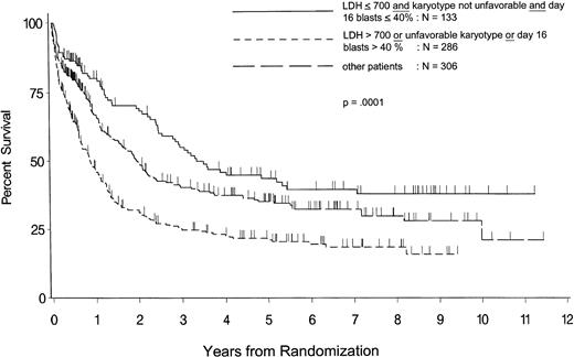
The impact of poor prognostic factors such as a high LDH (>700 U/L), unfavorable karyotype, or day-16 bone marrow blasts greater than 40% on overall survival is shown in Fig 4. Similar effects were seen on event-free survival (P = . 0001), survival of responders (P = .0096), and relapse-free interval (P = .0004). Of the 286 patients representing the poor prognostic group, 140 were poor risk for LDH alone, 81 for day-16 blasts alone, 26 for karyotype alone, 20 for both LDH and blasts, 7 for LDH and karyotype, 9 for blasts and karyotype, and 3 for all three features.
Overall survival from randomization of all eligible and evaluable patients in three risk groups according to initial LDH, unfavorable karyotype, and day-16 bone marrow blasts. “Other patients” include the rest of the patients whose risk is not defined by the criteria listed above. Tick marks indicate patients alive and patients censored at the time of allogeneic bone marrow tranplantation.
Overall survival from randomization of all eligible and evaluable patients in three risk groups according to initial LDH, unfavorable karyotype, and day-16 bone marrow blasts. “Other patients” include the rest of the patients whose risk is not defined by the criteria listed above. Tick marks indicate patients alive and patients censored at the time of allogeneic bone marrow tranplantation.
Outcome by treatment in prognostic groups.
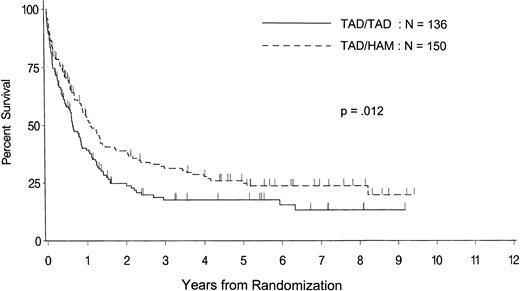
Table 5 shows the therapeutic outcome between the two treatment arms in patients exhibiting poor-risk features such as a high LDH, an unfavorable karyotype, or a high percentage of day 16-bone marrow blasts compared with patients having none of the three criteria and with the rest of the patients whose risk remained undefined by these criteria. Within the poor-risk patients, there were no differences between the two treatment arms in the distribution of FAB types or in the mean age, WBC counts, LDH, and day-16 blasts, respectively. Overall survival by treatment arm is shown in Fig 5 for poor risk according to LDH or karyotype or day 16 blasts. In keeping with the entire group of patients with poor prognosis, high-dose cytarabine in double induction was also superior for each of the single poor-risk features. Thus, in the high LDH population, a superior remission rate (P = .031), event-free survival (P = .036), and overall survival (P = .036) was achieved and again in those with an unfavorable karyotype (P = .011, .053, and .090, respectively) and (at least for the remission rate; P = .028) in the day-16 blasts greater than 40% subgroup.
Treatment Outcome by Randomization and Prognostic Factors
| . | LDH >700 U or Unfavorable Karyotype or Day-16 Blasts >40% . | LDH ≤700 U and Not-Unfavorable Karyotype and Day-16 Blasts ≤40% . | Other Patients . | ||||||
|---|---|---|---|---|---|---|---|---|---|
| TAD-TAD . | TAD-HAM . | P . | TAD-TAD . | TAD-HAM . | P . | TAD-TAD . | TAD-HAM . | P . | |
| No. of patients | 136 | 150 | 70 | 63 | 154 | 152 | |||
| CR | 49 (40-58) | 65 (56-73) | .004 | 81 (70-90) | 78 (65-88) | NS | 72 (64-79) | 74 (65-81) | NS |
| Event-free survival5-150 | .0125 | NS | NS | ||||||
| Prob. 5 yrs | 12 (6-18) | 17 (10-24) | 34 (22-47) | 34 (21-48) | 19 (11-26) | 29 (21-36) | |||
| Overall survival5-150 | .0118 | NS | NS | ||||||
| Prob. 5 yrs | 18 (10-25) | 25 (17-32) | 46 (32-59) | 41 (27-56) | 35 (25-44) | 36 (26-44) | |||
| RFS5-150 | NS | NS | NS | ||||||
| Prob. 5 yrs | 25 (13-37) | 26 (16-36) | 40 (25-54) | 44 (28-56) | 26 (16-35) | 38 (28-48) | |||
Values are percentages. In parentheses are 95% CI.
Abbreviation: NS, not significant.
From the date of the day-16 blast count.
Overall survival from randomization in the two randomized treatment arms for all patients entering the trial and showing poor risk according to LDH, karyotype, or day-16 bone marrow blasts. Tick marks indicate patients alive and patients censored at the time of allogeneic bone marrow transplantation.
Overall survival from randomization in the two randomized treatment arms for all patients entering the trial and showing poor risk according to LDH, karyotype, or day-16 bone marrow blasts. Tick marks indicate patients alive and patients censored at the time of allogeneic bone marrow transplantation.
DISCUSSION
In the present trial, starting in March 1985, the German AML Cooperative Group investigated the effects of intensification of induction treatment in patients 16 to 60 years of age. Intensification was approached in two ways: (1) by the introduction of double induction and (2) by the incorporation of high-dose cytarabine into induction treatment. Double induction is a strategy of very early intensification by starting a second induction course on day 21 of treatment irrespective of the presence or absence of residual leukemic blasts in the bone marrow after the first induction course. One of the two aims of this strategy was to administer a second course immediately to patients who would not have attained CR with 1 course alone. The second aim is to administer additional antileukemic treatment to patients who would not receive a second induction course if the normal convention for the majority of patients was followed. The enhanced cytotoxic activity provided by the second course aims at further reducing minimal residual disease to improve the long-term outcome of patients. At the start of the second course on day 21, CR by any criteria has not yet been reached. Thus, the routine second course becomes a part of induction treatment by definition. In contrast to conventional induction therapy,5,6,8,10,21 22 in which decisions on additional induction courses are made individually, double induction introduces a more standardized approach that achieves greater homogeneity in the quantity of induction treatment. Thus, double induction provides a basis for the second approach, namely the incorporation of high-dose cytarabine into induction treatment as a component of the second course by randomization.
Double induction in the present trial proved a useful and safe strategy for the study population of patients up to 60 years of age. It was found helpful that the decision about a second induction course was not individualized and that extensions of the risk period by postponement of this decision were largely avoided. This may explain why the combined early and hypoplastic death rate of 16%, which includes death in hypoplasia as late as 100 days from the start of double induction, remains similar to the 13%5 and 15%8 values obtained in recent reports from non–double induction regimens. However, in comparing treatment-related mortality, some differences in the definitions cannot be excluded; other publications may not even be comparable, because different definitions are used or data are lacking.6,10,21,22 As far as response is concerned, the 68% CRs from double induction compares with the highest values of 71%5 and 72%8 and even with younger populations in which 66%,6 58%,10 and 70%22 CR rates have been reported using similar criteria.
Because double induction fulfilled safety and efficacy standards in the remission induction phase, the question arises as to how this kind of very early intensification affected the long-term outcome of patients. In its original form, only using standard-dose cytarabine, double induction followed by standard consolidation and maintenance can be compared with the historical control of the preceding trial of the AML Cooperative Group, in which the same chemotherapy courses were used in a non–double induction fashion.2 With a median of 23 versus 14 months and 29% versus 22% at 5 years (P = .091), the RFS shows a tendency in favor of double induction. This finding has to be interpreted with caution, because improvements in supportive care may have contributed to the improved results, although they should have affected response rates rather than long-term outcome.
The contribution of high-dose cytarabine to the double induction effect has been investigated in the present trial by randomization for the second induction course between standard-dose cytarabine with daunorubicin and 6-thioguanine (TAD) or high-dose cytarabine with mitoxantrone (HAM). High-dose cytarabine, as in HAM adminstered in the present trial at 3 g/m2 twice daily on 3 days, has also been used recently for the intensification of postremission treatment.5 High-dose cytarabine in this dose range previously had been found successful in refractory AML.23-27 A single drug salvage effect had also been shown for mitoxantrone.28-30 These experiences led to the combination of high-dose cytarabine with mitoxantrone in the HAM regimen that, by its special timing, also uses a conditioning effect of cytarabine on subsequently administered anthracyclines.31The HAM regimen proved highly effective by inducing 53% CR in patients with refractory AML by rigid criteria.15When incorporated into first-line induction therapy as the second course, HAM provides two new components: (1) high-dose cytarabine at a dosage 12.9 times as high as the standard-dose cytarabine included in the preceding TAD induction course, which represents an intensification that potentially overcomes cellular resistance to standard-dose cytarabine32-34; and (2) mitoxantrone as a non–cross-resistant drug replacing the daunorubicin administered in the first course.
Potentiation of the antileukemic effect by HAM was not confirmed in the present trial for the entire target group of patients randomized. Whereas the CR rate was insignificantly higher in the HAM arm than in the standard-dose arm (71% v 65%), the long-term outcome was almost identical in the two arms. When applying explorative subgroup analyses, HAM appeared superior by producing significantly higher remission rates in special poor-risk groups such as patients with more than 40% residual blasts on day 16, patients with unfavorable karyotype, and patients with highly elevated LDH. In the entire group of 286 patients with poor-risk disease, TAD-HAM double induction resulted in 65% CR versus 49% in the TAD-TAD arm (P = .004). This also extended to a superior overall survival (P= .009) and event-free survival (P = .012). These data are in contrast to two other reports on subgroup analyses in which the curative effects of postremission high-dose cytarabine chemotherapy11 and of autologous bone marrow transplantation9 were associated with favorable and not with unfavorable karyotypes.9,11 The different timing of intensification between the induction phase of this trial and the postremission phase of others9,11 may partly account for the conflicting results. Furthermore, the endpoints were different; we have shown significant improvement in response, survival, and event-free survival in contrast to RFS in the other studies.9 11 Our data therefore suggest that a patient with poor prognostic criteria may have an improved outlook after very early intensification treatment.
An unfavorable karyotype and a highly elevated LDH in serum were the two most important independent poor prognostic factors in present trial. In common with other comparable trials, unfavorable karyotypes included losses or deletions of chromosomes 5 or 7, abnormalities involving 11q23, or complex karyotypes.11,20 LDH, which is considered as an index of the extent of the disease and cell turnover,35 was found to be the strongest laboratory parameter, predicting the length of response in AML in a large series35 and, more recently, also predicting survival of patients with aggressive non-Hodgkin’s lymphoma.36 The present trial therefore provides the first evidence that high-dose cytarabine with mitoxantrone in induction treatment may overcome, in part, cellular resistance in a high-risk group of patients that represents about 40% of all patients.
The intensification by HAM resulted in a prolongation in the median recovery time of neutrophils and platelets by 4 days (P= .0001) but did not increase the early and hypoplastic death rate (14% HAM v 18% TAD). Likewise, a delayed mortality from HAM is not seen in the event-free survival, RFS, and overall survival. Thus, the increased myelotoxicity of HAM may also increase the antileukemic cytotoxicity without increasing the therapeutic risk.
Patients assigned to high-dose cytarabine and mitoxantrone and evaluated on an intention to treat basis have a probability of RFS at 5 years, for the 260 patients in the HAM arm, of 35%. This compares with 41% for 106 patients in the high-dose cytarabine induction treatment arm of the Australian study8 and with 43% for 156 patients5 and 35% for 117 patients22 in the high-dose cytarabine postremission arms of the CALGB5 and Intergroup22 studies, respectively, with the latter two studies excluding patients from randomization in remission. Double induction including HAM followed by standard consolidation and maintenance thus contributed one of the most favorable long-term results in AML.
Thus, double induction has been established by the present trial to be a new way of delivering very early intensification. The results are comparable with leading recent reports of high-dose cytarabine in postremission5 and induction8 treatment. As suggested by explorative subgroup analyses, the incorporation of high-dose cytarabine with mitoxantrone as the second course in a double induction strategy may add to its effects in patients with unfavorable disease biology where it appeared to improve response and survival. Substantiation of this effect in a separate prospective study is clearly warranted.
ACKNOWLEDGMENT
The authors are indebted to Dr Wolfgang Köpcke (University of Münster, Münster, Germany) for his biostatistical advice and to Dr John Rees (University of Cambridge, Cambridge, UK) for his critical review of this report. We thank Sandra Cebulla for assistance in the preparation of the manuscript. We are grateful to the clinicians who entered their patients into this trial. The following institutions participated: Hospital Moabit Berlin (K.P. Hellriegel, H. Fülle); Municipal Hospital Neukölln Berlin (A. Grüneisen); St. Hedwig Hospital Berlin (C. Boewer); University Hospital Benjamin Franklin Berlin (E. Thiel, M. Notter); University Hospital Charité Berlin (A. Trittin); University Hospital Charlottenburg Berlin (H. Baurmann, R. Zimmermann); University Hospital Robert-Rössle Berlin (G. Maschmeyer, W.-D. Ludwig); Municipal Hospital St. Jürgen-Strasse Bremen (H. Rasche, A. Peyn); University Hospital Cologne (V. Diehl, B. Lathan); St. Johannes Hospital Dortmund (H. Pielken, B. Pahnke); Municipal Hospital Düren (J. Karow); University Hospital Düsseldorf (C. Aul, A. Heyll); St. Johannes Hospital Duisburg (R. Donhuijsen-Ant. C. Schadeck-Gressel); University Hospital Erlangen (J.R. Kalden); St. Antonius Hospital Eschweiler (R. Fuchs, A. Thomalla); University Hospital, Department of Hematology Essen (H.J. König, B. Ottinger); University Hospital, Tumor Research Essen (R. Becher, M.R. Nowrousian); Evangelian Hospital Essen-Werden (W. Heit, C. Tirier); Municipal Hospital Flensburg (L. Nowicki); University Hospital Göttingen (W. Hiddemann, B. Wörmann, D. Haase, C. Schoch); Municipal Hospital Hagen (H. Eimermacher); General Hospital Altona Hamburg (K. Mainzer, D. Braumann); St. George Hospital Hamburg (R. Kuse); University Hospital Hamburg (D.K. Hossfeld); Evangelian Hospital Hamm (L. Balleisen); District Hospital Herford (U. Schmitz-Hübner, G. Just); St. Bernward Hospital Hildesheim (D. Urbanitz, D. Bartholomäus); Municipal Hospital Kaiserslautern (A. Leimer, B. Völler); Municipal Hospital Karlsruhe (J. Fischer); University Hospital Kiel (H. Löffler, W. Gassmann, T. Haferlach); Municipal Hospital Krefeld (K. Becker, M. Planker); University Hospital Lübeck (I. Dörges); Municipal Hospital South Lübeck (H. Bartels); University Hospital Lübeck (A. Harms); Municipal Hospital Ludwigshafen (M. Uppenkamp, M. Baldus); University Hospital Mainz (C. Huber); University Hospital Mannheim (R. Hehlmann, E. Lengfelder); Maria-Hilf-Hospital Mönchengladbach (H.W. Reis, B. Trenn); Technical University Hospital Munich (A. Reichle); University Clinic Munich (B. Emmerich, R. Dengler, B. Schlag); University Hospital Münster (T. Büchner); Department of Biostatistics, University of Münster (A. Heinecke, M.C. Sauerland); St. Josef-Hospital Potsdam (A. Rupprecht); Johanniter-Hospital Rheinhausen-Duisburg (K. Ziegert, A. Lang); Department of Cell Biology, University of Vienna, Austria (C. Fonatsch); Municipal Hospital Wiesbaden (H.G. Fuhr); St. Willehad-Hospital Wilhelmshaven (W. Augener); and Heinrich-Braun-Hospital Zwickau (G. Schott, S. Sommer)
Supported by Grant No. 01ZP8701 of German Federal Minister for Research and Technology.
The publication costs of this article were defrayed in part by page charge payment. This article must therefore be hereby marked “advertisement” in accordance with 18 U.S.C. section 1734 solely to indicate this fact.
REFERENCES
Author notes
Address reprint requests to Thomas Büchner, MD, University Medical Center, Department of Medicine, Hematology/Oncology, Albert-Schweitzer-Str. 33, D-48129 Münster, Germany.

