Juvenile myelomonocytic leukemia (JMML) is a pediatric myelodysplastic syndrome that is associated with neurofibromatosis, type 1 (NF1). The NF1 tumor suppressor gene encodes neurofibromin, which regulates the growth of immature myeloid cells by accelerating guanosine triphosphate hydrolysis on Ras proteins. The purpose of this study was to determine if the NF1gene was involved in the pathogenesis of JMML in children without a clinical diagnosis of NF1. An in vitro transcription and translation system was used to screen JMML marrows from 20 children for NF1mutations that resulted in a truncated protein. Single-stranded conformational polymorphism analysis was used to detect RASpoint mutations in these samples. We confirmed mutations of NF1in three leukemias, one of which also showed loss of the normalNF1 allele. An NF1 mutation was detected in normal tissue from the only patient tested and this suggests that JMML may be the presenting feature of NF1 in some children. Activating RASmutations were found in four patients; as expected, none of these samples harbored NF1 mutations. Because 10% to 14% of children with JMML have a clinical diagnosis of NF1, these data are consistent with the existence of NF1 mutations in approximately 30% of JMML cases.
ALTHOUGH MYELODYSPLASTIC syndromes (MDS) account for only 3% of all hematologic malignancies in children, these disorders are of interest because they display unique clinical and biological features and have been associated with a variety of inherited predispositions including neurofibromatosis type 1 (NF1), Fanconi anemia, Schwachman-Diamond syndrome, and congenital neutropenia.1-3 Juvenile chronic myelogenous leukemia, now termed juvenile myelomonocytic leukemia (JMML), is the subtype of MDS most frequently observed in young children.4 This disorder is characterized by a male predilection, hepatosplenomegaly, leukocytosis, absence of the BCR-ABL fusion translocation, and a poor prognosis.4-6 In a recent large series of children with JMML, 14% were found to have NF1.7
NF1 is a common autosomal dominant disorder with an incidence of 1 in 3,500 population.8 Affected individuals are predisposed to developing specific benign and malignant neoplasms that primarily arise in cells derived from the embryonic neural crest. These tumors include neurofibromas, fibrosarcomas, optic gliomas, and pheochromocytomas.8 Young children with NF1 have a 200- to 500-fold increase in the risk of developing malignant myeloid disorders, particularly JMML, whereas adults with NF1 do not show an increased susceptibility to leukemia.3,9-11 The NF1gene encodes neurofibromin, a 327-kD protein with a domain which functions as a GTPase activating protein (GAP) for p21ras(Ras) family members.12 13
Genetic and biochemical data from children with JMML and from lines of knockout mice strongly support the hypothesis that NF1 acts as a tumor suppressor gene in immature myeloid cells by negatively regulating Ras output.14-18 The observation that oncogenicRAS point mutations and inactivation of NF1 are detected in separate subsets of children with JMML19suggest that deregulation of the Ras signaling pathway is a central event in the pathogenesis of this disorder; however, alterations of either NF1 or RAS are observed in less than half of the cases. Clinical stigmata of NF1 such as cafe au lait macules, dermal neurofibromas, and Lisch nodules may not be apparent in the first few years of life when most cases of JMML are diagnosed, and phenotypic expression of NF1 gene mutations is variable, even within families.20-23 The incidence of spontaneous NF1mutations during gametogenesis is high (1 in 10,000 gametes per generation),24 such that 50% of cases of NF1 are sporadic rather than familial.25 In contrast, our published17,18 26 and unpublished data show that 17 of 24 children (71%) with NF1 and de novo myeloid disorders referred for molecular investigation had a parent with NF1. This suggested that NF1 might be underdiagnosed in patients with JMML in the absence of a positive family history. We therefore used an in vitro transcription and translation system (IVTT) to study 20 children with JMML, without a diagnosis or family history of NF1, for mutations in the NF1gene.
MATERIALS AND METHODS
Patients.
We investigated bone marrow samples from 20 unselected young children with JMML from whom sufficient frozen material (at least 1 × 10⁷ mononuclear cells) was available for RNA extraction. Samples were referred to research laboratories at either the University of California at San Francisco or the University of Alabama at Birmingham. The experimental procedures were approved by the institutional review boards of the University of California, San Francisco, and the University of Alabama at Birmingham, and informed consent was obtained from the families who participated.
DNA and RNA isolation.
Total cellular RNA was extracted from bone marrow mononuclear cells by a single-step RNA isolation method using a monophasic solution of phenol and guanidium isothiocyanate (TRIzol reagent; GIBCO-BRL, Gaithersburg, MD). Genomic DNA was isolated using either TRIzol or standard methods.27
Investigating JMML samples for allelic loss at NF1.
Our methodology and primer sequences have been described in detail.18 26 This analysis was complicated by the fact that normal tissue from the same patients was not available to determine if bone marrow samples showed loss of constitutional heterozygosity (LOH) in the NF1 region, except in one case where DNA was extracted from an Epstein-Barr virus (EBV)-transformed lymphoblastoid cell line not involved in the JMML clone. Briefly, we performed polymerase chain reaction (PCR) amplification of DNA segments that contain a variable number of short nucleotide repeats with flanking oligonucleotide primers. Radiolabeled PCR products were separated on denaturing polyacrylamide gels and subjected to autoradiography. JMML samples without LOH at NF1 showed two DNA bands on gel electrophoresis in cases where our polymorphic markers were informative.
IVTT.
We used general experimental conditions and oligonucleotide primers as described elsewhere for IVTT.17,28 IVTT analysis detects nonsense or frameshift mutations by transcribing amplified cDNA into mRNA and translating mRNA into protein in a single reaction. Truncating mutations are represented by smaller radiolabeled peptides compared with the normal gene product on gel electrophoresis. First strand cDNA was synthesized from total cellular RNA using random hexamers. Reverse transcriptase-PCR (RT-PCR) amplification was performed in duplicate with five oligonucleotide primer pairs which amplify the entire NF1 coding sequence in five overlapping segments of approximately 2 kb each.28 The forward primer contained a T7 RNA polymerase promoter sequence as well as a translation initiation site. A 2-μL aliquot of PCR product and 10 μCi of L-35S-methionine (EXPRE35S35S; DuPont NEN, Wilmington, DE) were added to a coupled transcription/translation system containing rabbit reticulocyte lysate (Promega, Madison, WI) and incubated at 30°C for 1 hour. The resulting peptides were resolved by electrophoresis on a 12.5% sodium dodecyl sulfate/polyacrylamide gel and detected by autoradiography.
Dideoxy sequencing of abnormal RT-PCR products.
RT-PCR products that gave rise to truncated proteins were cloned using the CloneAmp vector system (GIBCO-BRL) as described elsewhere.17,19 Plasmid DNA was extracted from individual transformed colonies after overnight culture and used as template for a second round of IVTT. Plasmid-derived IVTT polypeptides were judged to comigrate with either the normal or truncated protein by gel electrophoresis, and only cDNA prepared from colonies giving rise to the latter were sequenced.17,29 Sequencing of cloned cDNA was performed by either automated methods using fluorescein-labeled dideoxy terminators (Applied Biosystems, Foster City, CA) or using Sequenase, version 2.0 (US Biochemical, Cleveland, OH). Mutations were confirmed in genomic DNA derived from JMML cells by amplifying the relevant exon using primers described elsewhere30 and performing cloning and sequencing reactions as above.
Single-strand conformational polymorphism analysis (SSCP) of leukemic DNA for RAS point mutations.
We used oligonucleotide primers described by Suzuki et al31to amplify NRAS and KRAS exons 1 and 2 for SSCP. DNA samples were amplified by PCR using reaction mixtures that contained 10 pmol each of sense and antisense primers; 50 to 100 ng of target genomic DNA; 1 U of Taq polymerase (AmpliTaq; Perkin Elmer-Cetus, Norwalk, CT); 100 μmol/L final concentrations of dCTP, dGTP, and dTTP; and 50 μmol/L dATP with 2 μCi of 33P α-dATP in a total volume of 25 μL. PCR amplification conditions were as described elsewhere.19 Radiolabeled amplified DNA fragments were resolved by nondenaturing polyacrylamide gel electrophoresis using an enhanced acrylamide solution (MDE; Hydrolink Inc, Malvern, PA) mixed in .6× Tris:Borate:EDTA. AmplifiedRAS fragments were diluted in a solution of 0.5 mol/L NaOH, 10 mmol/L EDTA, and 90% deionized formamide, then denatured at 95°C for 5 minutes before loading. Gel electrophoresis was performed at 4 to 7 W of constant power for 16 hours, followed by autoradiography. Abnormal SSCP fragments were cloned and sequenced exactly as described previously to confirm the presence of activating RAS point mutations.19
RESULTS
Clinical data from the 20 children with JMML are presented in Table1. The group comprised 16 boys and 4 girls, reflecting the strong male predilection observed in JMML,1,5,32 and the median age at disease onset was 13 months. Two of the patients (nos. 17 and 18, Table 1) were monozygotic twins and have been reported previously.33 Five patients had cytogenetic abnormalities of chromosome 7 at diagnosis, and 1 developed monosomy 7 (Mo 7) during the course of his disease (patient 2, Table 1). It is unclear whether JMML and Mo 7 syndrome are distinct clinical entities, as was previously thought,3,34 and there is considerable overlap between these two conditions.6 Like JMML, Mo 7 syndrome of infancy has been associated with NF1.11 In all 20 cases included in this analysis the referring clinician had made a diagnosis of JMML according to accepted criteria including (1) leukocytosis with an absolute monocytosis, (2) immature myeloid precursors present in the peripheral blood, (3) a bone marrow aspirate containing fewer than 30% blast cells, and (4) the absence of a Philadelphia chromosome.4,26,32 None of the patients had familial NF1; one patient (no. 6) was noted to have cafe au lait macules but did not fulfill consensus diagnostic criteria for NF1.20
Characteristics of 20 Children With JMML Analyzed forNF1 Gene Mutations
| Patient No. . | Age . | Sex . | Diagnosis . | LOH at NF1 . |
|---|---|---|---|---|
| 1 | 10 mo | M | JMML | HZ |
| 2 | 5 y | M | JMML/Mo 7 | HZ |
| 3 | 7 wk | M | JMML | HZ |
| 4 | 3 mo | M | JMML | HZ |
| 5 | 18 mo | M | JMML | HZ |
| 6 | 19 mo | M | JMML/Mo 7 | LOH |
| 7 | 32 mo | M | JMML | HZ |
| 8 | 4 y | F | JMML/Mo 7 | HZ |
| 9 | 13 mo | M | JMML | HZ |
| 10 | 18 mo | M | JMML | HZ |
| 11 | 6 mo | M | JMML | HZ |
| 12 | 4 mo | F | JMML | HZ |
| 13 | 21 mo | M | JMML | HZ |
| 14 | 3 y | M | JMML | HZ |
| 15 | 2 mo | F | JMML/Mo 7 | HZ |
| 16 | 23 mo | M | JMML | HZ |
| 17 | 7 mo | M | JMML/Mo 7 | HZ |
| 18 | 32 mo | M | JMML/Mo 7 | HZ |
| 19 | 3 mo | M | JMML | HZ |
| 20 | 12 mo | F | JMML | NI |
Abbreviations: M, male; F, female; HZ, retains heterozygosity; NI, not informative with NF1 markers tested.
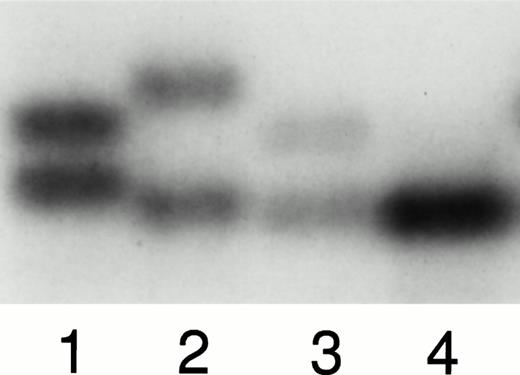
Eighteen of the 20 JMML bone marrow samples studied (90%) showed the presence of both NF1 alleles by PCR-based polymorphism analysis using three highly informative intragenic markers35-37(Table 1). Of the 2 JMML samples showing only a single NF1allele with all three markers, one (patient 20) was not informative because we had no sources of normal or parental tissue for analysis. In patient 6 we detected LOH at NF1 using the polymorphic marker described by Andersen et al.36 In this case we used DNA extracted from an EBV-transformed cell line for comparison with bone marrow DNA (Fig 1A). Because this child had Mo 7, we were able to establish that the EBV line was not part of the JMML clone as it retained both copies of chromosome 7 by PCR-based polymorphism analysis (data not shown). Four of the 20 children showed activating RAS point mutations in their bone marrows and these data are summarized in Table 2. Three cases had mutations in the KRAS protooncogene and one inNRAS. None of these 4 patients had LOH at NF1 or truncating NF1 mutations.
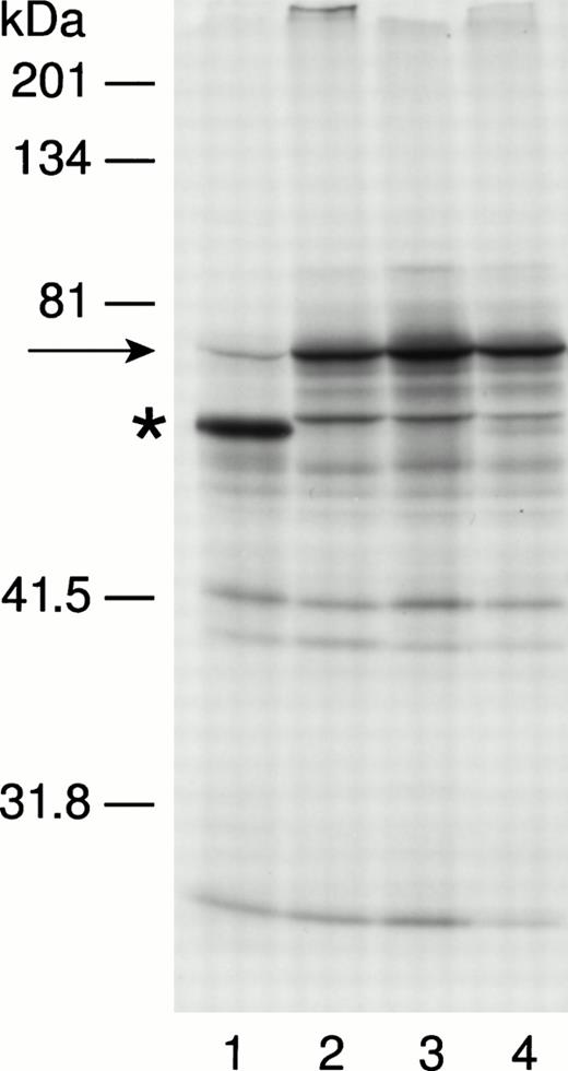
(A). Analysis of bone marrow and EBV cell-line DNA from patient 6 and DNA extracted from the blood of his parents. Lanes 1 through 4 were analyzed using the polymorphic nucleotide repeat described by Andersen et al.36 Lane 1 shows maternal DNA, lane 2 shows paternal DNA, lane 3 shows EBV-line DNA, and lane 4 shows DNA extracted from leukemic bone marrow. The patient's EBV line, which is not involved in the leukemic clone, retains both parentalNF1 alleles, whereas the affected bone marrow sample shows a single allele consistent with LOH at NF1 in this leukemia. (B). Results of the IVTT assay in patient 6. Lanes 1 to 4 show polypeptides synthesized from RT-PCR product corresponding to exons 28 through 38 of the NF1 coding sequence. The sample in lane 1 is derived from patient 6; samples in lanes 2 through 4 show a normal protein pattern. The full-length polypeptide is indicated by an arrow. The truncated protein in lane 1, marked by an asterisk, represents the 6579 + 18 G to A mutation seen in the splice consensus sequence flanking exon 34 ofNF1 in this patient. The full-length IVTT polypeptide in this patient is represented by a fainter band than is seen in lanes 2 to 4, consistent with loss of the normal NF1 allele in this leukemia.
(A). Analysis of bone marrow and EBV cell-line DNA from patient 6 and DNA extracted from the blood of his parents. Lanes 1 through 4 were analyzed using the polymorphic nucleotide repeat described by Andersen et al.36 Lane 1 shows maternal DNA, lane 2 shows paternal DNA, lane 3 shows EBV-line DNA, and lane 4 shows DNA extracted from leukemic bone marrow. The patient's EBV line, which is not involved in the leukemic clone, retains both parentalNF1 alleles, whereas the affected bone marrow sample shows a single allele consistent with LOH at NF1 in this leukemia. (B). Results of the IVTT assay in patient 6. Lanes 1 to 4 show polypeptides synthesized from RT-PCR product corresponding to exons 28 through 38 of the NF1 coding sequence. The sample in lane 1 is derived from patient 6; samples in lanes 2 through 4 show a normal protein pattern. The full-length polypeptide is indicated by an arrow. The truncated protein in lane 1, marked by an asterisk, represents the 6579 + 18 G to A mutation seen in the splice consensus sequence flanking exon 34 ofNF1 in this patient. The full-length IVTT polypeptide in this patient is represented by a fainter band than is seen in lanes 2 to 4, consistent with loss of the normal NF1 allele in this leukemia.
Activating RAS Point Mutations Among 20 Children With JMML
| Patient No. . | RAS Gene and Codon . | Alteration in DNA Sequence . | Effect on Protein . |
|---|---|---|---|
| 2 | NRAS 12 | GGT to GCT | Glycine to alanine |
| 5 | KRAS 13 | GGC to GAC | Glycine to aspartic acid |
| 13 | KRAS 12 | GGT to TGT | Glycine to cysteine |
| 16 | KRAS 12 | GGT to GTT | Glycine to valine |
All five NF1 segments were amplified successfully by RT-PCR from each sample. In three cases, one or more abnormal peptide bands were detected in one of the five NF1 segments. Data from patient 6 are shown in Fig 1B. RT-PCR products that gave rise to truncated proteins were cloned and subjected to a second round of IVTT as described elsewhere17 29 and cDNA clones that yielded an abnormal peptide by second-stage IVTT were sequenced. Truncating mutations of NF1 were confirmed in both cloned cDNA and genomic DNA and are summarized in Table 3. Patient 14 showed a nonsense mutation in exon 27a of NF1. In two cases (patients 6 and 11), direct sequencing of cloned cDNA revealed aberrant splicing resulting in a shift in the reading frame. Genomic DNA from patient 6 showed an alteration (6579 + 18 G to A) in the splice donor consensus sequence flanking exon 34. This mutation introduced an additional 17 nucleotides containing a novel BglI restriction enzyme site into the patient's cDNA. We were able to show the presence of this restriction site in amplified cDNA derived from this patient's EBV cell-line RNA, thus confirming that this mutation existed in the germline (data not shown). Cloned cDNA from patient 11 showed abnormal splicing of 7 nucleotides between exons 10c and 11. We have previously found the same mutation in a child with familial NF1 and MDS17; genomic DNA sequence showed an abnormal splice acceptor sequence upstream of exon 11 (1642 − 8 A to G) creating a cryptic splice site and consequent frameshift and premature stop codon.
Truncating Mutations of NF1 Found in 20 Children With JMML
| Patient No. . | Mutation Site . | Alteration in cDNA Sequence . | Effect on Protein . |
|---|---|---|---|
| 6 | Intron 34, splice donor sequence | 17 nucleotides spliced between exons 34 and 35 | Truncation at codon 2195 |
| 11 | Intron 10c, splice acceptor sequence | 7 nucleotides spliced between exons 10c and 11 | Truncation at codon 555 |
| 14 | Exon 27a | 4614 G to A | Tryptophan to stop at codon 1538 |
DISCUSSION
Studies performed in children with NF1 and in lines of knockout mice strongly support the hypothesis that NF1 functions as a tumor-suppressor gene in immature hematopoietic cells. Bone marrows from children with NF1 and malignant myeloid disorders (including JMML) frequently show loss of the NF1 allele inherited from the unaffected parent,18,26 and we have recently shown inactivation of both NF1 alleles in myeloid leukemias from a number of unrelated patients17 (and our unpublished data, April 1997). Similarly, mice that are heterozygous for a targeted disruption of Nf1 are predisposed to a myeloproliferative disorder that is reminiscent of JMML and is associated with somatic inactivation of the wild-type Nf1allele.15 Finally, although murine Nf1−/− embryos are nonviable because of complex cardiac anomalies, hematopoietic cells derived from Nf1−/− fetal livers induce a JMML-like disorder in irradiated recipients.16,38,39 The tumor-suppressor function of NF1 in hematopoietic cells seems to be related to its ability to accelerate guanosine triphosphate (GTP) hydrolysis on Ras. Primary JMML cells from children with NF1 show a reduction in neurofibromin-associated GAP activity and a modest elevation in the level of active GTP-bound Ras.14 Myb-transformed Nf1−/− hematopoietic cells derived from murine fetal liver show higher peak levels of Ras-GTP with prolonged activation after stimulation with granulocyte-macrophage colony stimulating factor (GM-CSF).16 Moreover, oncogenic RAS mutations are detected in the bone marrows of 20% to 30% of children with MDS (including JMML) but are restricted to patients who do not have NF1, presumably because a RAS mutation would confer no additional selective advantage on clones that have deregulated Ras signaling by inactivating NF1.19
Taken together, children with a diagnosis of NF1 and cases associated with RAS mutations account for approximately 40% of all patients with JMML.1,19 In this study, we tested the hypothesis that leukemic cells from children with JMML who had neither clinical stigmata nor family history of NF1 would harbor NF1mutations. Indeed, analysis of bone marrow cells from 20 young children with JMML revealed 3 with NF1 mutations that led to premature termination of protein translation. The finding of LOH at NF1in 1 of these samples (patient 6) indicates that both alleles are functionally inactivated in this JMML clone, in keeping with its role as a tumor-suppressor. The other 2 JMML bone marrows (patients 11 and 14) retained both NF1 alleles by PCR-based polymorphism analysis although only one allele harbored a truncating mutation. We have previously observed that approximately 50% of leukemic specimens from children who have germline mutations of NF1 do not show LOH at the NF1 locus.17,18 26 It is likely that the second NF1 allele in these bone marrow samples is inactivated by mechanisms not detectable by IVTT, such as missense or promoter mutations, or alterations in the 3′ untranslated region of the gene which might potentially destabilize the mRNA. Four other children in this series (20%) showed activating RAS point mutations in their JMML clones. As expected, none of these patients had alterations of NF1 in their bone marrow samples.
We have shown NF1 mutations in 15% of JMML bone marrow specimens obtained from children without clinical evidence ofNF1. In conjunction with previous studies showing that 10% to 14% of patients with JMML have NF1,1,3,7 and that an additional 20% to 30% of cases are associated with somaticRAS mutations,19 our data provide molecular evidence for altered Ras signaling in up to 60% of JMML clones. If hyperactive Ras is a general feature of JMML, these data also raise the possibility that alternative genetic events might deregulate the Ras pathway in the remaining 40% of cases. We have previously used a32P orthophosphate labeling technique to analyze primary leukemic cells from children with NF1 and JMML for increased Ras-GTP:GDP ratios commensurate with Ras activation.14 However, we were unable to perform this assay as part of the present study because we did not have access to fresh bone marrow cells from any of the patients. None of our JMML samples showed the presence of the Philadelphia chromosome or the t(5;12)(q33;p13) translocation found predominantly in adult chronic myelomonocytic leukemia; the latter is thought to perturb Ras signaling through activation of the platelet-derived growth factor receptor-β tyrosine kinase.40,41 Other negative regulators of Ras proteins in normal cells include p120 GAP (also known as RasGAP).42 Mutations in the catalytic domain of the p120 GAP gene (GAP) have not been shown in human cancer, including adult MDS,43,44 and we have previously shown normal levels of p120 GAP activity in leukemic cells from patients with and without NF1.14 Similarly, a cancer predisposition has not been described in mice heterozygous for a targeted disruption in the murineGAP homologue.45 Although JMML cells consistently show selective hypersensitivity to GM-CSF in vitro,46 no pathogenic GM-CSF receptor mutations have been found in the JMML samples studied to date.47 Recent evidence suggests that levels of the p85α regulatory subunit of phosphatidylinositol 3-OH kinase (PI 3-kinase) are elevated in unstimulated JMML cell lysates.48 The PI 3-kinase complex binds to the Ras effector domain in a GTP-dependent manner, resulting in increased levels of PI 3-phosphorylated lipid targets.49 It is possible that PI 3-kinase expression is higher in JMML cells without mutations of RAS or NF1, although how this might mediate leukemogenesis is at present unclear.
A recent large study of JMML showed that the median age of children without NF1 at presentation was 1.8 years, whereas NF1 was more common in children who had been diagnosed after the age of 5 years.7 Because 40% of children with JMML present before the age of 1 year and 60% are under 2 years,4 it seems likely that some children with germline NF1 mutations are not diagnosed because of subtle phenotypes or young age. This idea is also consistent with our experience that the proportion of children with MDS who have familial (v sporadic) NF1 is higher than expected.17,18,26 Patient 6 was a boy diagnosed as having JMML at 19 months of age who was noted to have cafe au lait macules but not other stigmata of NF1. Analysis of EBV cell-line RNA confirmed the presence of a germline NF1 mutation in this child although he did not fulfill consensus diagnostic criteria for NF1.20 No source of normal tissue was available from cases 11 and 14 to establish if the abnormalities existed in the germline, or represented somaticNF1 mutations restricted to the leukemic clone. Somatic inactivation of both NF1 alleles has not been shown in primary human cancer cells to date, although large tumor-specific deletions involving a single allele are seen in a number of NF1-associated malignancies.18 50-52 Three somatic NF1 mutations in cancers in non-NF1 patients have been described in the literature, in a colonic adenocarcinoma, an anaplastic astrocytoma, and an adult MDS53; these were missense mutations involving one allele. Thus, the role of somatic NF1 mutations in tumorigenesis remains unresolved.
The results of this study and the clinical incidence of NF1 among JMML patients suggest that the proportion of JMML clones harboringNF1 mutations could be as high as 30%. We conclude that JMML may be the initial presenting feature of NF1 in young children. Further investigation is necessary to determine whether the NF1mutations detected in leukemias that arise in children without clinical evidence of neurofibromatosis represent germline or somatic alterations, and to define other genetic events which may be responsible for Ras deregulation in JMML cells.
ACKNOWLEDGMENT
We thank Drs Vietta Vereen, Ronald Kline, and Ronald Oseas (Sunrise Hospital, Las Vegas, NV); Dr T.B. Moore (Department of Pediatrics, Division of Hematology/Oncology, University of California, Los Angeles); Dr David L. Baker (Princess Margaret Hospital for Children, Perth, Australia); and Dr Arnold J. Altman (Department of Pediatric Hematology/Oncology, University of Connecticut Health Center) for samples. We also thank all the physicians who have referred patients to us for analysis, and the families who participated in this study. This work was facilitated by a collaboration with the Children's Cancer Group (Study Number B24).
Supported in part by National Institutes of Health (NIH) grants CA72614 and by grants from the Concern 2 Foundation and the Frank A. Campini Foundation (K.M.S.); by fellowships from the Sir Halley Stewart Trust and the Lady Tata Memorial Trust (L.E.S.); and by NIH grant CA 60407 (P.D.E.).
Address reprint requests to Kevin M. Shannon, MD, Box 0519, University of California, 513 Parnassus Ave, San Francisco, CA 94143-0519; e-mail:kevins@itsa.ucsf.edu.
The publication costs of this article were defrayed in part by page charge payment. This article must therefore be hereby marked "advertisement" is accordance with 18 U.S.C. section 1734 solely to indicate this fact.


This feature is available to Subscribers Only
Sign In or Create an Account Close Modal