Abstract
Peripheral blood progenitor cells (PBPCs) are increasingly used instead of bone marrow for autologous or allogeneic transplantation. In this study PBPCs mobilized in cancer patients by chemotherapy and granulocyte-colony stimulating factor were collected by apheresis and first enriched by immunoaffinity removal of lineage positive cells. When these cells were exposed to both cyclophosphamide and taxol or cultured for 7 days in the presence of 5-fluorouracil, stem cell factor, and interleukin-3, 88% to 93% of the enriched PBPCs were killed and short-term clonogenic capacity in methylcellulose assays was lost, but week-5 cobblestone area-forming cell (CAFC) enrichment was higher than 10-fold in comparison to enriched PBPCs and higher than 700-fold in comparison to unmanipulated apheresis cells. After drug exposure, most of the progenitors displayed a CD34+, CD38−, multidrug-resistance (MDR+), Rhodamine 123 low, Hoechst 33342 low phenotype, and as few as 180 of these drug-resistant cells were able to generate a stable multilineage human hematopoiesis in sublethally irradiated immunodeficient mice. In these animals, the level of human hematopoietic engraftment was significantly increased by cotransplantation of irradiated cells from the human L87/4 stromal cell line. These observations are consistent with the functional isolation of a population of very early hematopoietic progenitors and might help to design new protocols for the removal of neoplastic cells from autografts.
CURRENTLY AVAILABLE methods for enrichment of human hematopoietic progenitors with long-term multilineage potential include immunoaffinity purification of CD34+/CD38− cells,1-3 staining with dyes such as Rhodamine (Rh) 123 and/or Hoechst (Ho) 33342 followed by cell sorting4-6 and culture in the presence of the antimetabolite 5-fluorouracil (5-FU), stem cell factor (SCF ), and interleukin-3 (IL-3).7 These methods have been recently used to purify a very small population including <0.1% of nucleated cells from human bone marrow (BM)1,3,4,6,7 or cord blood (CB)1,2 or from murine BM.5 Because mobilized peripheral blood progenitor cells (PBPCs) are increasingly used instead of BM for both autologous8 and allogeneic9 transplantation, we were interested in enrichment and characterization of PBPCs. In this field, Sutherland et al10 have recently indicated that CD34+Thy-1+Lin− PBPCs can engraft a preimmune fetal sheep. Because cancer relapse appears to be the major cause of death in neoplastic patients undergoing autologous transplantation, and purging of neoplastic cells by means of different drugs seems to be feasible,11,12 we were also interested in defining PBPC resistance to major anticancer drugs such as cyclophosphamide, taxol, and 5-FU. In this study we evaluated the long-term multilineage potential of hematopoietic progenitors exposed to (1) both cyclophosphamide and taxol and (2) 5-FU, SCF, and IL-3 as described by Berardi et al.7 PBPCs from mobilized cancer patients were collected by apheresis, enriched by immunoaffinity removal of lineage-positive cells, exposed to drugs, and finally evaluated by means of long-term stroma culture and transplantation in sublethally irradiated Non Obese Diabetic/LtSz-scid/scid (NOD/Scid) mice.
MATERIALS AND METHODS
In vitro studies: PBPC collection and enrichment. The approval for use of cells from human subjects was obtained by the Institutional Review Board, and cells were obtained after signed informed consent. Cancer patients undergoing PBPC autologous transplantation were treated with 7 g/m2 cyclophosphamide and subsequent administration of 5 μg/kg/d granulocyte colony-stimulating factor (G-CSF ). PBPC collection by apheresis was started when the postnadir CD34+ cell count was >50/μL. An aliquot of less than 5% of the apheresis product was processed by Ficoll (1.077 g/dL; Cedar Lane, Hornby, Ontario, Canada), and the Ficoll fraction was further processed for PBPC enrichment by means of immunoaffinity removal of lineage positive cells. Briefly, cells were resuspended in phosphate-buffered saline (PBS) without Ca+Mg+ supplemented by 5% autologous serum and incubated in ice with a mixture of antiglycophorin A, -CD3, -CD2, -CD56, -CD24, -CD19, -CD14, CD16, and -CD66b tetrameric antibody complexes (StemCell Technologies, Vancouver, British Columbia, Canada). These tetrameric antibody complexes are bispecific cross-linkers that bind the described antigen and dextran. After 30 minutes of incubation, 20 nm magnetic colloidal iron/dextran particles were added, and after a further 30 minutes incubation cells were processed through the StemSep TM device for depletion of targeted cells according to the manufacturer's instructions (StemCell Technologies). At the end of the procedure, CD34+ cell recovery was 53% to 83% and purity in the range 68% to 96%.
Flow cytometry. We used phycoerythrin (PE)-, fluorescein isothiocyanate (FITC)-, or Cy-Chrome (Cy-C)–labeled monoclonal antibodies against the external epitope of human P-glycoprotein (Signet, Dedham, MA), CD13, CD19, CD34, CD38, CD45, CD61, and glycophorin A (Becton Dickinson, Mountain View, CA and PharMingen, San Diego, CA). A total of 100 to 500 × 103 cells were incubated at 22°C for 30 minutes in PBS 1% bovine serum albumin (BSA) with monoclonal antibodies. On some occasions, cells were also stained with Rh 123 or Ho 33342 as described by Leemhuis et al.4 By means of flow cytometry (FACScan, Becton Dickinson), the percent of stained cells was determined as compared with PE-, FITC- or Cy-C–conjugated mouse isotypic control. A portion of each sample was incubated with the appropriate isotype control antibodies to establish the background level of nonspecific staining, and positivity was defined as being greater than nonspecific background staining. Dead cells and cells undergoing apoptosis were enumerated and excluded by 7-amino actinomycin D (7AAD) staining by the method recently described by Philpott et al.13 Finally, G0 -G1 cells were separated from S-G2M cells and enumerated following the method described by Goodell et al.5
PBPC exposure to mafosfamide and taxol. Mafosfamide (an active analogue of cyclophosphamide made by Asta Medica, Frankfurt, Germany) was a kind gift of Dr Carlo-Stella (University of Parma, Parma, Italy), taxol was generously provided by Dr Renzo Canetta (Bristol-Myers Squibb Pharmaceutical Research Institute, Wallingford, CT). Both were obtained as pure white powder, resuspended in RPMI-10% fetal calf serum (FCS) and sterilized by 0.25 μm filtration before use. A total of 10 to 20 × 106 PBPCs enriched as described previously were resuspended at 2 × 10⁶/mL and exposed to doses of 10 to 200 μg/mL mafosfamide and 1 to 10 ng/mL taxol. On some occasions, 50 μmol/L verapamil (Sigma, St Louis, MO) was added together with taxol to inhibit P-glycoprotein activity. Control cells were exposed to RPMI-10% FCS alone. Cells were incubated at 37°C for 30 minutes. After this period, cells were cooled by the addition of 4°C RPMI-10% FCS and washed twice with the same medium. Cell viability was evaluated by flow cytometry and 7AAD.
PBPC exposure to 5-FU, SCF, and IL-3. A total of 10 to 20 × 106 PBPCs enriched as described previously were resuspended at 2 × 10⁶/mL in Iscove's modified Dulbecco's medium (IMDM) supplemented on day 0 by 100 ng/mL SCF (Amgen, Thousand Oaks, CA), 100 ng/mL IL-3 (Peprotech, London, UK or R&D, Abingdon, UK), 0.6 mg/mL 5-FU (Sigma), and 10% FBS. Cultures were supplemented daily by 5-FU to enhance its effect. On day 7, cells were washed, and viability was evaluated by flow cytometry and 7AAD.
Evaluation of colony-forming units and CAFCs. Colony-forming units (CFUs) and cobblestone area-forming cells (CAFCs) were enumerated before and after PBPC enrichment and drug exposure.
A standard CFU assay was performed in duplicate by seeding 500 PBPCs enriched as described previously in 1 mL of a commercial medium containing 0.8% methylcellulose, 30% FCS, 1% BSA, 10−4 mol/L 2-mercaptoethanol, 3 U/mL erythropoietin, 50 ng/mL SCF, 10 ng/mL granulocyte-macrophage colony-stimulating factor (GM-CSF ), and 100 ng/mL IL-3 (StemCell Technologies). The number of seeded cells was chosen to be maximally informative, yielding 20 to 40 colonies for plate when cells were seeded before drug exposure. After 14 days of 37°C culture in 5% CO2 , colonies larger than 50 cells were scored by microscopy as colony-forming unit–granulocyte-macrophage (CFU-GM, containing granulocytes and macrophages), burst-forming unit-erythroid (BFU-E, containing erythroid cells) and colony-forming unit granulocyte, erythroid, monocyte, megakaryocyte (CFU-GEMM, containing myeloid cells, erythroid cells, and megakaryocytes).
To evaluate CAFC frequency, we followed the limiting dilution analysis (LDA) assays described by the Ploemacher14 and by the Dexter15 groups with few modifications. A layer of irradiated (80 Gy) M2-10B4 murine stromal cells was generated in 96-well plates in the presence of commercial long-term MyeloCult Medium (Stem Cell Technologies). A minimum of 1 and maximum of 10 × 103 viable PBPCs enriched as described previously were seeded in 10 different dilutions in 10 replicate wells each. The dilutions were chosen to be maximally informative on week 5 of the culture, yielding 10% to 37% negative wells for cobblestone areas. Cultures were feeded with MyeloCult medium weekly, and even more frequently if the pH indicator in the medium signaled a low pH. Every 7 days and at the end of the 5-week culture, wells were evaluated as positive or negative for the presence of cobblestone areas, defined as clusters of small, tightly packed cells that were nonrefractory when viewed under a phase contrast microscope and originated from a CAFC. Wells with cobblestone areas of greater than 15 cells or 3 separate foci of more than 5 cells were scored as positive. On some experiments, clonogenic assays were also performed at week 5 after CAFC reading on each replicate well. The adherent layer was washed and trypsinized. After centrifugation, cells were seeded in the commercial methycellulose medium described previously, and after 14-day culture at least one CFU was found to be generated from all CAFC-positive wells.
Liquid culture in the presence of cytokines. The proliferation potential of PBPCs enriched by immunoaffinity removal of lineage-positive cells was evaluated before and after drug exposure. A total of 100 × 103 cells were seeded in IMDM-10% FCS supplemented by 100 ng/mL each of SCF (kindly provided by Amgen), Flt3-ligand (FL was a gift from Immunex, Seattle, WA), IL-3 (R&D) and Megakaryocyte Growth and Development Factor (MGDF, a pegylated, truncated molecule related to thrombopoietin; kindly provided by Amgen). This cytokine combination is known to induce proliferation of very early hematopoietic progenitors such as BM-derived long-term culture-initiating cells (LTC-ICs).16 On some occasions, liquid cultures were supplemented by 30% stroma supernatant obtained from L87/4 human cell line cultured in IMDM.17-19
In vivo studies: Human PBPC engraftment in NOD/Scid mice. In vivo studies were approved by the Institutional Review Board. Sublethally irradiated (350 cGy), 5- to 6-week-old NOD/Scid mice were transplanted intravenously (IV) with 0.1 to 100 × 103 human PBPCs processed as described previously. On some occasions, mice were transplanted with PBPCs and 1 × 10⁶ human irradiated (20 Gy) L87/4 stromal cells, known by polymerase chain reaction (PCR) analysis to generate human SCF, G-CSF, GM-CSF, IL-1β, IL-6, IL-7, IL-8, IL-11, leukemia inhibitory factor (LIF), macrophage inflammatory protein (MIP)-1α, transforming growth factor-β (TGF-β), and tumor necrosis factor α (TNF-α).17-19 Irradiated stromal cells were transplanted to provide human cytokines and to enhance the number of transplanted cells which, in turn, reduce the risk of PBPC phagocytosis. Mice were studied on week 6 (peripheral blood [PB]) and on week 10 or 15 (BM) after transplantation to evaluate the presence of human cells by flow cytometry, CAFC, and CFU assay in the presence of human cytokines as described previously and PCR.
Evaluation of human PBPC engraftment in NOD/Scid mice by PCR. Human SEX gene expression was evaluated in PB and BM of transplanted mice by PCR.20 Total cellular DNA was prepared by using a QIAamp Tissue Kit (Quiagen, Hilden, Germany). PCR was performed in a DNA thermal cycler (Robocycler; Stratagene, La Jolla, CA) in a final volume of 25 μL with 1 μg of DNA and 1 U of Taq DNA polymerase (Boehringer Mannheim, Mannheim, Germany). The samples were subjected 35 times in succession to the following thermal cycle: 1 minute denaturation at 94°C, 1 minute annealing at 61°C, and 1 minute polymerization at 72°C. Primers (sense 5′ - CAT TCC GTA CTC CCA GCG TCC - 3′ positions 139600 to 139620 and antisense 5′ - ACA CCT ACC GTA GCG GCT GAG - 3′ positions 140200 to 140220 accession no. L44140 at Genbank) were added at 25 pmol each per reaction. Blood of untransplanted mice was used to show that primers for human SEX gene do not amplificate any mouse gene; water negative control, containing all the components of the PCR reaction but no target DNA, was also included. The PCR product (band expected 620 bp) was separated on 2% agarose gel and stained with ethidium bromide.
Data analysis. CAFC frequency was evaluated by using the maximum likelihood solution.21 Statistical comparisons were performed by Primer (McGraw Hill, New York, NY) and StatWiew (Abacus Concepts, Berkeley, CA) softwares using the paired t-test, the Bonferroni t-test, and analysis of variance (ANOVA) in paired studies and the nonparametric analyses of Mann-Whitney, Wilcoxon, and Kruskal-Wallis in nonpaired studies. Values of P < .05 were considered statistically significant.
RESULTS
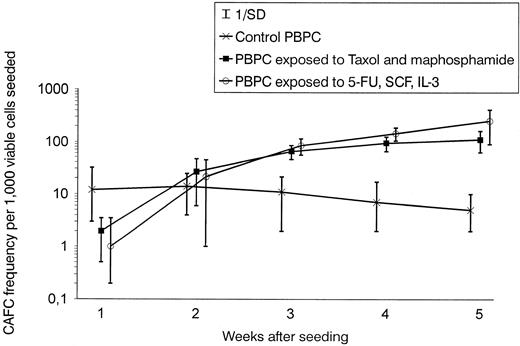
In vitro studies: CAFC enrichment by drug exposure. We first evaluated the sensitivity of enriched PBPCs to taxol and mafosfamide and determined that PBPC exposure to more than 100 μg/mL mafosfamide and more than 7.5 ng/mL taxol was associated with a significant loss of CAFC viability (data not shown). As reported in Table 1, exposure to 7.5 ng/mL taxol alone or to 100 μg/mL mafosfamide alone killed 56% to 68% of cells and resulted in twofold to threefold CAFC enrichment. When PBPCs were exposed to both taxol and mafosfamide, 88% ± 12% of the enriched PBPCs were killed, and CAFC enrichment was higher than 10-fold in comparison with enriched PBPCs and higher than 700-fold in comparison with unmanipulated apheresis cells. Similar cell killing and CAFC enrichment were observed when PBPCs were cultured for 7 days in the presence of 5-FU, SCF, and IL-3. When verapamil was added together with taxol, cell killing rose to 98% ± 1%, suggesting that in PBPCs most of the taxol resistance was caused by P-glycoprotein activity. Exposure to both mafosfamide and taxol and culture in the presence of 5-FU, SCF, and IL-3 resulted in the killing of virtually all CFU-GM, BFU-E, and CFU-GEMM, because methylcellulose 2-week cultures were negative. As shown in Fig 1, the evaluation of CAFC frequency during 5 weeks of culture indicated that early-appearing CAFCs were significantly less drug-resistant than late CAFCs and suggested that drug exposure enriched for a population of quiescent hematopoietic progenitors. Throughout 5-week culture, CAFC frequency was not significantly different in PBPCs exposed to both mafosfamide and taxol and in PBPCs cultured for 7 days with 5-FU, SCF, and IL-3.
CAFC Enrichment After PBPC Drug Exposure
| . | Percent of Cells Killed . | CAFC Frequency per Viable Cell Seeded . |
|---|---|---|
| Unmanipulated apheresis cells | 0 | 1/10,280 (±1,042) |
| Control cells (enriched PBPCs) | 0 | 1/203 (±62) |
| Taxol (7.5 ng/mL) | 56 ± 9 | 1/89 (±33) |
| Mafosfamide (100 μg/mL) | 68 ± 11 | 1/69 (±27) |
| Taxol and mafosfamide | 88 ± 12 | 1/18 (±10) |
| 5FU, IL-3, SCF | 93 ± 9 | 1/14 (±7) |
PBPCs were collected by apheresis from mobilized cancer patients and first enriched by immunoaffinity removal of lineage-positive cells. At the end of the enrichment procedure, CD34+ cell recovery was 53% to 83%, and purity in the range 68% to 96%. The percent of CD34+ cells killed by drug exposure was evaluated by flow cytometry, CAFC frequency was evaluated on week 5 by LDA. Results are expressed as mean (±SD), n = 7.
Abbreviations: CAFC, cobblestone area-forming cell; PBPC, peripheral blood progenitor cell; FU, Fluorouracil; IL, interleukin; SCF, stem cell factor; LDA, limiting dilution analysis; SD, Standard deviation.
CAFC week types significantly differ in their drug resistance. The proporton of CAFC week types in PBPCs enriched by immunoaffinity removal of lineage-positive cells and further exposed to different drugs was evaluated by 5-week culture over irradiated M2-10B4 feeder (n = 7). This assay allows week-by-week enumeration of CAFC frequency and indicated that early-appearing (and possibly less quiescent) CAFCs were significantly less drug resistant than late CAFCs (P < .01 by ANOVA). Moreover, CAFC frequency was similarly enriched in PBPCs exposed to both mafosfamide and taxol and in PBPCs cultured for 7 days with 5-FU, SCF, and IL-3.
CAFC week types significantly differ in their drug resistance. The proporton of CAFC week types in PBPCs enriched by immunoaffinity removal of lineage-positive cells and further exposed to different drugs was evaluated by 5-week culture over irradiated M2-10B4 feeder (n = 7). This assay allows week-by-week enumeration of CAFC frequency and indicated that early-appearing (and possibly less quiescent) CAFCs were significantly less drug resistant than late CAFCs (P < .01 by ANOVA). Moreover, CAFC frequency was similarly enriched in PBPCs exposed to both mafosfamide and taxol and in PBPCs cultured for 7 days with 5-FU, SCF, and IL-3.
Cell phenotype before and after drug exposure. Figure 2 shows the phenotype of PBPCs (previously enriched by immunoaffinity removal of lineage-positive cells) before and after drug exposure (n = 7). Before drug exposure, 68% to 96% of the cells were CD34+, and in the CD34+ cell population 2% to 8% were CD38−, 2% to 7% were MDR positive (ie, expressing high levels of P-glycoprotein), 8% to 15% were Rh 123 low, and 7% to 18% were Ho 33342 low. After exposure to both mafosfamide and taxol or 7-day culture with 5-FU, SCF, and IL-3, 96% to 98% of the cells were CD34+, 78% to 93% were CD38−, 81% to 92% were MDR positive, 83% to 97% were Rh 123 low, and 87% to 94% were Ho 33342 low.
Phenotype of PBPCs enriched by immunoaffinity removal of lineage-postitive cells, evaluated before and after drug exposure by means of two color staining with FITC- or PE-labeled anti-CD34 antibodies (vertical axis) and PE-labeled anti-CD38 or FITC-labeled anti–P-glycoprotein antibodies, Rh 123, or Ho 33342 (horizontal axis). A total of 18 × 106 PBPCs enriched as described in the Materials and Methods section were resuspended at 2 × 10⁶/mL in IMDM supplemented on day 0 by 100 ng/mL SCF, 100 ng/mL IL-3, 0.6 mg/mL 5-FU and 10% FBS. Cultures were supplemented daily by 5-FU. On day 7, cells were washed, and viability was evaluated by flow cytometry and 7AAD. About 0.7 × 10⁶ cells were still viable after drug exposure (ie, 4% of seeded cells). A total of 5,000 events were collected for each flow cytometry analysis, and the cell populations analyzed were included in the gates showed in the FSC/SSC panels. The fluorescence detectors were set up to have similar gates in all stains. Dead cells were excluded by their intense staining with 7AAD, and the percentage of positive cells per each quadrant indicated. The flow cytometry analysis shows that before drug selection (top panels) 51% to 64% of PBPCs were CD34+, CD38+, MDR−, Rh123 bright, and Ho33342 bright. After drug exposure (bottom panels), 88% to 94% of the cells were CD34+,CD38−, MDR+, Rh123 low, and Ho33342 low. Interestingly, after drug exposure the CD38−, MDR+, Rh123 low, and Ho33342 low phenotype was present also in a small (2% to 3%) but constantly present subset of CD34− cells. Differences between PBPCs exposed to both mafosfamide and taxol or cultured for 7 days in the presence of 5-FU, SCF, and IL-3 were not significant. The results of one representative experiment of 7 are shown.
Phenotype of PBPCs enriched by immunoaffinity removal of lineage-postitive cells, evaluated before and after drug exposure by means of two color staining with FITC- or PE-labeled anti-CD34 antibodies (vertical axis) and PE-labeled anti-CD38 or FITC-labeled anti–P-glycoprotein antibodies, Rh 123, or Ho 33342 (horizontal axis). A total of 18 × 106 PBPCs enriched as described in the Materials and Methods section were resuspended at 2 × 10⁶/mL in IMDM supplemented on day 0 by 100 ng/mL SCF, 100 ng/mL IL-3, 0.6 mg/mL 5-FU and 10% FBS. Cultures were supplemented daily by 5-FU. On day 7, cells were washed, and viability was evaluated by flow cytometry and 7AAD. About 0.7 × 10⁶ cells were still viable after drug exposure (ie, 4% of seeded cells). A total of 5,000 events were collected for each flow cytometry analysis, and the cell populations analyzed were included in the gates showed in the FSC/SSC panels. The fluorescence detectors were set up to have similar gates in all stains. Dead cells were excluded by their intense staining with 7AAD, and the percentage of positive cells per each quadrant indicated. The flow cytometry analysis shows that before drug selection (top panels) 51% to 64% of PBPCs were CD34+, CD38+, MDR−, Rh123 bright, and Ho33342 bright. After drug exposure (bottom panels), 88% to 94% of the cells were CD34+,CD38−, MDR+, Rh123 low, and Ho33342 low. Interestingly, after drug exposure the CD38−, MDR+, Rh123 low, and Ho33342 low phenotype was present also in a small (2% to 3%) but constantly present subset of CD34− cells. Differences between PBPCs exposed to both mafosfamide and taxol or cultured for 7 days in the presence of 5-FU, SCF, and IL-3 were not significant. The results of one representative experiment of 7 are shown.
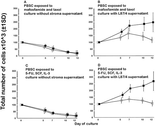
Liquid cultures in the presence of cytokines. As shown in Fig 3 (n = 5), PBPCs exposed to both mafosfamide and taxol and PBPCs cultured for 7 days in the presence of 5-FU, SCF, and IL-3 did not proliferate in stroma-free cultures in the presence of SCF, FL, IL-3, and MGDF, ie, a cytokine combination previously known to induce LTC-IC proliferation in liquid cultures of CB- or BM-derived CD34+CD38− purified cells.16 On the other hand, when 30% stroma supernatant obtained from L87/4 human cell line was present in the cultures in addition to SCF, FL, IL-3, and MGDF, a 1.5 to 2-fold expansion of the total number of CD34+ cells was observed. Under these conditions, CD34+ cell proliferation was similar in cultures of PBPCs exposed to both mafosfamide and taxol and in cultures of PBPCs exposed for 7 days to 5-FU, SCF, and IL-3.
Liquid culture in the presence of SCF, FL, IL-3, and MGDF of PBPCs enriched by immunoaffinity removal of lineage-positive cells and further exposed to mafosfamide and taxol or cultured for 7 days in the presence of 5-FU, SCF, and IL-3 (n = 5). At the indicated time interval the total number of CD34+ cells (open square) and the total number of nucleated cells (solid square) was calculated from the cell counts and the percentage of viable cells measured by 7AAD staining and flow cytometry. (A and C) Cells cultured in the presence of cytokines but without stroma supernatant did not proliferate. Conversely, a 1.5- 2-fold expansion of the total number of CD34+ cells was observed when drug-exposed PBPCs were cultured in the presence of the same cytokine combination supplemented by 30% supernatant from the human L87/4 stromal line (B and D).
Liquid culture in the presence of SCF, FL, IL-3, and MGDF of PBPCs enriched by immunoaffinity removal of lineage-positive cells and further exposed to mafosfamide and taxol or cultured for 7 days in the presence of 5-FU, SCF, and IL-3 (n = 5). At the indicated time interval the total number of CD34+ cells (open square) and the total number of nucleated cells (solid square) was calculated from the cell counts and the percentage of viable cells measured by 7AAD staining and flow cytometry. (A and C) Cells cultured in the presence of cytokines but without stroma supernatant did not proliferate. Conversely, a 1.5- 2-fold expansion of the total number of CD34+ cells was observed when drug-exposed PBPCs were cultured in the presence of the same cytokine combination supplemented by 30% supernatant from the human L87/4 stromal line (B and D).
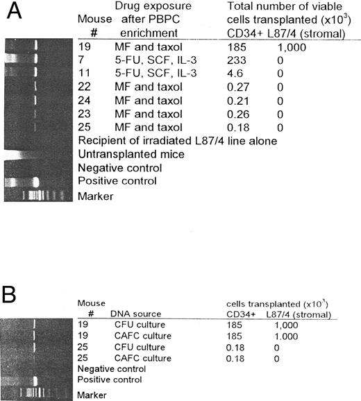
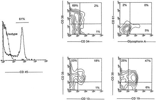
In vivo studies. Table 2 describes the phenotype of human hematopoietic cells engrafted in NOD/scid mice 6 to 10 weeks after transplantation, and Table 3 shows the frequency of NOD/scid mice engrafted and not engrafted at various cell doses. By means of flow cytometry, it was possible to detect myeloid (CD13+) erythroid (glycophorin A+), megakaryocytic (CD61+), and lymphoid (CD19+) cells in mice transplanted with PBPCs enriched as described previously containing 1 × 10⁶ CD34+ cells (control), in mice transplanted with a minimum of 1.3 × 10³ PBPCs viable after 7-day culture in the presence of 5-FU, SCF, and IL-3 (96%-98% CD34+) and in mice transplanted with a minimum of 0.18 × 10³ PBPCs viable after exposure to both mafosfamide and taxol (96%-98% CD34+). As shown in Fig 4A, human engraftment was further confirmed by PCR detection of human SEX gene in mice transplanted with drug-resistant PBPCs and not cotransplanted with human L87/4 stromal cells. Regarding multilineage human cell engraftment, a similar pattern was observed in mice transplanted with control PBPCs and drug-exposed PBPCs. Overall, 0.2% to 4% of human cells were myeloid, 3% to 34% lymphoid, 0.1% to 1.2% megakaryocytic, 0.1% to 3% erythroid and 0.1% to 1.8% expressed the human CD34 antigen (Fig 5).
Phenotype of Human Hematopoietic Cells Engrafted in NOD/Scid Mice
| Mouse No. . | Drug Exposure After PBPC Enrichment . | Total Number of Viable Cells Transplanted (×103) . | % of Human Cells 6-10 Weeks After Transplantation . | |||||||
|---|---|---|---|---|---|---|---|---|---|---|
| . | . | CD34+ . | L87/4 (stromal) . | CD45+ . | CD13+ . | CD19+ . | CD34+ . | CD38+ . | CD61+ . | GPA+ . |
| 1 | no | 1,000 | — | 21 | 2.2 | 19 | 1.2 | ND | 0.3 | 0.2 |
| 2 | no | 1,000 | — | 17 | 1.6 | 15 | 1.1 | ND | 0.2 | 0.3 |
| 3 | no | 1,000 | — | 29 | 1.8 | 22 | 1.5 | ND | 0.3 | 0.5 |
| 4 | no | 1,000 | — | 17 | 0.9 | 15 | 1.4 | ND | 0.2 | 0.3 |
| 5 | no | 1,000 | — | 28 | 2.7 | 19 | 1.4 | ND | 0.5 | 0.4 |
| 6 | 5-FU, SCF, IL-3 | 446 | — | 9.5 | 0.7 | 7.7 | 1.1 | ND | 0.6 | 0.1 |
| 7 | 5-FU, SCF, IL-3 | 233 | — | 32 | 1.9 | 28 | 1.3 | ND | 0.2 | 0.3 |
| 8 | 5-FU, SCF, IL-3 | 75 | — | 7.8 | 1.1 | 5.2 | 0.5 | ND | 0.1 | 0.3 |
| 9 | 5-FU, SCF, IL-3 | 22 | — | 14 | 0.8 | 13 | 0.7 | ND | 0.2 | 0.3 |
| 10 | 5-FU, SCF, IL-3 | 5.8 | — | 5.5 | 0.9 | 4.1 | 0.3 | ND | 0.1 | 0.2 |
| 11 | 5-FU, SCF, IL-3 | 4.6 | — | 23 | 1.8 | 18 | 1.0 | 22 | 0.2 | 0.1 |
| 12 | 5-FU, SCF, IL-3 | 4.6 | — | 9.6 | 0.2 | 8.0 | 0.4 | 8.4 | 0.1 | 0.1 |
| 13 | 5-FU, SCF, IL-3 | 2.4 | — | 17 | 1.1 | 15 | 1.2 | 16 | 0.1 | 0.1 |
| 14 | 5-FU, SCF, IL-3 | 2.0 | — | 3.7 | 0.2 | 2.9 | 0.6 | 3.1 | 0.2 | 0.1 |
| 15 | 5-FU, SCF, IL-3 | 1.3 | 5.3 | 0.3 | 4.8 | 1.0 | 5.1 | 0.9 | 0.6 | |
| 16 | MF and taxol | 616 | 1,000 | 58 | 14 | 34 | 1.3 | 55 | 0.8 | 2.6 |
| 17 | MF and taxol | 399 | 1,000 | 61 | 11 | 32 | 1.8 | 43 | 1.2 | 3.0 |
| 18 | MF and taxol | 223 | 1,000 | 33 | 3.7 | 17 | 0.3 | 31 | 0.1 | 1.0 |
| 19 | MF and taxol | 185 | 1,000 | 21 | 1.3 | 16 | 0.5 | 19 | 0.2 | 0.7 |
| 20 | MF and taxol | 87 | 1,000 | 37 | 4.2 | 28 | 1.2 | 27 | 0.4 | 1.2 |
| 21 | MF and taxol | 0.28 | — | 5.4 | 0.2 | 4.1 | 0.1 | 4.8 | 0.1 | 0.2 |
| 22 | MF and taxol | 0.27 | — | 12 | 0.5 | 7.3 | 0.3 | 11 | 0.2 | 0.1 |
| 23 | MF and taxol | 0.26 | — | 3.6 | 0.1 | 2.7 | 0.2 | 2.9 | 0.1 | 0.1 |
| 24 | MF and taxol | 0.21 | — | 14 | 0.9 | 8.4 | 0.8 | 10 | 0.2 | 0.2 |
| 25 | MF and taxol | 0.18 | — | 6.0 | 0.5 | 5.6 | 1.3 | 5.5 | 0.1 | 0.1 |
| 26 | MF and taxol | 0.19 | 1,000 | 24 | 1.9 | 19 | 1.1 | 22 | 1.1 | 0.2 |
| 27 | MF and taxol | 0.16 | 1,000 | 17 | 1.4 | 16 | 1.3 | 15 | 0.2 | 0.3 |
| 28 | MF and taxol | 0.14 | 1,000 | 13 | 1.1 | 13 | 0.7 | 12 | 0.1 | 0.1 |
| 29 | MF and taxol | 0.13 | 1,000 | 27 | 2.8 | 24 | 1.3 | 25 | 0.6 | 0.1 |
| 30 | MF and taxol | 0.12 | 1,000 | 18 | 1.2 | 16 | 1.7 | 16 | 0.9 | 0.3 |
Sublethally irradiated animals received mobilized peripheral blood hematopoietic progenitors enriched by immunoaffinity removal of lineage-positive cells. In some studies, cells were exposed to both mafosfamide and taxol or cultured for 7 days in the presence of 5-FU, SCF, and IL-3. On some occasions, animals received also irradiated L87/4 stromal cells.
Abbreviations: PBPC, peripheral blood progenitor cell; GPA, glycophorin A; ND, not done; FU, fluorouracil; SCF, stem cell factor; IL, interleukin; MF, mafosfamide.
Frequency of NOD/Scid Mice Engrafted and Not Engrafted at Various Doses of Human Hematopoietic Cells
| CD34+ Cell Dose × 103 . | Drug Exposure After PBPC Enrichment . | No. of Positive Mice . | No. of Negative Mice . |
|---|---|---|---|
| 1,000 | no | 5 | 1 |
| 1-1,000 | 5-FU, SCF, IL-3 | 10 | 3 |
| 0.1-1 | 5-FU, SCF, IL-3 | 0 | 2 |
| 1-1,000 | mafosfamide and taxol | 5 | 0 |
| 0.1-1 | mafosfamide and taxol | 10 | 9 |
Mice were considered to be engrafted if human cells were detected by flow cytometry and a band was detectable by PCR analysis 6 to 10 weeks after transplantation.
Abbreviations: PBPC, peripheral blood progenitor cell; FU, fluorouracil; SCF, stem cell factor; IL, interleukin; PCR, polymerase chain reaction.
Drug-resistant human PBPCs can engraft NOD/Scid mice (A). The human SEX gene was shown by PCR in the BM of NOD/Scid mice engrafted with as few as 180 to 270 PBPCs enriched by immunoaffinity removal of lineage-positive cells and still viable after exposure to both mafosfamide and taxol (mice 22-25). Higher levels of engraftment were found in the BM of NOD/Scid mouse transplanted with 185 × 103 PBPCs enriched by immunoaffinity removal of lineage-positive cells and still viable after exposure to both mafosfamide and taxol (mouse 19) and in the BM of mice transplanted with 4 to 233 × 103 PBPCs enriched by immunoaffinity removal of lineage-positive cells and still viable after 7 days culture in the presence of 5-FU, SCF, and IL-3 (mice 7 and 11). Human SEX gene expression from cells other than human drug-resistant hematopoietic progenitors was excluded, because untransplanted mice and mice transplanted with irradiated L87/4 cells alone were negative. (B) Human SEX gene expression in CFU and CAFC cultures from the BM of mice transplanted with 0.18 to 185 × 103 PBPCs enriched by immunoaffinity removal of lineage-positive cells and still viable after exposure to both mafosfamide and taxol (mice 19 and 25).
Drug-resistant human PBPCs can engraft NOD/Scid mice (A). The human SEX gene was shown by PCR in the BM of NOD/Scid mice engrafted with as few as 180 to 270 PBPCs enriched by immunoaffinity removal of lineage-positive cells and still viable after exposure to both mafosfamide and taxol (mice 22-25). Higher levels of engraftment were found in the BM of NOD/Scid mouse transplanted with 185 × 103 PBPCs enriched by immunoaffinity removal of lineage-positive cells and still viable after exposure to both mafosfamide and taxol (mouse 19) and in the BM of mice transplanted with 4 to 233 × 103 PBPCs enriched by immunoaffinity removal of lineage-positive cells and still viable after 7 days culture in the presence of 5-FU, SCF, and IL-3 (mice 7 and 11). Human SEX gene expression from cells other than human drug-resistant hematopoietic progenitors was excluded, because untransplanted mice and mice transplanted with irradiated L87/4 cells alone were negative. (B) Human SEX gene expression in CFU and CAFC cultures from the BM of mice transplanted with 0.18 to 185 × 103 PBPCs enriched by immunoaffinity removal of lineage-positive cells and still viable after exposure to both mafosfamide and taxol (mice 19 and 25).
Engraftment of drug-resistant human PBPCs in NOD/Scid mice. A representative highly engrafted NOD/Scid mouse transplanted with 400 × 103 drug-resistant PBPCs and 1 × 10⁶ L87/4 stromal cells was analyzed 6 weeks after transplantation by three-color flow cytometry. The percentage of positive cells per each quadrant is indicated. Cells were stained with Cy-C–labeled anti-human CD45 antibody and compared with cells stained with Cy-C isotype control. Dead cells were excluded by their intense 7AAD staining, and viable cells were gated according to their human CD45 expression (horizontal line in the histogram). Sixty-one percent of CD45+ human cells were present in the BM of this mice. As shown in the contour plots, among these CD45+ cells 19% (corresponding to 11% of all cells) were CD13+ (myeloid), 53% (corresponding to 32% of all cells) were CD19+ (lymphoid), 1% were CD34+/CD38− (early hematopoietic progenitors), 2% were CD61+ (megakaryocytic), and 5% were glycophorin A+ (erythroid).
Engraftment of drug-resistant human PBPCs in NOD/Scid mice. A representative highly engrafted NOD/Scid mouse transplanted with 400 × 103 drug-resistant PBPCs and 1 × 10⁶ L87/4 stromal cells was analyzed 6 weeks after transplantation by three-color flow cytometry. The percentage of positive cells per each quadrant is indicated. Cells were stained with Cy-C–labeled anti-human CD45 antibody and compared with cells stained with Cy-C isotype control. Dead cells were excluded by their intense 7AAD staining, and viable cells were gated according to their human CD45 expression (horizontal line in the histogram). Sixty-one percent of CD45+ human cells were present in the BM of this mice. As shown in the contour plots, among these CD45+ cells 19% (corresponding to 11% of all cells) were CD13+ (myeloid), 53% (corresponding to 32% of all cells) were CD19+ (lymphoid), 1% were CD34+/CD38− (early hematopoietic progenitors), 2% were CD61+ (megakaryocytic), and 5% were glycophorin A+ (erythroid).
In 5 mice engrafted with 1 × 10⁶ control CD34+ cells, 22% ± 5% of BM cells expressed the human CD45 antigen. Similarly, a mean of 12% ± 9% of BM cells expressed the human CD45 antigen in 10 mice engrafted with 1 to 400 × 103 PBPCs still viable after 7-day culture in the presence of 5-FU, SCF, and IL-3 (P > .05 v control by ANOVA). A total of 15 mice were engrafted with PBPCs exposed to both mafosfamide and taxol. Five of these mice received as few as 180 to 280 PBPCs still viable after exposure to both mafosfamide and taxol, and 8% ± 4% of BM cells expressed the human CD45 antigen 6 to 10 weeks after transplantation. Five more of these mice received as few as 120 to 190 PBPCs still viable after exposure to both mafosfamide and taxol together with 1 × 10⁶ L87/4 stromal cells expressing human cytokines, and in these mice 20% ± 5% of BM cells expressed the human CD45 antigen (P < .05 v mice transplanted with a similar amount of drug-resistant PBPCs but without L87/4 stromal cells). Finally, 5 more of these mice received a higher amount (80 to 600 × 103) of PBPCs still viable after exposure to both mafosfamide and taxol plus 1 × 10⁶ L87/4 stromal cells, and the BM of these mice expressed the highest (42% ± 17%, P < .05) levels of human CD45 antigen.
By means of PCR we detected the human SEX gene in both CFUs and CAFC cultures from the BM of NOD/Scid mice engrafted with human PBPCs (Fig 4B). Human CFUs were in the range 1 to 190 × 105 mice BM cells. At the end of a 5-week culture on murine M2-10B4 stromal line, the frequency of human CAFCs was found to be in the range 1 to 87 × 106 mice BM cells.
DISCUSSION
In this work we investigated PBPC sensitivity to major anticancer drugs such as cyclophosphamide (mafosfamide), taxol, and 5-FU and showed that PBPC exposure to (1) both mafosfamide and taxol or (2) 5-FU, SCF, and IL-3 highly enriched for a population of very early hematopoietic progenitors with striking multilineage engraftment potential. However, PBPC resistance to these drugs appears to be caused by different mechanisms. As indicated by the work of Berardi et al,7 5-FU activity seems to be strictly cell-cycle–dependent. In the progenitor cell purification procedure based on culture in the presence of 5-FU, SCF, and IL-3, committed cells expressing the IL-3 receptor are induced to proliferate and forced to a 5-FU–dependent metabolic death, whereas very early and quiescent progenitors were preserved from apoptosis by the presence of SCF. Conversely, taxol and mafosfamide killing is apparently not dependent on cell cycle. In fact, when, in our studies, mafosfamide or taxol were used together with SCF and IL-3 to induce PBPCs to proliferate, cell killing was not significantly modified (data not shown). The cytosolic enzyme aldehyde-dehydrogenase (ALDH) is currently thought to be involved in the mechanism of cellular resistance to cyclophosphamide,22 and a correlation has been shown between cyclophosphamide resistance and ALDH levels during hematopoietic stem cell differentiation.23 In our studies, PBPC resistance to taxol was possibly caused by P-glycoprotein activity, because PBPC exposure to taxol highly enriched for a MDR+ cell population and taxol resistance declined in the presence of verapamil. Similarly, Goodell et al5 recently indicated that in the mouse model a Ho 33342 stained side population (SP) was at least 1,000-fold enriched for in vivo reconstitution ability and multilineage proliferation potential. The Ho33342 SP profile was blocked when staining was performed in the presence of verapamil, indicating that the distinctly low staining pattern of these cells is probably due to P-glycoprotein or MDR-like mediated efflux of the dye from the cells. Another crucial observation from the study of Goodell et al5 is that at least 1% to 3% of Ho33342 SP cells were in S-G2M, and that the reconstitution capacity of these cells was similar to that of quiescent Ho33342 SP cells. In our study, 7-day culture in the presence of 5-FU, SCF, and IL-3 virtually eliminated all PBPCs that were in S-G2M, whereas after PBPC exposure to both mafosfamide and taxol 0.7% to 2.1% of viable cells were in S-G2M (Table 4). This was the most significant difference we found between the two PBPC enrichment procedures described in the study, and we are currently evaluating the engraftment potential of this small S-G2M cell population, removed by culture in the presence of 5-FU, SCF, and IL-3 but spared by exposure to both mafosfamide and taxol.
Frequency of S-G2M Cells in PBPC Enriched by Immunoaffinity Removal of Lineage-Positive Cells and Still Viable After Exposure to Both Mafosfamide and Taxol or to 5-FU, SCF, and IL-3
| Experiment No. . | Frequency of S-G2M Cells (% of all cells) . | |
|---|---|---|
| . | Drug Exposure After PBPC Enrichment . | 5-FU, SCF, and IL-3 . |
| . | Mafosfamide and Taxol . | . |
| 1 | 1.7 | 0 |
| 2 | 1.3 | 0 |
| 3 | 1.2 | 0.1 |
| 4 | 0.7 | 0 |
| 5 | 2.1 | 0 |
| 6 | 1.1 | 0 |
| 7 | 1.4 | 0.1 |
Abbreviations: PBPC, peripheral blood progenitor cell; FU, fluorouracil; SCF, stem cell factor; IL, interleukin.
Rh 123 and Ho 33342 have been recently used by other investigators to enrich for hematopoietic progenitors. In the studies by Uchida et al,6 the efflux of Rh 123 was most rapid from Thy-1+ CD34+ cells and could be inhibited by verapamil, again suggesting that also Rh 123 efflux from hematopoietic cells might primarily be caused by the P-glycoprotein. CD34+Lin− cells were deposited in culture as single cells, and only Rh123 low cells were able to generate CAFCs after 5 to 6 weeks, despite the fact that they were proliferating more slowly as we also observed in our studies. In SCID-hu mice and in chimeric sheep after in utero transplantation, all the engraftment potential resided within the Rh 123 low-mid but not Rh 123 high subset. In the studies by Leemhuis et al,4 Ho 33342 and Rh 123 were used to define a population of both quiescent (Ho 33342 low) and metabolically inactive (Rh 123 low) CD34+ cells. Limiting dilution analysis showed that the frequency of LTC-ICs was higher in CD34+, Ho 33342 low, Rh 123 low cells than in CD34+ cells expressing higher staining for Ho 33342 and Rh 123 and that mouse CD34+, Ho 33342 low, Rh 123 low cells were able to fully reconstitute other lethally irradiated mice. On the other hand, it has been recently suggested in mouse studies that CD34-negative hematopoietic progenitors might exist. In the recent paper by Jones et al,24 10 mouse BM cells, expressing high levels of aldehyde dehydrogenase and mafosfamide resistance, but lacking Thy-1, (Sca-1), c-kit, or CD34 expression, generated all blood cell lineages for the lifetime of the animal. However, these very primitive cells failed to support short-term engraftment (mice radioprotection), thus suggesting the presence of two different classes of hematopoietic stem cells: one that generates both short-term and long-term engraftment and one that produces delayed but durable engraftment. This hypothesis is further supported by recent evidence by Osawa et al,25 indicating that in the mouse injection of one single CD34low/−, c-Kit+, Sca-1+, Lin− cell results in long-term reconstitution of lethally irradiated mice, provided that committed cells are cotransplanted to support short-term engraftment of the animal. Although the search for a CD34-negative human hematopoietic progenitor has been so far elusive, it should be remembered that in our PBPC enrichment procedure we first removed lineage-positive cells by immunoaffinity and then exposed the cells to drugs so that, at the end of these procedures, 2% to 4% of our cells were CD34− and CD38−, MDR+, Rh 123 low, and Ho 33343 low. Further studies are now under way to evaluate whether this small CD34-negative cell population contains hematopoietic progenitors.
In the present study the NOD/Scid model was used to define the multilineage engraftment potential of drug resistant hematopoietic progenitors. In the recent study from Larochelle et al,2 injection of human BM or CB progenitors in the same NOD/scid mouse model resulted in engraftment of a small number of human primitive cells that proliferated and differentiated in the murine BM producing large numbers of early hematopoietic progenitors (such as LTC-ICs) enumerated by gene marking. The human primitive hematopoietic cells that initiated the graft were operationally defined as SCID-repopulating cells (SRCs) and were present only in the CD34+CD38− fraction and were highly quiescent as judged by low retrovirus-mediated gene transfer efficiency. Hao et al1 also recently studied the hematopoietic potential of human CD34+CD38− cells from BM or CB. These sorted cells were are able to generate CFUs for at least 100 days, ie, beyond the LTC-IC standard. These “extended” LTC-ICs (ELTC-ICs) are functionally distinct from LTC-ICs in timing of initial cloning proliferation and generative capacity, and again, the significantly less efficient retrovirus-mediated gene marking of ELTC-ICs compared with LTC-ICs is consistent with a more quiescent progenitor population. The BM CD34+CD38− cell population was recently studied also by Civin et al.3 In their hands, the CD34+/CD38− subset, but not the CD34+CD38+ subset, generated sustained, retransplantable and multilineage hematopoiesis in the preimmune fetal sheep model. In the present study, most of the drug-resistant hematopoietic progenitors were CD34+CD38−, but, differently from sorted BM or CB CD34+CD38− cells,16 drug-resistant PBPCs were unable to proliferate in stroma-free culture in the presence of SCF, FL, MGDF, and IL-3, unless stroma supernatant was provided. In this context, it is of interest that drug-resistant, MDR+, Rh 123 low, and Ho 33343 low CD34+CD38− cells are a subset of all CD34+CD38− PBPCs (data not shown). This subset might be very immature, enriched for SRCs and poorly responsive to cytokine-induced proliferation signals, as suggested by gene-marking studies indicating that SRCs are very difficult to transduce.2 Further studies are now under way to define whether this phenomenon is present also in drug-resistant progenitors from CB and BM.
Regarding human hematopoietic engraftment in NOD/Scid mice, we have found that as few as 180 PBPCs exposed to both mafosfamide and taxol were sufficient to engraft NOD/Scid mice. According to Larochelle et al,2 who recently indicated the SRC assay as a novel, useful tool to study human hematopoiesis, as few as 500 CD34+CD38− cells can engraft NOD/Scid mice. In our hands, successful engraftment of such a small number of human cells was possible only when other cells (either irradiated stromal cells or, as in this case, drug-sensitive PBPCs killed by drug exposure) were cotransplanted to preserve hematopoietic progenitors from recipient mice phagocytes, possibly still active after sublethally irradiation. We have observed in the NOD/Scid mice model a high level of human lymphoid engraftment that correlates well to what has been described previously by Larochelle et al2 and Turner et al.26 Here we also report the novel observation that human hematopoietic progenitors underwent significantly greater proliferation when irradiated cells from the L87/4 stromal line were cotransplanted in the recipient mice to produce human cytokines and, possibly, other unknown factors able to support human hematopoiesis. In this context, Goan et al27 recently transplanted SCID mice with unmanipulated PBSCs and found that cotransplantation of a rat fibroblast cell line expressing human IL-3 significantly accelerated engraftment. In their hands, cotransplants of allogeneic human BM stromal cells from long-term cultures resulted in comparable, though less prominent, support of engraftment.
In conclusion, we have shown that (1) a population of human PBPCs with striking multilineage engraftment potential is resistant to both mafosfamide and taxol, (2) as judged by CAFC week type, this drug-resistant population is more quiescent than drug-sensitive PBPCs, and (3) drug-resistant PBPCs can proliferate in liquid culture only if stroma supernatant is provided. A number of investigators have described the use of mafosfamide to purge BM autografts.11,12 The present work is, to our knowledge, the first to report about PBPC drug resistance and the first to confirm in human cells recent data by Blau et al,28 who showed in the mice that taxol in vivo treatment is a method for enriching for early hematopoietic cells. Data from the present study might be used to design new protocols to purge PBPC autografts, possibly by means of drug exposure together with immunoaffinity removal of neoplastic cells by monoclonal antibodies. Moreover, the enumeration of drug-resistant hematopoietic progenitors could be useful to predict the PBPC engraftment potential, as recently suggested by Kirkland et al29 who indicated that the total number of BM-derived mafosfamide-resistant progenitors infused to chronic myeloid leukemia patients undergoing allogeneic transplantation predicted the rate of neutrophil engraftment, whereas the total number of CFU-GM did not correlate with engraftment potential.
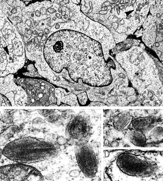
A 29-year-old man presented with a 20-pound weight loss and was found to have a large retroperitoneal mass. Electron microscopy was diagnostic for metastatic amelanotic melanoma, characterized by large tumor cells with prominent nucleoli and focal clusters of membrane-bound, ellipsoid granules (arrowheads in A). Higher magnification of these organelles showed that they were premelanosomes containing perpendicular striated filaments (B and C) or parallel lamellar arrays (D). Extremely electron-dense melanin was absent from the premelanosomes. Immunoperoxidase stains of tumor cells were positive for the melanoma-associated antigen HMB-45 and negative for leukocyte common antigen, B-cell antigen (CD20 and CD19), T-cell antigens (CD2, CD3, CD4, CD5, and CD8), and lymphoid activation antigens (CD30 and CD15). Retrospectively, the patient was stated to have fair skin, numerous freckles, and red hair; no history of excesssive sunburns or skin lesions was elicited. Original magnifications: (A) ×8,000; (B) ×90,000; (C) ×71,500; and (D) ×91,000. (Courtesy of Ann M. Dvorak, MD, Department of Pathology, Beth Israel Deaconess Medical Center, 330 Brookline Ave, Boston, MA 02215.)
A 29-year-old man presented with a 20-pound weight loss and was found to have a large retroperitoneal mass. Electron microscopy was diagnostic for metastatic amelanotic melanoma, characterized by large tumor cells with prominent nucleoli and focal clusters of membrane-bound, ellipsoid granules (arrowheads in A). Higher magnification of these organelles showed that they were premelanosomes containing perpendicular striated filaments (B and C) or parallel lamellar arrays (D). Extremely electron-dense melanin was absent from the premelanosomes. Immunoperoxidase stains of tumor cells were positive for the melanoma-associated antigen HMB-45 and negative for leukocyte common antigen, B-cell antigen (CD20 and CD19), T-cell antigens (CD2, CD3, CD4, CD5, and CD8), and lymphoid activation antigens (CD30 and CD15). Retrospectively, the patient was stated to have fair skin, numerous freckles, and red hair; no history of excesssive sunburns or skin lesions was elicited. Original magnifications: (A) ×8,000; (B) ×90,000; (C) ×71,500; and (D) ×91,000. (Courtesy of Ann M. Dvorak, MD, Department of Pathology, Beth Israel Deaconess Medical Center, 330 Brookline Ave, Boston, MA 02215.)
ACKNOWLEDGMENT
The authors thank Dr Carmelo Carlo-Stella (University of Parma, Parma, Italy) for useful discussions and the gift of mafosfamide, Dr Renzo Canetta (Bristol-Myers Squibb Pharmaceutical Research Institute) for taxol, Dr Karin Thalmeier (GSF-Institute for Experimental Hematology, München, Germany) for the availability of the L87/4 cell line, Dr Connie Eaves (Terry Fox Laboratories, Vancouver, Canada) for the availability of M2-10B4 cells, and Amgen and Immunex for cytokines used in in vitro studies.
Supported in part by a United States National Blood Foundation grant and by European Union Concerted Action Biomed 2 “Eurocord.” FB is a scholar of the US National Blood Foundation.
Address reprint requests to Francesco Bertolini, MD, PhD, Division of Medical Oncology, IRCCS Maugeri Foundation, Pavia Medical Center, via Ferrata 8, 27100 Pavia, Italy.


This feature is available to Subscribers Only
Sign In or Create an Account Close Modal