Abstract
Clinical studies of bone marrow transplantation (BMT) suggest that the immune system contributes to the eradication of acute myeloid leukemia (AML). A recent study also showed that the Fas (CD95/APO1) mediates apoptotic signal from cytotoxic T lymphocytes. Sixty-four patients with AML were studied for the expression of Fas in the context of CD34 and CD38 coexpression. The clinical relevance of Fas expression and function on AML was also investigated. Fas was expressed on 2% to 98% of AML cells (2% to 20% in 11 patients, 20% to 50% in 20 patients, 50% to 80% in 24 patients, and 80% to 98% in nine patients). Only 44.4% of patients with AML M1 (French-American-British [FAB] classification) were Fas+ (≥20% of leukemia cells expressed Fas), whereas 89.1% of patients with AML M2, M3, M4, M5 were Fas+ (P < .01). Among 43 CD34+ patients (≥20% leukemia cells were CD34+), 34 were Fas+, and 19 of 21 CD34− patients were Fas+ (P = NS). Thirteen cases were studied for their expression of Fas in the context of CD34 and CD38 using three-color analysis. Fas is expressed at a high level in the gated CD34+CD38± and CD34+CD38+ population. In 10 AML samples, Fas was expressed at a higher level in CD34+/CD38+ population than in CD34+/CD38± or CD34− cell populations. Fas-induced apoptosis by anti-Fas monoclonal antibody (MoAb) was determined by morphologic features and colorimetric DNA fragmentation assay. Induction of apoptosis was found in 14 of 24 cases. However, no statistically significant correlation was observed between Fas expression and induction of apoptosis. Leukemia colony-forming unit assays suggested that in some cases, Fas-induced apoptosis occurred in the clonogenic cell populations. Parameters such as laboratory and clinical data at initial diagnosis were correlated with Fas expression and only response to initial induction chemotherapy showed significant correlation with Fas expression (P < .05). We conclude that the majority of AML cells exhibit variable expression of Fas, and apoptosis could be induced by anti-Fas MoAb in some cases. Our results suggest the Fas-mediated apoptosis may be clinically relevant, whereas the issue of clonogenic leukemia cells and Fas expression needs further studies.
CLINICAL EXPERIENCE FROM allogeneic bone marrow transplantation (BMT) for acute myeloid leukemia (AML) has suggested that immune mechanisms contribute to the eradication of leukemia.1 Occurrence of acute or chronic graft-versus-host disease (GVHD) reduced the risk of relapse after BMT to a relative risk of 0.78 and 0.48, respectively. Furthermore T-cell depletion in allogeneic BMT resulted in higher relapse rates. It has been well documented that in some cases recurrence of AML after BMT can be treated using donor lymphocyte infusions (DLI).2 In the autologous setting, in vivo and in vitro administration of interleukin (IL)-2 induces stimulation of cellular immunity to kill tumor cells by generating lymphokine-activated killer cells.3 Various types of hematopoietic cells are involved in the immunosurveillance against leukemia cells.
Recently, the interaction between Fas receptor (Fas/CD95/APO1) on the target cell and Fas ligand (FasL) on cytotoxic T lymphocytes (CTLs) has been shown to be an important mechanism of cell killing.4 Fas is a cell surface molecule that directly mediates apoptosis.5 Fas was first identified when a monoclonal antibody (MoAb) (anti-Fas) was developed that had cytolytic activity toward several Fas+ cell lines. Molecular cloning of the cDNAs for Fas showed that they are the same molecule that has also been designated as CD95.6 Fas is a member of the nerve growth factor/tumor necrosis factor (TNF ) receptor superfamily that is characterized by cysteine-rich extracellular domains. Fas and TNF receptor also contain a unique cytoplasmic region, termed the “death domain” that is essential for initiating a cytolytic response.7 The expression and the function of Fas on virus-infected hematopoietic cells or hematopoietic malignant cells such as multiple myeloma or adult T-cell leukemia (ATL) has been reported.8 9
Several studies have suggested that AML is maintained in vivo by the small population of clonogenic leukemia cells that possess substantial proliferative capacity and the ability to undergo differentiation into nondividing cells.10 Clonogenic capacity is generally considered to be associated with an immature phenotype. Lapidot et al reported that clonogenic potential of myeloid leukemia cells in vitro is restricted to leukemia that had CD34+CD38− phenotype.11-14
In the current study, we analyzed the expression of Fas on AML cells from 64 cases in the context of CD34 and CD38 coexpression. In addition, we investigated clinical relevance of Fas expression and the function of Fas.
MATERIALS AND METHODS
Clinical samples. Sixty-four newly diagnosed patients with AML were included in this study. All patients received standard induction-therapy just after the diagnosis. The diagnosis of AML was established using classical morphologic and immunologic criteria. At diagnosis, more than 90% of mononuclear cells (MNCs) were leukemia cells. Bone marrow (BM) aspirates were obtained from patients at the initial diagnosis and healthy volunteers after signing informed consent. The studies were performed on randomly selected cryopreserved leukemia blast cells and freshly isolated cells. All patients have their own unique patient number (UPN, FAB-number).
Cell preparations. MNCs were isolated using standard procedures. Briefly heparinized cell samples were separated by Ficoll-Hypaque density centrifugation at the time of collection to obtain highly purified mononuclear cell fractions consisting mostly of leukemia blasts. The purified fractions were washed in phosphate-buffered saline (PBS). Nineteen samples were used immediately after isolation and 45 samples were cryopreserved in RPMI 1640 (GIBCO, Grand Island, NY) with 10% fetal calf serum (FCS) and 10% dimethyl sulfoxide at −196°C. On thawing, blasts were resuspended in culture medium and nonviable cells were removed by Ficoll centrifugation.
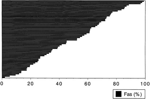
Fas expression on leukemia cells from 64 patients with AML by flow cytometric analysis. Each column shows the percentage of Fas+ cells in leukemia cells from respective patients at initial diagnosis. Wide range of percentage (2% to 98%) and equal distribution are observed. Most leukemia cases are Fas+ (more than 20 % in cells tested express Fas).
Fas expression on leukemia cells from 64 patients with AML by flow cytometric analysis. Each column shows the percentage of Fas+ cells in leukemia cells from respective patients at initial diagnosis. Wide range of percentage (2% to 98%) and equal distribution are observed. Most leukemia cases are Fas+ (more than 20 % in cells tested express Fas).
Cell culture. All cell cultures were performed in RPMI 1640 with 10% FCS (culture medium) at 37°C in a humidified 5% CO2 atmosphere. Leukemia cells at a concentration of 1 × 10⁶/mL were incubated for 20 hours in 24-well plate (MULTIWELL, Becton Dickinson, Lincoln Park, NJ) using medium with various factors as indicated below. For Fas induction, cells were incubated with human natural interferon (IFN)α (Sumiferon, Sumitomo Pharmaceutical Co, Osaka, Japan) or recombinant human IFNγ (provided by Shionogi Pharmaceutical Co, Osaka, Japan) at the concentration of 40, 200, and 1,000 IU/mL. For the induction of apoptosis, anti-Fas MoAb (CH11, IgM k, Medical & Biological Laboratory, MBL, Nagoya, Japan), which is known to induce apoptosis mimicking the FasL, was used at the concentration of 1 or 0.2 μg/mL.
Flow cytometric analysis. To analyze for the expression of Fas and CD34 on AML cells, the following MoAbs were used: fluorescein isothiocyanate (FITC)-conjugated anti-Fas MoAb (UB-2, IgG1, MBL, Nagaoya, Japan) and phycoerythrin (PE)-conjugated MoAb to CD34 (HPCA-2, IgG1, Becton Dickinson, Mountain View, CA) and FITC- or PE- conjugated IgG1 MoAbs for isotype-matched nonspecific control antibodies (Becton Dickinson). Fas expression is weak, in general, and we tested three anti-Fas MoAb, FITC-conjugated (MBL), PE-conjugated (Pharmingen, San Diego, CA), and PE-conjugated (MBL). There was no significant difference between MoAbs. For flow cytometric analysis, 5 × 10⁵ CD34 cells or BM MNCs were washed twice with ice cold dilution buffer (PBS containing 0.1% sodium azide, 5 mmol/L EDTA and 2% bovine serum albumin [BSA]). Cells were incubated in dilution buffer for 30 minutes at 4°C with two isotypic control antibodies, PE-conjugated anti-CD34 MoAb/FITC isotypic control, FITC-conjugated anti-Fas MoAb/PE isotypic control, and PE-conjugated anti-CD34 MoAb and FITC conjugated anti-Fas MoAb. After incubation, cells were washed twice and immediately analyzed using a flow-cytometer (EPICS EXL, Coulter Co, Hialeah, FL). The results were obtained using two-color flow cytometric techniques. Samples scored as positive contained more than 20% of Fas-expressing cells. Relative fluorescence intensity (RFI = mean fluorescence intensity of cell stained with anti-Fas MoAb/mean fluorescence intensity of cells stained with control MoAb) was used to compare the level of Fas expression. No difference in fluorescence data between fresh samples and cryopreserved samples was observed by analyses of several paired cell samples (data not shown).
Three color flow-cytometric analysis was performed with FITC-conjugated anti-Fas MoAb, PE-conjugated anti-CD38 MoAb, and R-phycoerythrin–cyanine 5 (PE-Cy5) conjugated anti-CD34 MoAb (clone 581, IgG1, MBL) and three control antibodies including PE-Cy5 isotypic antibody (IgG1, MBL).
Detection of apoptosis. After incubation, 1 × 10⁵ cells were applied to cytospin slides and stained by May-Giemsa solution for morphologic analysis. Cells showing morphologic features of apoptosis such as condensed nucleus and relative condensed cytoplasm, were judged as undergoing apoptosis. Apoptosis was determined by analyzing 100 cells microscopically. The ratio of cells undergoing apoptosis was calculated as percentage of apoptosis. Viability of cultured cells was also tested by Trypan-Blue. For analysis of DNA fragmentation, 5 × 10⁵ cells were mixed with 100 μL of 10 mmol/L Tris-HCl (pH7.4), 10 mmol/L EDTA and 0.5% Triton-X (Wako Pure Chemical Industries, Tokyo, Japan). The lysate was incubated for 10 minutes at 4°C and then spun at 13,000g for 20 minutes. The supernatant was incubated with ribonuclease A (RNase A; Sigma Chemical Co, St Louis, MO) for 1 hour at 37°C and then Proteinase K was added to remove proteins. DNA was precipitated in 1 vol isopropanol and 0.5 mol/L of NaCl, centrifuged, and suspended in Tris-EDTA (TE) buffer. Extracted DNA solution was applied to a 2% agarose gel, electrophoresed, and stained by ethidium bromide.
FAB Classification and Fas Expression
| FAB . | No. of Patients . | Positive* (%) . | % Median (range) . | RFI (SD) . |
|---|---|---|---|---|
| M1 | 9 | 4/9 (44.4) | 19.5 (5.3-85.3) | 3.33 (1.64) |
| M2 | 26 | 23/26 (88.5) | 56.7 (1.9-98.9) | 6.31 (5.30) |
| M3 | 6 | 6/6 (100) | 67.0 (23.1-77.1) | 4.70 (1.50) |
| M4 | 14 | 12/14 (85.7) | 48.7 (13.3-85.7) | 4.73 (1.95) |
| M5 | 6 | 5/6 (82.5) | 52.3 (19.3-75.3) | 4.34 (1.76) |
Positive, >20% cells of leukemia express Fas.
Abbreviations: RFI, relative fluorescence intensity; SD, standard deviation.
Significant difference was observed between FAB M1 and other types (P < .01).
Apoptosis was measured by Colorimetric DNA-fragmentation assay to calculate the numbers of apoptotic DNA nuclei. The cultured cells were pelleted, resuspended in 400 μL PBS (0.1% sodium citrate, 0.2% NP-40, 5 mmol/L EDTA) containing 5 μg/mL propidium iodide (PI; Calbiochem, La Jolla, CA), 10 μg/mL RNase A (Sigma) and incubated at 4°C for 30 minutes. AML cells were analyzed by EXL immediately. Data were expressed as the percentage of the apoptotic DNA nuclei fraction.
Leukemia colony-forming unit (AML-CFU) assay. Leukemia cells (2 × 10⁵) were plated in methylcellulose culture with IL-3 (10 ng/mL), granulocyte colony-stimulating factor (G-CSF; 20 ng/mL; Chugai Pharmaceutical Co, Tokyo, Japan), granulocyte-macrophage colony stimulating factor (GM-CSF; 50 ng/mL; KIRIN Brewery Co, Tokyo, Japan), erythropoietin (Epo; 2 U/mL; KIRIN), and stem cell factor (SCF; 50 ng/mL; KIRIN). After 7 days, colonies containing more than 20 cells were counted under an inverted microscope. In each experiment, five colonies were removed from the plate and analyzed for their morphology. All cultures were performed in duplicate.
Statistics. For factors that were discrete, such as sex or classification of AML, the χ2 test was used to determine the significance of the relationship. Factors that were continuous, such as laboratory data, were examined using the Student's t-test to compare the results.
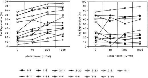
Induction of Fas expression on leukemia cells after the incubation with interferon γ and interferon α at the concentration of 40, 200, and 1,000 IU/mL. Each symbol denotes one patient whose UPN is shown. UPN x-y; × represents the FAB classification of the patient.
Induction of Fas expression on leukemia cells after the incubation with interferon γ and interferon α at the concentration of 40, 200, and 1,000 IU/mL. Each symbol denotes one patient whose UPN is shown. UPN x-y; × represents the FAB classification of the patient.
Fas Expression and Clinical Data of 64 Patients With AML
| . | Total . | Fas <50% . | Fas ≥50% . | P Value . |
|---|---|---|---|---|
| Total | 64 | 31 | 33 | |
| Sex (M/F) | 28/36 | 15/16 | 13/20 | NS |
| Age (yr), range (median) | 15-84 (53) | 15-79 (49) | 21-84 (55) | NS |
| Initial laboratory data, range (median) | ||||
| Leukocyte (109/L) | 0.7-460.0 (38.0) | 2.3-406.0 (54.0) | 0.7-310.0 (25.9) | NS* |
| Erythrocyte (109/L) | 840-4330 (2480) | 1450-4200 (2480) | 840-4330 (2470) | NS |
| Platelet (109/L) | 7-867 (55) | 7-254 (47.5) | 11-867 (52) | NS |
| LDH (IU/dL) | 199-9932 (1142) | 289-9932 (1167) | 199-2840 (1128) | NS |
| Remission after initial induction (%) | 36/55 (65.5) | 14/27 (51.8) | 22/28 (78.6) | <.05 |
Abbreviations: NS, not significant; LDH, lactate dehydrogenase.
P value = .063.
RESULTS
Fas expression on AML cells and its relationship with clinical features. Sixty-four AML samples diagnosed as AML were analyzed for their expression of Fas. As shown in Fig 1, the percentage of Fas positivity ranged from 2% to 98%. Eleven cases expressed Fas in less than 20% of leukemia cells (Fas−) and 20 cases expressed Fas in 20% to 50% of leukemia cells (Fas weak positive). Twenty-four cases showed Fas expression in 50% to 80% of leukemia cells (Fas intermediate positive), and nine expressed in more than 80% of leukemia cells (Fas strong positive). From these data, it is obvious that Fas was expressed in all cases, but the expression level varied widely.
Secondly, FAB classification and Fas expression was studied (Table 1). Median percentages of Fas positivity in leukemia cells of each patient were 19.5% in M1 (n = 9), 56.7% in M2 (n = 26), 67.0% in M3 (n = 6), 48.7% in M4 (n = 14), and 52.3% in M5 (n = 6) (Fig 2). RFI showed similar results (Table 1). Only 4 of 9 (44.4%) patients with AML M1 were Fas+ (≥20% of leukemia cell expressed Fas), whereas 49 of 55 (89.1%) patients with M2-5 cases were Fas+ (P < .01).
Clinical parameters such as sex, age, leukocyte/erythrocyte/platelet count at initial diagnosis, as well as response to the induction chemotherapy, were correlated with Fas expression: Fas ≥50% group and Fas <50% group (Table 2). Response to initial induction chemotherapy showed significant correlation with Fas expression (P < .05). Only 14 of 27 (51.8%) patients in Fas <50% group had complete remission after initial induction therapy, whereas 22 of 28 (78.6%) patients in Fas ≥0% group had it. Leukocyte count at initial diagnosis was higher in Fas <50% group than Fas ≥50% group with borderline significance (P = .063)
Induction of Fas by IFN. To study the effect of IFN, AML cells were cultured with IFNγ and α at the concentration of 40, 200, and 1,000 IU/mL. The results are shown in Fig 2. In most cases, a dose-dependent effect was observed in IFNγ. However, IFNα induced Fas in only three cases. In the remaining samples, IFNα failed to induce suppression of proliferation.
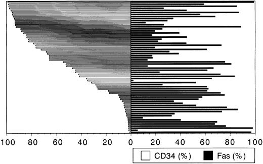
Relation between Fas expression and CD34 expression. The results of Fas and CD34 expression measured by two-color flow cytometry analysis are shown in Fig 3. Among 43 cases with CD34+ leukemia (≥20% leukemia cells were CD34+), 34 were Fas+ (≥20% cells Fas+) and 19 of 21 CD34− (<20% of cells were CD34+) were Fas+. Similar data is shown in Table 3. At any definition, there was no significant relationship between Fas expression and CD34 positivity (P = NS)
Fas and CD34 expression on cells from 64 patients with AML. The left half shows the percentage of CD34+ cell and the right half shows the percentage of Fas+ cell corresponding with the left column. No correlation was found between Fas expression and CD34 expression.
Fas and CD34 expression on cells from 64 patients with AML. The left half shows the percentage of CD34+ cell and the right half shows the percentage of Fas+ cell corresponding with the left column. No correlation was found between Fas expression and CD34 expression.
Fas Expression in the Context of CD34 Expression
| . | CD34− . | CD34+ . |
|---|---|---|
| Fas ≥20% | 19/21 (90.5%) | 34/43 (79.1%) |
| Fas ≥50% | 14/21 (66.7%) | 19/43 (44.2%) |
| Fas RFI | 1.28-14.66 (4.66) | 1.33-25.27 (4.33) |
| . | CD34− . | CD34+ . |
|---|---|---|
| Fas ≥20% | 19/21 (90.5%) | 34/43 (79.1%) |
| Fas ≥50% | 14/21 (66.7%) | 19/43 (44.2%) |
| Fas RFI | 1.28-14.66 (4.66) | 1.33-25.27 (4.33) |
Among 43 cases with CD34+ leukemia (CD34+, ≥20% leukemia cells were CD34+) 34 expressed Fas in ≥20% of cells and 19 expressed Fas in ≥50% cells. Among 21 CD34− (CD34−) cases, 19 and 14 expressed Fas in ≥20% and 50% of leukemia cells, respectively. Range and median are shown.
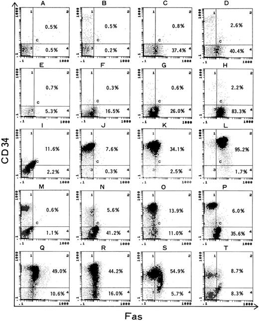
Figure 4 shows representative results of flow cytometric analysis of normal BM MNCs and leukemia cells. The results of two-color flow cytometric analysis was divided into three groups: the single population group (SP; 34 cases, E-L), the two-population group (TP; 22 cases, M-N), and the wide distributed group (WD; 8 cases, Q-T). The SP group (E-L) was divided into three subgroups according to the intensity of CD34 expression; 15 negative subgroup (0.6% to 8.1%, E-H) and 12 were CD34 strongly positive (J-L, 83.5% to 97.7% of population were CD34+). It is interesting that only three cases expressed CD34 intermediately (34.5%, 47.2%, and 66.1% of population were CD34+, I). Among CD34 strongly positive cases, Fas was expressed in 7.0% to 95.2% (median, 42.0%; RFI = 4.33) of total cells. Similarly, among CD34− cases, Fas was expressed in 7.3% to 98.3% (median, 54.4%; RFI = 4.66) of total cells. Analysis of CD34+ fraction of normal volunteers' bone marrow showed that Fas was expressed on only 12% of cells (median of 5 samples, D; data not shown). In the analysis of the TP group, the minor contamination with normal hematopoietic cells was theoretically possible. To avoid this possibility, if the minor population (<10%) was in the CD34− fraction, the case was excluded. A total of 21 cases met these criteria. The intensity of Fas expression was compared between two populations; Fas expression is stronger in the CD34− cell population than in the CD34+ one (CD34+ < CD34−) in 13 cases, CD34+ = CD34− in 8 cases, and CD34+ > CD34− in no cases. In the WD group, various patterns were observed (Q-T). One case (Q) presented that Fas expression was dominant in the CD34+ fraction. Three cases showed Fas expression was equivalent in both CD34+ and CD34− fraction (R).
Normal BM MNC (A-D) and representative data of 16 AML cells (E-T) of two-color flow cytometric analysis of Fas expression and CD34 expression. (A) Two isotypic control antibodies, (B) FITC control antibody and PE-conjugated anti-CD34 MoAb, (C) FITC-conjugated anti-Fas MoAb and PE control antibody, (D-T) FITC-conjugated anti-Fas MoAb and PE-conjugated anti-CD34 MoAb. In contrast to normal BM MNC (D) whose CD34+ cells are Fas−, some CD34+ cell in AML express Fas. Various patterns of distribution are shown; the single population group (SP, E-L), the two-population group (TP, M-N), and the wide distributed group (WD, Q-T).
Normal BM MNC (A-D) and representative data of 16 AML cells (E-T) of two-color flow cytometric analysis of Fas expression and CD34 expression. (A) Two isotypic control antibodies, (B) FITC control antibody and PE-conjugated anti-CD34 MoAb, (C) FITC-conjugated anti-Fas MoAb and PE control antibody, (D-T) FITC-conjugated anti-Fas MoAb and PE-conjugated anti-CD34 MoAb. In contrast to normal BM MNC (D) whose CD34+ cells are Fas−, some CD34+ cell in AML express Fas. Various patterns of distribution are shown; the single population group (SP, E-L), the two-population group (TP, M-N), and the wide distributed group (WD, Q-T).
Fas expression in context of CD34 and CD38. Three-color flow cytometric analysis was performed using 13 AML samples to determine the Fas expression in the gated population of CD34+/CD38 weak (CD34+CD38±, gate T), CD34+/CD38+ (CD34+CD38+, gate S), and CD34− (CD34−, gate R), which reflect the normal hematopoietic progenitor's developing pathway. Figure 5 demonstrates typical features of three-color analysis. Figure 6 shows the entire results of 13 AML cases. In most AML cells, both gated population CD34+CD38± cells and CD34+CD38+ cells express a high level of Fas. Fas was expressed at the highest level in CD34+CD38+ gated population in nine cases. The other four cases demonstrate that Fas expression become enhanced according to the developing pathway of normal hematopoiesis.
Representative data of three-color analysis of three AML cells. Cells were stained with anti-Fas–FITC, anti-CD38–PE, and anti-CD34–PE-Cy5. The left side shows a two-dimensional picture of CD34 and CD38. Electric gates were set to contain CD34− (R), CD34+/CD38+ (S) and CD34+/CD38± (T) cells. On the right, the histograms showing Fas expression on the three gated populations are presented.
Representative data of three-color analysis of three AML cells. Cells were stained with anti-Fas–FITC, anti-CD38–PE, and anti-CD34–PE-Cy5. The left side shows a two-dimensional picture of CD34 and CD38. Electric gates were set to contain CD34− (R), CD34+/CD38+ (S) and CD34+/CD38± (T) cells. On the right, the histograms showing Fas expression on the three gated populations are presented.
Three-color analysis of 13 leukemia samples. Fas expression (%) of each gated population (CD34+/CD38±, CD34+/CD38+ and CD34−) is demonstrated. Each symbol denotes each patient whose UPN is written below; see legend for Fig 2.
Three-color analysis of 13 leukemia samples. Fas expression (%) of each gated population (CD34+/CD38±, CD34+/CD38+ and CD34−) is demonstrated. Each symbol denotes each patient whose UPN is written below; see legend for Fig 2.
Induction of apoptosis by anti-Fas IgM. The summary of the induction of apoptosis by anti-Fas IgM in 24 cases is shown in Table 4. The morphologic apoptotic change (ΔN ≥ 5%) was observed in eight of 17 cases. DNA ladder experiments of these eight cases showed stronger intensity of ethidium fragment in all cases as compared with control (data not shown).
Fas Expression and Induction of Apoptosis
| UPN . | Fas (%) . | ΔN (%) . | Δ200 (%) . | Δ1000 (%) . | CFU-AML (−) . | CFU-AML (+) . |
|---|---|---|---|---|---|---|
| 2-18 | 11.8 | 4 | 4.1 | 15.2 | 0 | 0 |
| 1-5 | 13.4 | 8 | −2.9 | 3.9 | 63 | 58.5 |
| 1-9 | 15.4 | ND | −1.5 | −1.3 | 0 | 0 |
| 4-14 | 17.1 | ND | 0.6 | 1.7 | 29.5 | 5 |
| 1-8 | 17.5 | ND | −3.5 | −2.1 | 0 | 0 |
| 4-13 | 22.2 | 7 | 7.0 | 18.9 | 113 | 132.5 |
| 2-21 | 24.7 | 8 | 10.4 | 19.8 | ND | ND |
| 2-14 | 26.6 | −5 | ND | 5.9 | ND | ND |
| 4-6 | 34.1 | −1 | 0 | 5.6 | 23.5 | 21 |
| 2-16 | 38.6 | 0 | 0.1 | 1.6 | ND | ND |
| 4-11 | 41.6 | ND | 1.5 | 2.8 | ND | ND |
| 3-6 | 41.6 | ND | ND | 1.1 | ND | ND |
| 5-8 | 45.0 | −1 | 0.6 | 1.3 | 0 | 0 |
| 2-9 | 45.1 | 1 | 7.5 | 24.9 | 20 | 6.5 |
| 4-4 | 54.4 | 37 | 13.6 | 19.5 | 7.5 | |
| 2-12 | 55.9 | 9 | ND | 14.7 | 247 | 5.5 |
| 5-10 | 61.2 | ND | 0.3 | 9.1 | 0 | 0 |
| 4-9 | 65.1 | 22 | ND | 14.6 | ND | ND |
| 2-26 | 72.1 | 7 | −0.6 | 9.0 | 81 | 4.5 |
| 5-3 | 72.8 | 8 | ND | 10.4 | 21 | 13 |
| 2-22 | 73.1 | 0 | 7.5 | 7.3 | ND | ND |
| 3-5 | 77.1 | ND | 0.6 | 2.2 | ND | ND |
| 2-23 | 83.4 | 1 | 0.8 | 2.0 | 0 | 0 |
| 2-7 | 85.9 | 1 | 1.5 | 5.4 | 7 | 3 |
Δ, difference between the number of cells (%) cultured with anti-Fas MoAb and those cultured with control MoAb; ΔN, counted by morphological method, Δ200 and Δ1000; determined by colorimetric DNA fragmentation assay by flow cytometric analysis after the culture with anti-Fas MoAb at the concentration of 200 and 1,000 ng/mL, respectively.
Abbreviation: ND, not done.
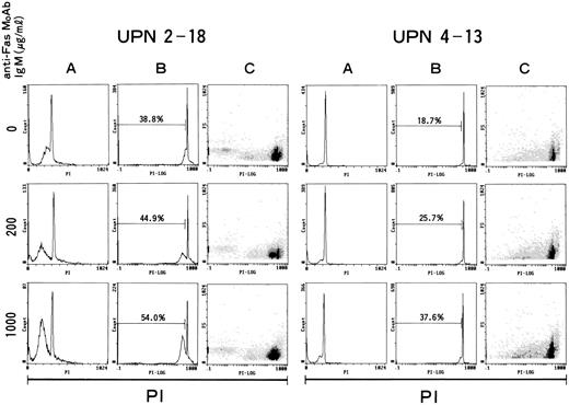
Representative results of three colorimetric DNA fragmentation assays. AML cells were incubated with anti-Fas MoAb control; top, 200 ng/mL; center, 1,000 ng/mL; bottom). X axis shows intensity of fluorescence caused by PI. (A) Count and linear PI, (B) count and logarithmic PI, and (C) FS and logarithmic PI. UPN, unique patient number. Dose-dependent induction of Fas-induced apoptosis is demonstrated.
Representative results of three colorimetric DNA fragmentation assays. AML cells were incubated with anti-Fas MoAb control; top, 200 ng/mL; center, 1,000 ng/mL; bottom). X axis shows intensity of fluorescence caused by PI. (A) Count and linear PI, (B) count and logarithmic PI, and (C) FS and logarithmic PI. UPN, unique patient number. Dose-dependent induction of Fas-induced apoptosis is demonstrated.
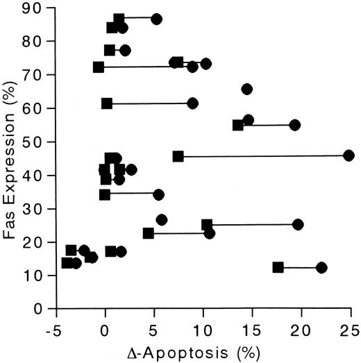
DNA fragmentation was also measured by flow cytometry in 24 leukemia samples. The representative results are shown in Fig 7. In 14 of 24 samples, more than 5% increase of the fraction, which indicates apoptosis after 20 hours incubation with anti-Fas IgM at the concentration of 1 μg/mL, was observed (Table 4). As shown in Fig 8 and Table 5, there was no clear relationship between the Fas expression and the induction of apoptosis. However, only one of five Fas− (Fas <20%) leukemias demonstrate induction of apoptosis, whereas 13 of 19 Fas+ leukemias showed borderline significance (χ2 test, P = .051).
Fas expression and the induction of apoptosis is shown. Δ-apoptosis (Δ200 and Δ1000; % increase of apoptosis after the incubation with anti-Fas MoAb at different concentration) is the percentage of increased number of apoptotic cells by colorimetric DNA fragmentaion assay. (▪ and • indicate the value after the culture with anti-Fas at the concentration of 200 and 1,000ng/mL, respectively.
Fas expression and the induction of apoptosis is shown. Δ-apoptosis (Δ200 and Δ1000; % increase of apoptosis after the incubation with anti-Fas MoAb at different concentration) is the percentage of increased number of apoptotic cells by colorimetric DNA fragmentaion assay. (▪ and • indicate the value after the culture with anti-Fas at the concentration of 200 and 1,000ng/mL, respectively.
Fas-Induced Apoptosis and Expression of Fas and CD34
| Δ1000 . | N . | Fas+ (%) . | CD34+ (%) . | Fas+CD34+ (%) . |
|---|---|---|---|---|
| <5% | 10 | 40.1 | 46.9 | 6.2 |
| ≥5% | 14 | 55.2 | 87.8 | 15.9 |
| P value5-150 | 0.27 | 0.07 | 0.03 |
| Δ1000 . | N . | Fas+ (%) . | CD34+ (%) . | Fas+CD34+ (%) . |
|---|---|---|---|---|
| <5% | 10 | 40.1 | 46.9 | 6.2 |
| ≥5% | 14 | 55.2 | 87.8 | 15.9 |
| P value5-150 | 0.27 | 0.07 | 0.03 |
Median percentage of Fas+ fraction, CD34+ fraction and double positive fraction in each group are shown.
t-test.
Next, we compared the CD34 expression with Fas-mediated apoptosis (Δ1000 ≥5%) using Student's t-test. As shown in Table 5, the expression of CD34 is higher in Δ1000 ≥5% group than in Δ1000 <5% group with borderline significance (P = .07) When the percentage of double positive fraction (CD34+Fas+) was compared, Δ1000 ≥5% group demonstrated a significantly greater percentage (P < .05).
CFU-AML assay was performed using 16 leukemias and among them, leukemia colonies were detected in only 10 cases. Colonies were separated and stained to confirm the malignant morphology. Using these 10 cases, the effect of anti-Fas MoAb was studied. The numbers of colonies decreased more than 20% in seven cases after the incubation with anti-Fas MoAb (1 μg/mL) for 16 hours (Table 4).
DISCUSSION
In the current study, we found that AML cells expressed Fas with variable intensity (2% to 98%) in all cases. Only a few studies have reported the expression of Fas on AML cells.15,16 Twenty-eight percent of AML cases were positive for Fas expression in one study,15 whereas Fas was expressed on the majority of AML cells in another study,16 which is in agreement with our results. These discrepancies may be explained by the different detection methods that were used, immunofluorescence assay with a fluorescence microscope and flow cytometric analysis, respectively.
Fas expression has been studied in various leukemias using flow cytometric analysis. For the expression in plasma cells from 28 multiple myeloma (MM) patients, 15 were positive.17 The Fas was expressed in a minority (5% to 41%; mean, 15.6%) of chronic lymphocytic leukemia (CLL) cells in 10 of 21 patients.18 The leukemia cells from all patients with ATL strongly expressed Fas.9 Compared with these studies, the intensity of Fas expression in AML was quite variable (2% to 98%), and this may reflect the heterogeneity of AML. In normal hematopoiesis, immature cells do not express a significant level of Fas.19 Fas expression becomes enhanced with the maturation pathway of myeloid series.20 Variable expression in AML may reflect only the difference in the maturation stage of the leukemia cell. Indeed, our study demonstrated very immature subtype; AML-M1 showed significantly weaker expression than the other subtypes (Table 1). Interestingly, some CD34+ AML cells express Fas at a high level despite the weak expression of Fas in normal CD34+ cells.19,21 Thus, an alternative interpretation is that the Fas might be induced via several pathways such as cytokines and cell-cell interactions. Recent studies have shown that IFNγ and TNFα can induce Fas on the surface of normal and tumor cells.8,19 22 It could be hypothesized that effector cells recognize AML cells and produce IFNγ or TNFα enhancing Fas expression. Thereafter, different effector cells (CTL) would attack the target cells using the Fas system. In the current study, we have confirmed the induction of Fas by IFNγ on AML cells (Fig 2). Preliminary study suggests that IFNγ enhanced the Fas-mediated apoptosis in addition to the Fas phenotype (data not shown). The possibility of aberrant expression of Fas in tumor cells could not be excluded.
Several investigators have studied whether Fas mediates apoptosis using the anti-Fas MoAb, which can act as Fas agonist.8,9,17,18 Leukemia cells from CLL were resistant to Fas-mediated apoptosis despite moderate expression of Fas. However, induction of apoptosis was apparently observed in ATL. In multiple myeloma, some cases also were susceptible to the treatment with anti-Fas MoAb. In a parallel study of CML cells performed in our laboratory, anti-Fas MoAb inhibited colony growth of leukemia cells (data not shown). The colorimetric DNA fragmentation assay also shows that 14 leukemia samples of 24 AML cases were susceptible to the treatment (Table 4). The amount of Fas did not directly correlate with the induction of apoptosis, as suggested by others. However, Fas+ (≥20%) leukemias proved to be more susceptible to the induction of apoptosis than Fas− (<20%) with borderline significance (P = .051). It is possible that apoptosis may be induced by the accumulation of various kinds of signals and the Fas pathway may be one important mechanism of apoptosis. In one study, the amount of bcl-2 expression was correlated with the resistance to Fas-induced apoptosis.23 24
From all these studies, we conclude that the induction of apoptosis by anti-Fas MoAb seems to be the strongest in CML and MM, weaker in AML, and had the least effect in CLL. It is interesting that these data are similar to the efficacy of DLI for relapse after BMT. Effectiveness of DLI was reported to be 60% in CML, 40% in MM, 15% in AML, and 20% in ALL.25 This evidence for the relation between Fas expression and cellular immunity is also supported by recent basic studies. One study showed that the CD4+ CTL killing was largely dependent on FasL and Fas system.26 Taken together, clinical observations and laboratory data suggest that the Fas system may be one of the pathways of cellular immunity for leukemia cells.
Although the number of patients was small, Fas expression was significantly associated with the results of initial induction therapy (P < .05). Synergistic effect of Fas-mediated apoptosis and antileukemia agents on apoptosis may be suggested. A recent study has reported that doxorubicin-induced apoptosis in T-cell line use FasL/Fas system.27 In another study, inhibitor of interleukin-1 converting enzyme prevented antitumor agent-induced apoptosis in human myeloid leukemia U937.28 Recently, one study concluded that selection of drug resistance in the cell lines results in a resistance to Fas-mediated apoptosis.29
The goal of this study was to establish whether clonogenic populations of AML expressed Fas and were functionally more susceptible to apoptosis. CD34 and CD38 expression are well studied as markers for normal hematopoietic progenitors. However, phenotype of clonogenic leukemia cells are still investigated. In the study using severe combined immunodeficiency (SCID) mice, the frequency of leukemia-initiating cells in the peripheral blood of AML patients was one in 250,000 cells and these clonogenic cells belonged to the CD34+ CD38− population.11 Similarly, Haase et al13 reported that malignant transformation, as well as disease progression, may occur at the level of CD34+/CD38− cells with multilineage potential. We investigated the Fas expression in the context of CD34 in 64 cases, yet we could not demonstrate differences between CD34+ fraction and CD34− population. For the detection of relatively small populations of clonogenic cells, CD34 positivity is too insensitive for precise study. Therefore, we have used the CD34+CD38±, CD34+CD38+ and CD34− populations for their expression of Fas using three-color flow cytometric analysis. In general, Fas is expressed at a high level both in the gated CD34+CD38± and CD34+CD38+ populations (Fig 6). In the majority of cases, Fas was expressed strongest in the CD34+CD38+ population. Recently, using more sensitive methods, expression of Fas on CD34+ hematopoietic progenitors isolated from human fetal liver was reported. The most immature subfractions CD34++CD38− and CD34++CD38+ cells (equivalent to CD34+CD38± in the current study) expressed Fas, whereas the more mature CD34++CD38++ and CD34+CD38++ cells (equivalent to CD34+CD38+ in the current study) displayed low Fas expression.30 The discrepancy between ours and this study may be explained by the different sources of hematopoietic progenitor cells (leukemia cells v fetal liver cells).
To study the Fas-mediated apoptosis in the clonogenic population, we performed CFU-AML assay using a relatively small number of samples. In some of the samples tested, a reduced number of colonies after the culture with anti-Fas MoAb could be observed. However, further study is needed to clarify the function of Fas in the clonogenic population of leukemia. Finally, the analysis of the expression and function of Fas on the leukemia stem cells will shed light on the mechanism of eradication of leukemia cells and the prognosis and the response to several kinds of strategies such as chemotherapy, radiotherapy, immunotherapy, and cytokine therapy.
ACKNOWLEDGMENT
This study was contributed by clinical samples from Drs Y. Morishita (Kouseiren Showa Hospital), Y. Kodera (Nagoya First Red Cross Hospital), S. Yokomaku (Sannomaru Hospital), K. Suzuki (Okazaki City Hospital), T. Takeo (Yokkaichi City Hospital), K. Yano (Hamamatsu Medical Center), A. Matsuoka (Anzyo Kosei Hospital) and T. Kataoka (Nagoya Memorial Hospital). We thank S. Suzuki and M. Suzuki for technical assistance. We are also grateful to Drs S. Yonehara (Kyoto Unviersity), K. Nishimura (Japan Tobacco Co), and K. Koike (Aichi Cancer Center Hospital) for fruitful discussions and to Dr Yokozawa for critical reading of the manuscript.
Supported by The Grant in Aids of Ministry of Health and Welfare Japan (Tokyo, Japan) and also by Aichi Blood Disease Research Foundation (Nagoya, Japan).
Address reprint requests to Koichi Miyamura MD, The First Department of Internal Medicine, Nagoya University School of Medicine, Tsurumai 65, Showa-ku, Nagoya 466, Japan.



This feature is available to Subscribers Only
Sign In or Create an Account Close Modal