Abstract
This study evaluated the effect of filgrastim (granulocyte colony-stimulating factor [G-CSF]) on the duration of granulocytopenia and thrombocytopenia after intensive consolidation therapy with diaziquone (AZQ) and mitroxantrone for patients less than 60 years of age with acute myeloid leukemia (AML) in complete remission. Patients less than 60 years of age with AML who achieved complete remission (CR) with daunorubicin and cytarabine induction therapy, were scheduled to receive three sequential courses of high-dose cytarabine, cyclophosphamide/etoposide, AZQ, and mitroxantrone in a pilot study to determine their tolerance of these three sequential consolidation regimens. The initial patients treated with AZQ and mitoxantrone experienced prolonged bone marrow suppression and, therefore, subsequent cohorts were treated with G-CSF, 5 μg/kg, beginning the day after completion of the third cycle of chemotherapy. There was a marked decrease in the duration of granulocytopenia less than 500/μL in two groups of patients receiving two different dose levels of AZQ and the same dose of mitoxantrone compared with patients not receiving the G-CSF. There was also a decrease in the need for hospitalization, as well as the duration of hospitalization. There was a trend towards shortening of the duration of thrombocytopenia, as well. The duration of complete remission and overall survival was similar in patients who received or did not receive G-CSF. G-CSF markedly shortened the duration of granulocytopenia in patients with AML receiving intensive postremission consolidation with AZQ and mitoxantrone. There was no adverse effect on CR duration or survival.
ACUTE MYELOID LEUKEMIA (AML) is treatable and potentially curable with intensive chemotherapy accompanied by recent improvements in supportive care. In a recent Cancer and Leukemia Group B (CALGB) study, 44% of patients less than 60 years of age who achieved complete remission (CR) with standard cytosine arabinoside (Ara-C) and daunorubicin chemotherapy and who subsequently received up to four courses of postremission consolidation chemotherapy with high-dose cytosine arabinoside (HIDAC), were estimated to remain free of disease at 5 years.1 This is comparable to results in similar patients treated with bone marrow transplantation in remission.2 Barriers to a higher rate of cure in AML include drug-resistant leukemia, extramedullary toxicity from chemotherapeutic drugs, and prolonged pancytopenia due to ablative chemotherapy.
Hematopoietic growth factors, including granulocyte-colony stimulating factor (G-CSF ) and granulocyte macrophage-colony stimulating factor (GM-CSF ), have been shown to accelerate granulocyte recovery following chemotherapy for patients with a variety of solid tumors, including small cell carcinoma of the lung, sarcomas, and bladder cancers.3,4 These agents have also been evaluated as a means of accelerating granulocyte recovery after completion of induction chemotherapy for AML, and as ‘priming’ agents to induce leukemia cell proliferation and increased sensitivity to chemotherapy. The role of G-CSF and GM-CSF before, during, and following a variety of Ara-C based induction chemotherapy regimens in adult AML has recently been reviewed.5,6 Randomized trials using GM-CSF after completion of induction therapy have been conducted in patients greater than 60 years of age, with one trial demonstrating a shortening of the duration of neutropenia and a possible decrease in the incidence of severe infections.7 A larger CALGB trial of similar design showed a small decrease in the length of neutropenia, but failed to show associated clinical benefit from the use of GM-CSF.8 Lastly, two recent trials using G-CSF after induction therapy also showed a 2- to 3-day decrease in severe neutropenia, again, however, without an improvement in CR rate or the incidence of severe infections.9 10 Most importantly, however, none of these studies suggested a significant rate of clinically detectable stimulation of leukemia growth, a theoretically critical concern when these agents are used in patients with myeloid malignancies.
CALGB protocol 9022 was a pilot study designed to evaluate the feasibility of administering sequential courses of potentially noncross resistant chemotherapy to patients with AML in CR. Patients were scheduled to receive three sequential courses of HIDAC, cyclophosphamide and etoposide (VP-16), and diaziquone (AZQ) and mitoxantrone. The doses used in these courses were determined by a series of Phase I/II trials previously reported by the CALGB or its member institutions, which demonstrated activity in patients with AML in relapse.11-14 The initial patients treated with AZQ and mitoxantrone on protocol 9022 experienced prolonged bone marrow suppression with delayed recovery of granulocytes and platelets, even after a decrease in the dose of AZQ. The protocol was, therefore, amended so that subsequent patients received G-CSF following completion of the AZQ/mitoxantrone therapy. We report a definitive decrease in the duration of severe granulocytopenia and, to a lesser degree, a decrease in the total number of days of thrombocytopenia with the use of G-CSF. This is the first report of a well-defined series of AML patients, treated with intensive consolidation chemotherapy in remission, demonstrating a definite decrease in the duration of bone marrow suppression with G-CSF.
MATERIALS AND METHODS
Eligibility.Eligible patients were between 15 and 60 years of age with newly diagnosed AML, morphologically defined as M0 to M7 by the French-American-British (FAB) classification of acute leukemias.15 Megakaryocytic leukemia was diagnosed by ultrastructural peroxidase or appropriate monoclonal antibodies (MoAbs). FAB M0 was defined in patients with morphologically undifferentiated leukemia if blasts were reactive with at least one of the ‘myeloid’ MoAbs against CD33, CD13, CD11b, or CD14, with all lymphoid markers required to be negative.16 At least 30% replacement of nonerythroid marrow elements by myeloid blasts was also required for eligibility. Patients were excluded for significant cardiac, hepatic, or pulmonary dysfunction not attributable to infiltration by leukemia. Patients were not eligible if they had received previous chemotherapy or radiation therapy or had any previous hematologic malignancy, myeloproliferative disorder, or myelodysplastic syndrome.
Treatment plan.This phase II study was designed to determine the feasibility of administering three sequential courses of intensive postremission therapy with HIDAC, cyclophosphamide and etoposide, and AZQ and mitoxantrone, to patients with AML in first remission after treatment with standard induction chemotherapy. Induction chemotherapy consisted of Ara-C 200 mg/m2 daily for 7 days by continuous infusion and daunorubicin 45 mg/m2/daily for 3 days by rapid intravenous infusion. A bone marrow aspirate was performed on day 14 to determine the degree of marrow hypoplasia. If more than 5% of remaining cells were leukemic and if the cellularity was 15% or greater, a second induction course was given consisting of 5 days of Ara-C and 2 days of daunorubicin in the same doses described above.
Patients achieving CR received three sequential courses of intensification therapy. The first consisted of HIDAC 3,000 mg/m2 given over 3 hours every 12 hours on days 1, 3, and 5 (for a total of six doses over 5 days). This was identical to the regimen used in an earlier CALGB study.1 Upon recovery of blood counts, a second intensification course was given with etoposide (1,800 mg/m2 by infusion over 25 to 26 hours) followed by cyclophosphamide (50 mg/kg given as a 2-hour infusion on each of days 2 and 3 for a total dose of 100 mg/kg). Following recovery from intensification cycle number 2, patients were to receive AZQ (NSC #182986) at 28 mg/m2 by continuous infusion each day for 3 days along with mitoxantrone (dihydroanthracenedione) 12 mg/m2 by slow intravenous push each day for 3 days.
The first 28 patients receiving the third intensification course experienced prolonged granulocytopenia. Because it was believed that this prolonged marrow suppression was most likely due to AZQ, the protocol was amended to reduce the dose of AZQ to 24 mg/m2 . The myelosuppression remained severe even after this dose reduction, and the protocol was amended further to allow the use of G-CSF. This resulted in more rapid myeloid recovery and the dose of AZQ then was increased to its original value. Hence, there were four treatment groups, resulting from modifications to the third intensification course, which will be described in more detail: Group 1, AZQ 28 mg/m2 , mitoxantrone 12 mg/m2 , no G-CSF; Group 2, AZQ 24 mg/m2 , mitoxantrone 12 mg/m2 , no G-CSF; Group 3, AZQ 24 mg/m2 , mitoxantrone 12 mg/m2 , G-CSF 5 μg/kg; Group 4, AZQ 28 mg/m2 , mitoxantrone 12 mg/m2 , G-CSF 5 μg/kg. G-CSF (Filgrastim, Amgen, Thousand Oaks, CA) was given beginning on day 4 of the AZQ/mitoxantrone course and continued until granulocyte recovery as defined below.
Investigational Review Board (IRB) approval was obtained at each participating institution and appropriate written informed consent was given by each patient. Members of the Data Audit committee of the CALGB visit all participating institutions at least once every 3 years to verify compliance with federal regulations and protocol requirements.17 The medical records of 25 of the 249 patients (10%) treated in this study, drawn from all 24 participating institutions and their affiliates, were randomly selected and audited.
Evaluation and response criteria.All patients were evaluated with detailed medical history and physical examination at the initiation of therapy, through the induction cycle(s), and during each intensification course. CR was documented when a bone marrow cellularity of at least 20% was achieved with normal maturation of all cell lines with less than 5% blast cells and the elimination of any cells considered to be leukemic (Auer rods, etc). The definition of CR by CALGB criteria also required an absolute granulocyte count ≥1,500/μL, platelet count ≥100,000/μL and the absence of any leukemic cells in the peripheral blood or extramedullary disease. These same criteria had to be met before each consolidation course. Patients were considered to have failed to respond to induction therapy if CR was not achieved after two cycles of therapy. Relapse was defined as marrow infiltration with 25% or greater leukemic cells in a patient with previous CR, the detection of leukemic cells in the central nervous system, or any other evidence of leukemic relapse.18
Granulocyte recovery.The duration of granulocytopenia was defined as the number of days from the date granulocytes decreased below 500/μL to the date of recovery of granulocytes ≥500/μL on 2 successive days. Therefore, this represented the time from the earliest demonstration of myelosuppression to the recovery of granulocytes. Because patients began each course of intensification therapy with normal blood counts and some were outpatients afterwards, if values were not available, it was assumed the granulocyte count remained above 500/μL for the first 5 days of the intensification course. If some values were missing on subsequent days, the data were handled as follows. If one of the known values was high (≥ 500/μL) and another was low, then the number of days of granulocytopenia was defined to be one half the number of days for which the values were missing, rounding up to the next highest number of days when fractions of days were considered.
Platelet recovery.The duration of thrombocytopenia was calculated from the date when the platelet count decreased below 20,000/μL to the date of recovery of platelets ≥ 20,000/μL on 2 successive days without platelet transfusion. Similar guidelines were used for missing data points as described above. The duration of granulocytopenia and thrombocytopenia were calculated separately for each patient.
Recovery of both counts.The time to recovery of both counts was taken to be the interval between the first day of myelosuppression (either absolute neutrophil count [ANC] < 500/μL or platelets < 20,000/μL) and the last day of myelosuppression (ANC ≥ 500/μL and platelets ≥ 20,000/μL).
Experimental design/statistical methods.The initial planned main outcome measure in this study was the proportion of patients completing all three courses of the postremission intensification therapy. It was anticipated that 60 patients would be required to meet this objective. Due to more than expected myelosuppression on the third course of intensification, three treatment modifications were made. A statistical amendment to the protocol specified that approximately 25 patients be entered into each of the modified treatment groups. The duration of granulocytopenia and thrombocytopenia were analyzed using the proportional hazards regression model.19 Specifically, the model consisted of a term for AZQ (28 or 24 mg/m2), and a term for G-CSF (0 or 5 μg/kg). To test whether G-CSF works differently depending on the dose of AZQ, a test of the interaction between these two treatments was performed using the likelihood ratio statistic.
Survival, defined to be the time from study entry to death from any cause, and CR duration, defined as the time from achieving CR to relapse (bone marrow or nonbone marrow) or death, were other endpoints used in this study. Time to recovery of blood counts, survival, and remission duration curves were based on Kaplan-Meier estimates, and differences between groups were tested using the log rank statistic, adjusting for multiple comparisons when appropriate.20-22 Ninety-five percent confidence intervals (CI) for medians were calculated by the method of Brookmeyer and Crowley.23 All reported P values are based on two-sided significance tests. The results are based on follow-up as of August 21, 1996.
RESULTS
CALGB study 9022 accrued 249 patients from 24 participating main member institutions and their affiliated hospitals between October 1, 1990 and March 31, 1992. The characteristics for these patients are given in Table 1. The median age was 41 years with a range of 16 to 59 years; 55% were women, and 81% were Caucasian. The histologic evaluation included 46% with an FAB classification of M1 or M2. In 24 patients who were considered to have AML, the FAB type could not be determined. On central review of histology, three cases could not be classified as AML and two cases were felt to have myelodysplasia. These five cases and one case that had central nervous system (CNS) disease at study entry were considered ineligible for the study and are excluded from the statistical analyses.
Characteristics at Time of Study Entry for 249 Patients Entered on CALGB 9022
| Age (yrs) | |
| Median, 41 (range, 16-59) | |
| Years | No. of Patients |
| <30 | 57 (23%) |
| 30-39 | 63 (25%) |
| 40-49 | 63 (25%) |
| 50-59 | 66 (27%) |
| Sex | |
| Males: | 113 (45%) |
| Females: | 136 (55%) |
| Race | |
| White: | 201 (81%) |
| Black: | 22 (9%) |
| Other: | 26 (10%) |
| Performance status | |
| 0 | 80 (32%) |
| 1 | 113 (45%) |
| 2 | 41 (16%) |
| 3 | 10 (4%) |
| 4 | 5 (2%) |
| FAB classification* | |
| M0 | 5 (2%) |
| M1 | 40 (16%) |
| M2 | 74 (30%) |
| M3 | 34 (14%) |
| M4 | 57 (23%) |
| M5 | 10 (4%) |
| M6 | 1 (<1%) |
| Mixed | 3 (1%) |
| AML, other | 24 (10%) |
| Hematologic values | |
| Hemoglobin (gm/dL; n = 245) | Median, 9.2 (range, 3.5-22.6) |
| Platelets (×109/L; n = 248) | Median, 58.0 (range, 10-514) |
| WBC (×109/L; n = 248) | Median, 11.1 (range, 0.2-318.4) |
82% were reviewed and classified in a CALGB central morphology laboratory; the remainder represent institutional assessments.
CR was achieved in 186 (77%) of the 243 eligible patients. Forty (16%) failed to respond and were followed off study for survival, while 17 (7%) of the patients treated died during induction. Of the 186 patients who achieved CR, 176 (95%) received the first course of intensification treatment. The reasons for not continuing therapy beyond induction included: residual toxicity from induction therapy or treatment discontinued (6 patients), early relapse (2 patients), patient withdrawal (2 patients). Induction and postremission treatment results are given in Table 2.
Induction and Postremission Treatment Results
| Induction | ||
| CR | 186 (77%) | |
| Failed induction | ||
| No response | 40 (16%) | |
| Died in induction | 17 (7%) | |
| Total | 243 | |
| Postremission intensification |
| Induction | ||
| CR | 186 (77%) | |
| Failed induction | ||
| No response | 40 (16%) | |
| Died in induction | 17 (7%) | |
| Total | 243 | |
| Postremission intensification |
| Course 1 | |||
| Course 2 | |||
| Course 3 | |||
| No. eligible | 186 | 170 | 153 |
| Started course | 176 | 157 | 127 |
| Did not start | |||
| Treatment discontinued | 6 | 1 | 4 |
| Withdrew | 2 | 10 | 15 |
| Relapse | 2 | 2 | 7 |
| Completed course | 170 | 153 | 112 |
| Did not finish | |||
| Treatment discontinued | 3 | 1 | 0 |
| Withdrew | 0 | 0 | 0 |
| Relapsed | 2 | 2 | 5 |
| Died | 1 | 1 | 6 |
| Lost to follow-up | 0 | 0 | 1* |
| Other treatment | 0 | 0 | 3* |
These four patients are not evaluable for recovery from myelosuppression. One patient in group 1 and two in group 2 received G-CSF during their course.
After the first course of postremission therapy, 157 patients began the second course, and 127 entered the third intensification course. Overall, 68% of patients achieving CR received three courses of intensification. The primary reasons for not reaching the third course of intensification were the physician's decision to discontinue treatment (9 patients [18%]), withdrawal or refusal by the patient to continue with further treatment (25 patients [51%]), relapse (13 patients [27%]), and death (2 patients [4%]) (Table 2).
Patients experienced various degrees of myelosuppression during the third intensification course, resulting in three dose modifications, two of which included the growth factor G-CSF. The 123 evaluable patients were assigned to the sequential treatment groups shown in Table 3. The demographics of these 123 patients were similar to those of the total group of patients entered on the study. The median interval between the date of CR and the time to start of the third course was 81, 105, 109, and 120 days for groups 1 through 4, respectively. Eleven patients failed to recover blood counts after course 3. Failure to recover was due to death in six patients and leukemic relapse in five. Hence, of 176 patients starting the postremission regimen, 112 (64%) completed all three courses of therapy. This is slightly less than in the previous CALGB trial where 69% of patients of similar age completed four courses of Ara-C treatment at doses of 400 mg/m2 or 3gm/m2 .1
Recovery of Blood Counts Following the Third Intensification Course by Treatment Group
| Group . | N . | AZQ3-150 (mg/m2) . | G-CSF . | Median Time to Recovery (days) and 95% CI . | Hospitalized . | Median (range) Duration of Hospitalization (days) . | ||
|---|---|---|---|---|---|---|---|---|
| . | . | . | (μ/kg) . | ANC ≥500 . | PLT ≥20,000 . | Both Counts3-151 . | . | . |
| 1 | 28 | 28 | 0 | 34.8 (30, 40) | 35.5 (28, 45) | 42.3 (30, 47) | 25 (96%)3-152 | 40 (11-91) |
| 2 | 34 | 24 | 0 | 30.6 (30, 34) | 27.7 (24, 32) | 33.7 (31, 41) | 31 (97%) | 30 (2-80) |
| 3 | 27 | 24 | 5 | 19.5 (18, 23) | 18.8 (17, 26) | 24.3 (22, 31) | 20 (83%) | 24 (6-44) |
| 4 | 34 | 28 | 5 | 21.9 (18, 28) | 31.9 (21, 41) | 35.6 (28, 43) | 27 (87%) | 20 (1-58) |
| 1 + 2 | 62 | 24/28 | 0 | 31.1 (31, 36) | 30.2 (26, 38) | 39.1 (32, 43) | 56 (97%) | — |
| 3 + 4 | 61 | 24/28 | 5 | 20.5 (19, 24) | 23.4 (19, 31) | 29.1 (25, 36) | 47 (85%) | — |
Abbreviation: N, number.
All patients received 12 mg/m2 of mitoxantrone for 3 doses.
Time to recovery of both ANC and platelets.
Percent is based on number of patients with complete data on hospitalization.
Table 3 summarizes the median number of days to recovery (and 95% CI) of blood counts for each of the treatment groups in the third intensification course. The proportional hazards model was used to help distinguish the effect of the two different doses of AZQ and the use of G-CSF on time to recovery of blood counts during the third intensification phase of treatment. A test for interaction between the two drugs was not statistically significant for any of the endpoints considered (P = .89 for recovery of ANC ≥ 500/μL, P = .27 for recovery of platelets ≥ 20,000/μL, and P = .28 for recovery of both counts). In addition, there was no statistically significant effect of the two different doses of AZQ for ANC (P = .34). For platelets and for both counts however, there was evidence of an effect of the lower dose of AZQ (P = .01 and .04, respectively).
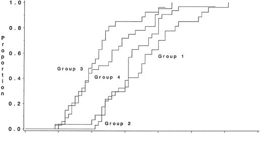
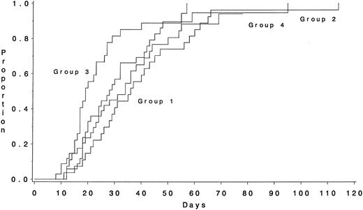
There was a statistically significant effect of G-CSF on time to recovery of granulocytes ≥ 500/μL (P < .001) (Fig 1), but not on time to recovery of platelets >20,000/μL (P = .12) or time to recovery of both counts (P = .07) (Fig 2). Patients receiving the growth factor (groups 3 and 4) had a median time to recovery of ≥ 500/μL granulocytes of 20.5 days, compared with 31 days for patients not treated with G-CSF (groups 1 and 2, Fig 1). The median time to recovery of both counts (first day of myelosuppression to recovery of ANC ≥ 500/μL and platelets ≥ 20,000/μL) was 29 days and 39 days, respectively, for these two groups of patients. When the two treatment groups that received 24 mg/m2 of AZQ were considered separately, however, there was still no significant difference in time to recovery of platelets > 20,000/μL between groups 2 and 3 (27.7 and 18.8 days, respectively; P = .13), after adjustment for multiple comparisons. Lastly, the groups that did not receive G-CSF had a median time to recovery of platelets ≥50,000/μL compared with 31.9 days for groups 3 and 4 (P = .16).
Time to granulocyte recovery (ANC ≥ 500/μL) for the four groups who received the third course of postremission intensification therapy. The median times to recovery were 30.6 days, 19.5 days, 19.0 days, and 23.3 days for groups 1, 2, 3, and 4, respectively. Patients receiving G-CSF (groups 3, 4) had significantly shorter times to recovery compared with those who did not receive growth factor (groups 1 and 2; P < .001).
Time to granulocyte recovery (ANC ≥ 500/μL) for the four groups who received the third course of postremission intensification therapy. The median times to recovery were 30.6 days, 19.5 days, 19.0 days, and 23.3 days for groups 1, 2, 3, and 4, respectively. Patients receiving G-CSF (groups 3, 4) had significantly shorter times to recovery compared with those who did not receive growth factor (groups 1 and 2; P < .001).
Time to platelet recovery (≥20,000/μL) for the four groups who received the third course of postremission intensification therapy. The median times to recovery were 35.5 days, 27.7 days, 18.8 days, and 31.9 days for groups 1, 2, 3, and 4, respectively. There was no significant difference between the two growth factor groups (P = .12).
Time to platelet recovery (≥20,000/μL) for the four groups who received the third course of postremission intensification therapy. The median times to recovery were 35.5 days, 27.7 days, 18.8 days, and 31.9 days for groups 1, 2, 3, and 4, respectively. There was no significant difference between the two growth factor groups (P = .12).
Importantly, the shortened duration of neutropenia was associated with decreased durations of hospitalization (Table 3). Although most patients in all four groups received antibiotics, fewer patients receiving G-CSF required hospitalization (approximately 85%) compared with the cohorts not receiving the growth factor (approximately 97%, P = .05) with a marked decrease in the length of hospitalization. There was also a decrease in the frequency of grade III or greater infections in the groups receiving G-CSF (71% and 75% incidence in groups 1 and 2 v 58% and 47% in groups 3 and 4, respectively). There were three deaths and three relapses in groups 1 and 2 with three deaths and four relapses in the patients receiving G-CSF.
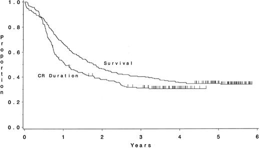
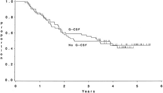
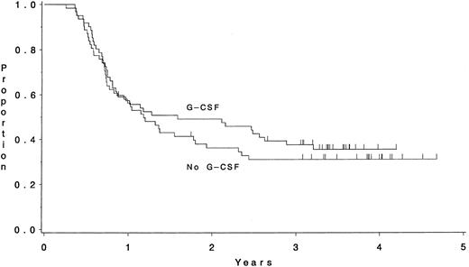
The median survival time for all eligible patients is estimated to be 21 months, while the median duration of CR is estimated to be 12 months (Fig 3). The median follow-up time was 63 months for surviving patients. Because of theoretic concern about stimulation of leukemic cell growth by the G-CSF, we compared patients in each group for overall survival and the duration of CR. Figure 4 displays the survival of all patients who received the third intensification by treatment category. Similarly, the duration of CR for these patients is shown in Fig 5. There were no significant differences between the groups with respect to these endpoints (P = .99, and P = .54, respectively).
Overall survival and CR duration for eligible and evaluable patients on CALGB 9022. The median survival time is 1.8 years and the median length of remission is 1 year.
Overall survival and CR duration for eligible and evaluable patients on CALGB 9022. The median survival time is 1.8 years and the median length of remission is 1 year.
Survival of patients who received the third course of postremission intensification therapy according to whether or not G-CSF was part of the regimen. There was no significant difference between the two groups (P = .99) with a median survival time of 2.4 years for patients who did not receive the growth factor (n = 62) and 3.4 years for patients who did receive it (n = 61).
Survival of patients who received the third course of postremission intensification therapy according to whether or not G-CSF was part of the regimen. There was no significant difference between the two groups (P = .99) with a median survival time of 2.4 years for patients who did not receive the growth factor (n = 62) and 3.4 years for patients who did receive it (n = 61).
CR duration of patients who received the third course of postremission intensification therapy according to whether or not G-CSF was part of the regimen. There was no significant difference between the two groups (P = .54) with a median length of remission of 1.2 years for patients who did not receive the growth factor and 1.4 years for patients who did receive it.
CR duration of patients who received the third course of postremission intensification therapy according to whether or not G-CSF was part of the regimen. There was no significant difference between the two groups (P = .54) with a median length of remission of 1.2 years for patients who did not receive the growth factor and 1.4 years for patients who did receive it.
DISCUSSION
Because myeloid leukemia cells express receptors for GM-CSF and G-CSF, many clinicians have been appropriately cautious in the use of this group of therapeutic agents in patients with AML.24 Concerns have included the possible stimulation of the growth of leukemia and protection of leukemic cells from destruction by chemotherapeutic agents.
Ohno et al25 first reported decreased time to granulocyte recovery in a heterogeneous group of patients with relapsed or refractory leukemia receiving intensive chemotherapy when G-CSF was begun after chemotherapy was completed and continued until granulocyte recovery. The incidence of documented infection was decreased, and there did not appear to be stimulation of leukemic growth. A subsequent retrospective review by Ohno et al26 described an additional 23 patients who received G-CSF after they developed life-threatening infections during induction (14 patients) or in one of three consolidation sequences. In this small nonrandomized study, G-CSF produced no toxicity, but was associated with an apparent decrease in the time to granulocyte recovery compared with those patients who did not receive G-CSF. There was no evidence of stimulation of leukemic regrowth or increased number of relapses in the group receiving G-CSF.
Based, in part, on these reports, as well as smaller studies from a number of institutions,27-30 large randomized trials were begun in newly diagnosed patients with AML using either G-CSF or GM-CSF as a means of attenuating neutropenia and decreasing infectious morbidity. Most of these studies focused on more elderly patients with AML because it is in this large subgroup of individuals that death from infection is a common cause of failure to achieve CR. Data from at least five large randomized trials have now been presented with, in general, remarkably similar conclusions.7-10,31 All studies demonstrated a statistically significant, but usually relatively modest, shortening of the duration of severe neutropenia, which in some studies, was accompanied by a decreased duration of hospitalization and need for parenteral antibiotics. Most studies did not demonstrate differences in the incidence of severe infection or response rate, which, in retrospect, was not surprising since most severe infections occur early in the course of induction therapy. Only one study noted an improved complete response rate and in this study (as in all of the others), there was no improvement in disease-free survival.31 Importantly, however, with the exception of a very small randomized study reported by the European Organization for Research and Treatment of Cancer (EORTC),32 there was no evidence of a deleterious effect of the growth factors as assessed by a lower CR rate or an appreciable, clinically detectable incidence of stimulation of leukemia cell growth. Thus, unfortunately, it appears that the use of myeloid growth factors as an adjunct to induction therapy, does not appear to have a substantial impact on improving response rate. The effect on the duration of neutropenia may represent a modest improvement in supportive care, but this needs to be balanced by the cost and side effects of the administration of the growth factor.6 There are few randomized data in younger patients with AML, although, given the high CR rate in such patients, it is unlikely that a significant improvement in CR rate could be detected.
Surprisingly, there have been few evaluations of the use of growth factors after the administration of intensive postremission consolidation therapy. Recent studies have strongly suggested intensive consolidation is of major benefit for at least subgroups of patients with AML and frequently, it is difficult to deliver repetitive courses of intensive therapy because of the development of significant infectious complications.1,33-35 Because such patients begin with normal blood counts and relatively normal marrow function, one might predict that benefit would be more easily demonstrated, somewhat analogous to the use of growth factors after high-dose chemotherapy in patients with nonhematologic cancer, than in the induction setting when patients are starting with abnormal blood counts and frequently with ongoing infection. The available results are somewhat conflicting. In the study reported by the Eastern Cooperative Oncology Group, which is considered to be the most “positive” of the studies of growth factors and induction therapy, there was no benefit in terms of shortening of the duration of neutropenia in the small group of 49 patients randomized to receive GM-CSF or placebo following a single course of consolidation with 12 doses of high-dose cytosine arabinoside.7 In this study, chemotherapy was administered for 6 days and the growth factor was begun on day 11. In contrast, Heil et al,9 have reported in abstract form that there was a 7-day shortening in the median duration of neutropenia following “standard dose consolidation” and 5.5 days following high-dose Ara-C consolidation in patients randomized to receive G-CSF. Details are not given, but there was also said to be decreases in duration of fever, parenteral antibiotic use, and hospitalization.
We report a group of 123 patients who entered four sequential treatment groups receiving intensely myelosuppressive postremission chemotherapy with AZQ and mitoxantrone. Of these, 112 patients completed all of the prescribed course of treatment. There were 61 patients who received G-CSF beginning the day after completion of chemotherapy. AZQ and mitoxantrone were known from the first two groups of patients to be associated with profound myelosuppression and prolonged granulocyte and platelet recovery times. The addition of G-CSF produced a decrease in the number of days of granulocytopenia (<500/μL) from 35 days and 31 days in groups 1 and 2, respectively, to 19 and 22 days in groups 3 and 4, respectively. This represents a dramatic decline in the duration of granulocytopenia and a coincident decrease in days of hospitalization, antibiotic therapy, and incidence of grade III/IV infection. A similar decrease in time to recovery of 1,000 granulocytes/μL was also observed.
Although this was not a randomized trial, these were consecutive groups of patients treated by the same group of physicians on the same protocol and over a relatively short duration of time. There were no apparent clinical differences among the four groups, and the overall results of this pilot study were similar in terms of CR rate and demographics to other CALGB studies of patients in this age group. Comparisons of groups 1 and 4, as well as 2 and 3, also suggest improvement in platelet recovery after G-CSF, although not nearly so dramatic as that seen with granulocytes. Although it is difficult to be certain, the amelioration of the thrombocytopenia did not appear to be due to resolution of infection by the rising neutrophil count with decreased consumption of platelets. There was no apparent deleterious effect of G-CSF on CR duration or survival, and G-CSF is now being used as an adjunct to AZQ/mitoxantrone in a randomized Phase III comparison of the three regimens given in this protocol with three courses of high-dose ARA-C.
It is of interest that G-CSF was so effective in this study, even following intensive therapy with agents suspected to have significant cytotoxic activity against hematopoietic stem cells. Higher doses of both mitoxantrone and AZQ have been associated with periods of prolonged marrow aplasia following treatment of patients with relapsed leukemia.11,13,14 Although more details are needed, the study by Heil et al9 suggests a similar effect of G-CSF with less marrow ablative regimens. The effect of growth factors administered after consolidation therapy for AML clearly deserves further study. Many postremission programs incorporate repetitive cycles of intensive therapy, and it is frequently difficult to administer all scheduled courses, either because of delays in marrow recovery or residual sequelae from infections encountered during the prior consolidation treatment. Clinical trials evaluating whether growth factors would permit safer administration of repetitive treatment with the end point of increasing the fraction of disease-free survivors, would be of considerable interest.
ACKNOWLEDGMENT
The authors are indebted to Erin Trikha and Bryan Blanton for central data management on this study.
APPENDIX
The following institutions participated in the study: Bowman Gray School of Medicine, Winston-Salem, NC; supported by CA03927; Central Massachusetts Oncology Group, Worcester, MA; Dana-Farber Cancer Institute, Boston, MA, supported by CA32291; Dartmouth Medical Center, Hanover, NH, supported by CA04326; Duke University Medical Center, Durham, NC, supported by CA47577; Finsen Institute, Copenhagen, Denmark; Long Island Jewish Medical Center, New Hyde Park, NY, supported by CA11028; Massachusetts General Hospital, Boston, MA, supported by CA12449; Mount Sinai Hospital, New York, NY, supported by CA04457; New York Hospital-Cornell Medical Center, New York, NY, supported by CA07968; Rhode Island Hospital, Providence, RI, supported by CA08025; Roswell Park Cancer Institute, Buffalo, NY, supported by CA59518; SUNY Health Science Center at Syracuse, Syracuse, NY, supported by CA21060; University of Alabama, Birmingham, AL, supported by CA47545; University of California at San Diego, San Diego, CA, supported by CA11789; University of Chicago Medical Center, Chicago, IL, supported by CA41287; University of Iowa Hospitals, Iowa City, IA, supported by CA47642; University of Maryland Cancer Center, Baltimore, MD, supported by CA31983; University of Minnesota, Minneapolis, MN, supported by CA16540; University of Missouri-Ellis Fischel Cancer Center, Columbia, MO, supported by CA12046; University of North Carolina at Chapel Hill, Chapel Hill, NC, supported by CA47559; University of Tennessee, Memphis, TN, supported by CA47555; Walter Reed Army Medical Center, Washington, DC, supported by CA26806; Washington University-Barnes Hospital, St Louis, MO, supported by CA47546.
Conducted by the Cancer and Leukemia Group B and supported by Public Health Service Grants from the National Cancer Institute (Bethesda), National Institutes of Health (Bethesda, MD), and the Department of Health and Human Services (Washington, DC). This research for CALGB 9022 was supported, in part, by grants from the National Cancer Instutitute (Grant No. CA31945) to the Cancer and Leukemia Group B (Richard L. Shilsky, Chairman).
Address reprint requests to Joseph O. Moore, MD, Division of Hematology/Oncology, Box 3536, Duke University Medical Center, Durham, NC 27710.

This feature is available to Subscribers Only
Sign In or Create an Account Close Modal