Abstract
This study identifies multiple pathways used by monocytes to adhere to 4-hour interleukin-1β stimulated human umbilical vein endothelial cells under flow conditions. Physiologic shear stresses were simulated in a flow chamber with parallel plate geometry; quantitation of primary adhesion, secondary adhesion, and transmigration was performed using phase contrast videomicroscopy. Neuraminidase treatment of monocytes reduced primary interaction by 50%, whereas blocking L-selectin or very late antigen-4 showed significant but smaller effects (∼30% inhibition). However, a combined treatment against all three pathways was able to reduce interaction by 80%. Blocking β2 and α4 integrin pathways together inhibited secondary/firm adhesion by 75%. Only 40% of firmly adherent monocytes transmigrated across the endothelial monolayer with significantly increased transmigration times when both β2 and α4 integrins were blocked. These results demonstrate that monocytes can use multiple receptors to interact with endothelial cells at both primary and secondary adhesion stages, and that these pathways have to be blocked simultaneously for maximum inhibition.
MONOCYTES HAVE BEEN shown to adhere to endothelial monolayers in vitro.1-8 Assays that simulate some aspects of physiologic shear reveal that endothelial monolayers can upregulate adhesive mechanisms capable of capturing flowing monocytes following stimulation with interleukin-4 (IL-4) or tumor necrosis factor α (TNFα).5,6 A dominant contribution of L-selectin on the monocyte was implicated by the use of blocking monoclonal antibodies (MoAbs), but the endothelial ligands for this adhesion molecule induced by the cytokines remain obscure. In addition, some contribution of endothelial P-selectin to monocyte capture under flow conditions has been apparent following TNFα stimulation,6 a result not inconsistent with studies of monocyte adhesion to venules in sections of rheumatoid synovium.9 E-selectin upregulated by TNFα, but not IL-4, stimulation of endothelium appears to be insignificant as a primary capture mechanism for monocytes.6 Very late antigen-4 (VLA-4), an integrin present on monocytes, interacts with vascular cell adhesion molecule-1 (VCAM-1) and has been reported to function as a primary capture mechanism under conditions of flow.10-12 Though IL-4 and TNFα both induce surface expression of endothelial VCAM-1, this pathway also seems insignificant for primary adhesion of monocytes under flow.5 6
We have chosen to determine whether endothelial monolayers can upregulate adhesive mechanisms, following stimulation with IL-1β, that are capable of capturing flowing monocytes, and to determine the adhesive molecules involved. Our results reveal that IL-1β stimulation will upregulate endothelial capture of flowing monocytes, but the adhesive mechanisms differ in some respects from those reported for treatment with IL-4 and TNFα.
MATERIALS AND METHODS
Isolation of Monocytes
Peripheral blood monocytes were isolated from citrate phosphate dextrose (CPD) anticoagulated buffy coat concentrates from normal donors (Gulf Coast Blood Center, Houston, TX) using counter current centrifugal elutriation in a Beckman J-6M centrifuge with a JE-5.0 elutriation rotor and a Sanderson chamber of 5.5 mL chamber capacity (Beckman Instruments, Palo Alto, CA). In brief, the buffy coat was diluted 5 times with Dulbecco's phosphate buffered saline (D-PBS) lacking Ca++ and Mg++ (Sigma, St Louis, MO) with 3 mmol/L disodium EDTA, layered on Ficoll-Paque (Pharmacia, Piscataway, NJ) and centrifuged at 600g for 40 minutes at 22°C. Mononuclear cells obtained from the interface were washed (100g, 10 minutes at 22°C) and resuspended in elutriation buffer consisting of Ca++ and Mg++ free D-PBS, 0.1% glucose (Sigma), 0.1% bovine serum albumin (BSA) (Sigma), and 3 mmol/L EDTA. Mononuclear cells (∼109) suspended in 10 mL elutriation buffer were loaded into the elutriation chamber at 14 mL/min with a rotor speed of 2,500 rpm at 4°C as described previously.4 5 Flow rate was gradually increased to 21 mL/min in steps of 1 mL/min. Most of the lymphocytes eluted out before 19 mL/min. Fractions collected at 19 mL/min, 20 mL/min, and 21 mL/min were pooled to give monocytes of purity (84% ± 2%, n = 19) with 10% lymphocytes and <2% granulocytes as determined with flow cytometry (FACScan; Becton Dickinson, San Jose, CA) using light scatter and dual color CD45/CD14 markers (Immunotech, Westbrook, ME). Platelet contamination was negligible and monocytes appeared to be spherical with no platelets attached on the cell surface as observed under 20× videomicroscopy. Monocytes were washed and resuspended at 4°C in Ca++ and Mg++ free D-PBS containing 0.1% BSA at 107 cells/mL. All buffers and reagents used were endotoxin tested (<0.01 ng/mL).
Cells and Cell Culture
Human umbilical vein endothelial cells (HUVECs) were isolated by collagenase treatment as described previously13 and seeded in fibronectin coated (2 μg/cm2 ) T-75 tissue culture flasks. Cells were passaged 4 to 7 days after initial seeding using 0.05% trypsin and 0.02% EDTA (GIBCO, Grand Island, NY) and plated at confluence on fibronectin coated 35-mm tissue culture dishes (Corning Glass Works, Corning, NY). HUVECs were stimulated with recombinant human IL-1β (0.1 ng/mL; R&D Systems Inc, Minneapolis, MN) for 4 hours. L-cells transfected with the seven domain form of VCAM-1 (referred to as L-VCAM-1) were generously provided by Dr Ted Yednock (Athena Neurosciences, South San Francisco, CA) and maintained by culture in RPMI 1640 containing 600 μg/mL G418, 10% fetal bovine serum (FBS), 3% penicillin-streptomycin and 1% glutamine (GIBCO). L-ELAM (L-E selectin) cells have been described previously.14
Flow Cytometry
Whole blood samples (100 μL), mononuclear cells or elutriated monocytes (106 cells) were incubated with 20 μL of fluorescein isothiocyanate (FITC)-labeled anti-CD62L (Immunotech, Inc) for 30 minutes at 4°C. Whole blood samples were lysed (erythrolyse RBC lysing buffer; Harlan Bioproducts for Science Inc, Indianapolis, IN) before analyzing. Specimens were diluted with D-PBS and immediately analyzed in FACscan flow cytometer. Monocytes were gated from other leukocyte subsets based on their forward and side scatter profiles and FITC fluorescence of 10,000 cells was measured.
Enzyme Treatment
Monocytes were incubated with 0.1 U/mL Vibrio cholera neuraminidase (Boehringer Mannheim, Indianapolis, IN) for 30 minutes at room temperature to remove cell surface sialyl acid residues. Control monocytes were treated similarly but in absence of the enzyme.
MoAbs
MoAb R15.7 (anti-CD18) was provided by Dr R. Rothlien (Boehringer Ingleheim Ltd Ridgefield, CT). MoAb GA6 (anti–P-selectin and function blocking) were obtained from Becton Dickinson (San Jose, CA). HP2/1 (anti-α4 ; Immunotech, Inc), CL2/6 (anti–E-selectin, CD62E), Dreg-56 (anti–L-selectin, CD62L) have been described previously.13 14 MoAb 4B9 (anti–VCAM-1) was obtained from Dr R. Lobb (Biogen, Inc, Cambridge, MA). All MoAbs were used at 20 μg/mL and were purified IgG. F(ab′ )2 fragments of MoAb CL2/6 were also used. In flow adhesion assays, L-cell and HUVEC monolayers were treated with MoAbs for 30 minutes at 37°C before use while monocytes were treated with MoAbs at 4°C for 30 minutes. Saturating concentrations of MoAbs were used for all incubations. During combined treatment with MoAbs HP2/1 and R15.7, saturating conditions were also maintained in the flow buffer. However, in the remaining treatments, no difference was observed whether or not saturating MoAb concentrations were maintained in the flow buffer.
Adhesion Assay
Monocyte interaction on IL-1β (4 h) stimulated endothelial cells or L-cell monolayers was quantitated under flow conditions using a parallel plate flow chamber as described in detail previously.12-14 Each experiment used monocytes prepared from different batches of buffy coats obtained from the Blood Center. Monocytes (106 cells/mL) in D-PBS/0.1% BSA were perfused through the chamber at a wall shear stress of 2 dynes/cm2. The flow system was maintained at 37°C in an air curtain incubator. Interactions between monocytes and the cell monolayer were visualized with videomicroscopy using phase contrast optics (Diaphot-TMD microscope, Nikon, Inc, Garden City, NY; Hamamatsu C1000 video camera, Hamamatsu Photonics, Bridgewater, NJ). A single field of view was monitored for the entire 10-minute duration of the experiment and videotaped for later analysis. Five quantities were measured in the analysis to characterize monocyte interaction: (1) Total interacting cells; defined as cells interacting with the monolayer for at least 2 seconds (included both rolling and immediately arrested cells) and the number per field of view (×20 objective; 0.274 mm2) were quantified by reviewing the videotapes for the entire 10-minute run. (2) Firmly adherent cells accumulated throughout the experiment and were analyzed at the end of each experiment. Monocytes adherent on the luminal surface of the monolayer (appeared as phase bright objects) and those that had transmigrated to the subluminal space (appeared as phase dark objects as documented before15 ) were included in the analysis. (3) Transmigrated cells (see above) were easily distinguishable from those on the lumen based on their phase dark and flattened appearance and were measured at the end of the 10-minute run. (4) Rolling velocities were calculated as previously described13 by acquiring 4-second maximization images using a digital image processing system (Sun Microsystems, Mountain View, CA; Inovision Corp, Durham, NC). Only monocytes that rolled without stopping during the entire 4-second acquisition period were included in the analysis. (5) Transmigration time was measured by following individual monocytes from the time they firmly attached on the monolayer (phase bright appearance) till they finally diapedesed and turned phase dark in appearance.
Statistics
For comparing multiple treatments, significance was determined with one-way analysis of variance followed by Fisher's PLSD post-hoc test using superANOVA 1.11 software. P values less than .05 were considered to be statistically significant. Results in the text and figures are represented as mean ± SEM.
RESULTS
Monocytes Retain L-Selectin Levels After Elutriation
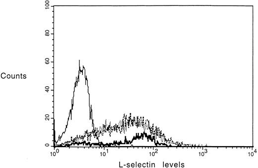
L-selectin levels were measured on the monocyte surface after the final elutriation step by flow cytometry to check for possible activation during the separation procedure. Results show minimal differences in the L-selectin levels of elutriated monocytes (mean fluorescence = 45) compared with monocytes in the mononuclear suspension (mean fluorescence = 47) obtained after Ficoll centrifugation and with monocytes in whole blood (mean fluorescence = 51). Chymotrypsin-treated monocytes were used as control for background fluorescence (mean fluorescence = 3) (Fig 1).
L-selectin levels of monocytes in whole blood, mononuclear/pre-elutriation fraction, and elutriated monocytes. Dreg-56 FITC was used to label L-selectin on monocytes. Monocytes treated with chymotrypsin (0.4 U/106 cells) for 10 minutes at 25°C were used for background fluorescence as shown by a solid line. L-selectin on monocytes in whole blood is indicated by a bold solid line. L-selectin levels on pre- (solid dashes) and post-elutriation monocyte fractions (dotted line) were almost identical.
L-selectin levels of monocytes in whole blood, mononuclear/pre-elutriation fraction, and elutriated monocytes. Dreg-56 FITC was used to label L-selectin on monocytes. Monocytes treated with chymotrypsin (0.4 U/106 cells) for 10 minutes at 25°C were used for background fluorescence as shown by a solid line. L-selectin on monocytes in whole blood is indicated by a bold solid line. L-selectin levels on pre- (solid dashes) and post-elutriation monocyte fractions (dotted line) were almost identical.
Monocyte Adhesion to IL-1β–Stimulated Endothelial Cells
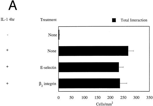
Stimulation of HUVECs with IL-1β for 4 hours promoted significant levels of monocyte interaction (273 ± 23 monocytes/mm2 ) compared with unstimulated endothelium under flow conditions (Fig 2A). The predominant pattern of monocyte interaction observed was that of immediate arrest on the HUVEC monolayer on intial contact as also observed previously.6 For most monocytes, immediate arrest resulted in stable adhesion. Some interacting monocytes after initial tethering displayed rolling behavior either before or after arrest.
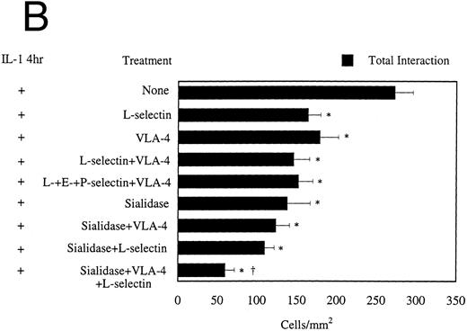
(A) Monocyte adhesion to HUVECs under flow conditions. Endothelial cells were stimulated with 0.1 ng/mL IL-1β and monocytes (106/mL) were perfused over the monolayer for 10 minutes at 2 dynes/cm2. Total interaction gives a measure of primary adhesion and includes all monocytes interacting with the monolayer for at least 2 seconds. On endothelial cells, MoAb CL2/6 was used to block E-selectin function, whereas MoAb R15.7 binds to the β2 subunit of CD18 integrins present on monocyte surface, and blocks its function (n = 3 to 19). (B) Adhesion of monocytes to (4 h) IL-1β–stimulated HUVECs at 2 dynes/cm2. Dreg-56 was used to block L-selectin function on monocytes. Data is shown as mean ± SEM for 3 to 19 experiments. MoAb Dreg-56 was used to block L-selectin function on monocytes. MoAb HP2/1 binds to VLA-4 on monocytes and is function blocking. To block E- and P-selectin, endothelial cells were treated with MoAbs CL2/6 Fab2 and GA6, respectively. Sialidase treatment included use of neuraminidase enzyme (0.1 U/mL, 30 minutes incubation at 25°C) to cleave sialyl lewis residues from the monocyte surface. *Statistical significance (P < .05) compared with control with no MoAb treatments.† Statistical significance (P < .05) compared with neuraminidase, anti–L-selectin, and anti–VLA-4 treatment.
(A) Monocyte adhesion to HUVECs under flow conditions. Endothelial cells were stimulated with 0.1 ng/mL IL-1β and monocytes (106/mL) were perfused over the monolayer for 10 minutes at 2 dynes/cm2. Total interaction gives a measure of primary adhesion and includes all monocytes interacting with the monolayer for at least 2 seconds. On endothelial cells, MoAb CL2/6 was used to block E-selectin function, whereas MoAb R15.7 binds to the β2 subunit of CD18 integrins present on monocyte surface, and blocks its function (n = 3 to 19). (B) Adhesion of monocytes to (4 h) IL-1β–stimulated HUVECs at 2 dynes/cm2. Dreg-56 was used to block L-selectin function on monocytes. Data is shown as mean ± SEM for 3 to 19 experiments. MoAb Dreg-56 was used to block L-selectin function on monocytes. MoAb HP2/1 binds to VLA-4 on monocytes and is function blocking. To block E- and P-selectin, endothelial cells were treated with MoAbs CL2/6 Fab2 and GA6, respectively. Sialidase treatment included use of neuraminidase enzyme (0.1 U/mL, 30 minutes incubation at 25°C) to cleave sialyl lewis residues from the monocyte surface. *Statistical significance (P < .05) compared with control with no MoAb treatments.† Statistical significance (P < .05) compared with neuraminidase, anti–L-selectin, and anti–VLA-4 treatment.
Primary adhesion.Total interaction gives a measure of primary adhesion since all monocytes interacting with the monolayer for 2 seconds are included. To investigate the molecular pathways involved in the primary adhesion, experiments were performed using a panel of functional blocking MoAbs against receptors on both monocytes and HUVECs (Fig 2, A and B). Blocking E-selectin on the endothelium (Fig 2A) with MoAb CL2/6 did not significantly reduce interaction although the expression of E-selectin is maximum after 4 hours of IL-1β stimulation. This is in excellent agreement with earlier work using both flow6 and static1,16 adhesion assays. P-selectin expression was also observed after TNF-α stimulation of HUVECs for 6 hours.6,17 A recent study,18 however, has shown no increase in P-selectin mRNA levels after IL-1β or TNF-α stimulation of endothelial cells. Consistent with this, no role for P-selectin was observed in our system as MoAb GA6 to P-selectin did not affect monocyte interaction (data not shown). MoAb R15.7 directed against β2 integrins (anti-CD18) had no significant effect (Fig 2A), which agrees with previous studies19 showing no role of β2 integrins in primary adhesion under conditions of flow. Treatment of monocytes with functional blocking MoAb Dreg-56 against L-selectin caused a significant inhibition of ∼30% in primary adhesion (Fig 2B). Similar inhibition was also observed after adding MoAb HP2/1 against VLA-4 on monocytes. Blocking both L-selectin and VLA-4 simultaneously did not result in an additive inhibitory effect (inhibition ∼40%, n = 7). Treatments with anti–L-, E-, P-selectin and anti–VLA-4 caused 40% ± 6.7% (n = 3) inhibition in primary adhesion, which was not significantly different from the inhibition observed with anti–L-selectin + anti–VLA-4 treatment. Neuraminidase treatment of monocytes to remove terminal sialic acid moieties from the cell surface glycoproteins blocked 50% of the primary interaction (Fig 2B). Addition of either Dreg-56 or HP2/1 to neuraminidase treated monocytes did not significantly further inhibit adhesion. Interestingly, a combination of Dreg-56 and HP2/1 MoAbs, when added to neuraminidase treated monocytes, resulted in almost 80% reduction of primary adhesion.
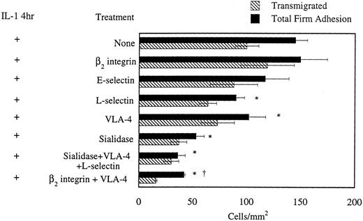
Secondary adhesion.Firmly adherent monocytes accumulated throughout the 10-minute experiment and indicated the extent of secondary adhesion. Figure 3 shows the effects of various treatments on firm adhesion of monocytes to IL-1β 4-hour stimulated HUVECs. MoAbs to E-selectin (CL2/6), L-selectin (Dreg-56), and VLA-4 (HP2/1) reduced firm adhesion by 20% to 25% compared with control. Also, removal of sialic acid residues on monocytes with neuraminidase caused 46% inhibition and the combination of neuraminidase, Dreg-56 and anti-α4 treatments resulted in 73% inhibition of firm adhesion. It was interesting to note that all the above treatments reduced secondary adhesion to approximately the same extent as they inhibited primary interaction (Fig 2A). Anti-CD18 MoAb treatment alone did not inhibit firm adhesion. Blocking β2 and α4 integrin pathways together with MoAbs R15.7 and HP2/1, respectively, dramatically changed the pattern of interaction of monocytes. Almost all (>90%) of the interacting monocytes exhibited rolling behavior (988 ± 193/mm2, n = 3), whereas only 15% of control (untreated) interacting monocytes rolled (47 ± 0/mm2 ) on IL-1β–stimulated endothelium. Using MoAbs to β2 and α4 integrins together reduced firm adhesion by 75% compared with firm adhesion observed with untreated monocytes (Fig 3). Further quantitation of adhesion data revealed that 60% of total interacting untreated (control) monocytes firmly adhered to IL-1–stimulated endothelium. However, with the β2 and α4 integrins blocked, only 4% of total interacting monocytes were able to engage in secondary adhesion. In the remaining treatments, 60% of interacting monocytes firmly adhered, which was similar to that seen with untreated monocytes. These results indicate that β2 and α4 integrin pathways when blocked together cause a significant reduction in secondary adhesion, but not when blocked individually.
Adhesion of monocytes to (4 h) IL-1β–stimulated HUVECs at a wall shear stress of 2 dynes/cm2: effects of MoAb and enzyme treatment on both firm adhesion and on monocyte transmigration, (n = 3 to 19 experiments). MoAbs CL2/6 Fab2 and Dreg-56 were used to inhibit E- and L-selectin function. MoAb HP2/1 was used to block VLA-4 on the monocyte surface while MoAb R15.7 was used against β2 integrins. Neuraminidase was used in the sialidase treatment. *Statistical significance (P < .05) compared with control with no MoAb treatments. †Statistical significance (P < .05) compared with anti–VLA-4 and anti-β2 integrin treatments.
Adhesion of monocytes to (4 h) IL-1β–stimulated HUVECs at a wall shear stress of 2 dynes/cm2: effects of MoAb and enzyme treatment on both firm adhesion and on monocyte transmigration, (n = 3 to 19 experiments). MoAbs CL2/6 Fab2 and Dreg-56 were used to inhibit E- and L-selectin function. MoAb HP2/1 was used to block VLA-4 on the monocyte surface while MoAb R15.7 was used against β2 integrins. Neuraminidase was used in the sialidase treatment. *Statistical significance (P < .05) compared with control with no MoAb treatments. †Statistical significance (P < .05) compared with anti–VLA-4 and anti-β2 integrin treatments.
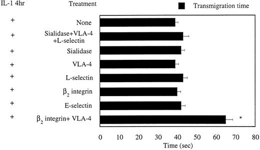
Transmigration and transmigration time.Approximately 70% of untreated (control) monocytes that firmly adhered were able to transmigrate across IL-1β–stimulated HUVECs (100 ± 11 monocytes/mm2 ) as evident in Fig 3. With both β2 and α4 integrins blocked, only 40% of firmly adherent monocytes were able to transmigrate, whereas in the remaining treatments, approximately 70% of firmly adherent monocytes transmigrated, which was similar to that seen with untreated monocytes. To elucidate the kinetics of transmigration, the time taken by firmly adherent monocytes to diapedese was measured. Untreated monocytes transmigrated across IL-1β–stimulated HUVECs in 38.9 ± 1.5 seconds as shown in Fig 4. Blocking β2 and α4 pathways together significantly prolonged the transmigration time (65 ± 3.6 sec). None of the remaining treatments had any significant effect on monocyte transmigration kinetics.
Effects of various treatments on monocyte transmigration kinetics across (4 h) IL-1β–stimulated HUVECs. *Statistically significant (P < .05) differences in transmigration times as compared with control, n = 20 to 30. Monocytes from different experiments were used to measure transmigration times.
Effects of various treatments on monocyte transmigration kinetics across (4 h) IL-1β–stimulated HUVECs. *Statistically significant (P < .05) differences in transmigration times as compared with control, n = 20 to 30. Monocytes from different experiments were used to measure transmigration times.
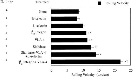
Monocyte rolling velocities.Figure 5 presents average rolling velocities of monocytes under various treatments. Untreated monocytes rolled with an average velocity of 8.6 ± 0.45 μm/sec and blocking L-selectin with Dreg-56 had no effect on the rolling velocity. Pretreatment of monocytes with neuraminidase did not abrogate rolling and monocytes rolled on the endothelium at a significantly higher velocities (12.5 ± 0.9 μm/sec) compared with untreated monocytes. Interestingly, blocking both L-selectin and VLA-4 on neuraminidase-treated monocytes still did not abolish rolling but further increased the rolling velocity to 14.1 ± 0.7 μm/sec. MoAbs to both β2 and α4 integrins also increased rolling velocity to 21 ± 0.81 μm/sec, which was significantly greater than all other treatments.
Average rolling velocities of monocytes on IL-1β–stimulated HUVECs at a wall shear stress of 2 dynes/cm2. Four-second maximization images were acquired to calculate rolling velocities of monocytes. *Statistically significant (P < .05) difference in rolling velocity as compared with control. †Statistical significance (P < .05) of anti–VLA-4 and anti-β2 integrin treatments compared with all other treatments (n = 25-35). Monocytes from different experiments were included for rolling velocity measurements.
Average rolling velocities of monocytes on IL-1β–stimulated HUVECs at a wall shear stress of 2 dynes/cm2. Four-second maximization images were acquired to calculate rolling velocities of monocytes. *Statistically significant (P < .05) difference in rolling velocity as compared with control. †Statistical significance (P < .05) of anti–VLA-4 and anti-β2 integrin treatments compared with all other treatments (n = 25-35). Monocytes from different experiments were included for rolling velocity measurements.
Monocyte Rolling on L-ELAM Monolayers
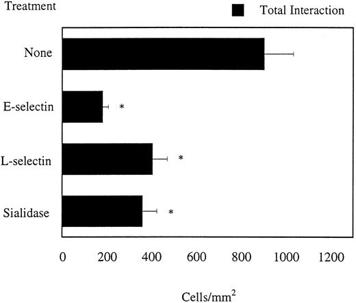
To further investigate the limited role played by E-selectin in IL-1β–stimulated endothelium, flow experiments were performed using E-selectin transfected monolayers. Extensive monocyte interaction was observed (904 ± 131 monocytes/mm2 ) as shown in Fig 6. MoAb CL2/6 reduced this interaction by 80%, while Dreg-56 blocked by 50%. Neuraminidase treatment also resulted in 60% inhibition. Qualitatively, almost all monocytes rolled on E-selectin with an average velocity of 8.12 ± 0.2 μm/sec. These results are consistent with previous results6 and show that E-selectin alone is capable of supporting monocyte rolling. Partial involvement of L-selectin and sialyl acid residues on the monocyte cell surface in binding to L-cells transfected with E-selectin was also observed, which is similar to results previously reported for neutrophils.14 20
Monocyte interaction on E-selectin transfected L-cell monolayers at a wall shear stress of 2 dynes/cm2. *Statistical significance (P < .05) with respect to untreated monocytes (n = 4-6 experiments).
Monocyte interaction on E-selectin transfected L-cell monolayers at a wall shear stress of 2 dynes/cm2. *Statistical significance (P < .05) with respect to untreated monocytes (n = 4-6 experiments).
Monocyte Interaction on L-VCAM Monolayers
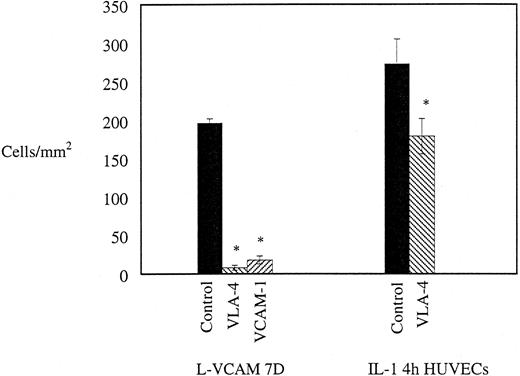
To assess the role of VLA-4/VCAM-1 pathway in mediating primary interaction, monocytes were perfused on L-VCAM (7D) monolayer at 2 dynes/cm2. As shown in Fig 7, L-VCAM monolayer supported monocyte interaction (190 monocytes/mm2 ) and the most obvious pattern of interaction, as seen in HUVECs, was that of immediate arrest. Both anti–VLA-4 and anti–VCAM-1 treatment eliminated this interaction while, as discussed before, anti–VLA-4 was only able to show an inhibition of 30% on IL-1β–stimulated HUVECs.
Monocyte interaction on L-VCAM (7D) monolayers at a wall shear stress of 2 dynes/cm2. Effects of blocking α4 β1 integrin using MoAb HP2/1 and VCAM-1 with MoAb 4B9 on monocyte interaction with L-VCAM and IL-1β–stimulated HUVEC monolayers. For L-VCAM-1 monolayer, n = 2-3 experiments; for HUVCEs, n = 12-19. *Statistical significance (P < .05) of each treatment compared to control on respective substrates.
Monocyte interaction on L-VCAM (7D) monolayers at a wall shear stress of 2 dynes/cm2. Effects of blocking α4 β1 integrin using MoAb HP2/1 and VCAM-1 with MoAb 4B9 on monocyte interaction with L-VCAM and IL-1β–stimulated HUVEC monolayers. For L-VCAM-1 monolayer, n = 2-3 experiments; for HUVCEs, n = 12-19. *Statistical significance (P < .05) of each treatment compared to control on respective substrates.
Monocyte Accumulation Kinetics on HUVEC and L-ELAM Monolayers
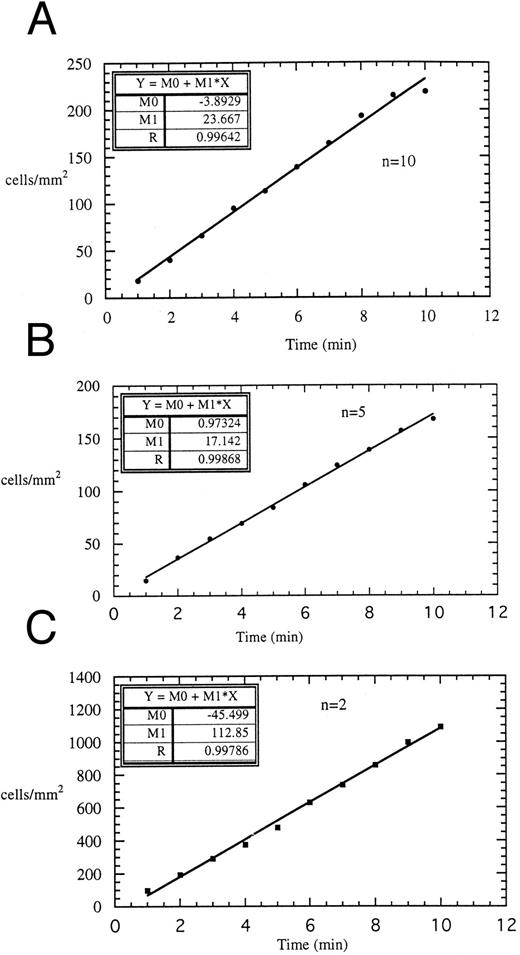
Figure 8 shows monocyte adhesion on (Fig 8A) 4-hour IL-1β–stimulated HUVECs and (Fig 8C) L-ELAM monolayer, quantitated over the 10-minute experiment. A linear trend in monocyte accumulation (R > .99) was observed on both substrates. Treating monocytes with Dreg-56 (Fig 8B) did not alter the linear profile (R > .99) on IL-1β–stimulated HUVECs. These results indicate a constant rate of accumulation for monocytes, independent of any possible L-selectin–mediated homotypic recruitment by previously arrested monocytes.
Accumulation kinetics of monocytes on (A) 4 h IL-1β–stimulated HUVEC monolayers (n = 10), (B) 4 h IL-1β–stimulated HUVEC monolayers in presence of Dreg-56 (n = 5) and (C) L-ELAM monolayers (n = 2). For all studies, 106/mL monocytes were perfused over the monolayers at a wall shear stress of 2 dynes/cm2. Accumulation of monocytes in all three cases were fitted to a linear equation where Y = accumulated monocytes (cells/mm2 ), X = time (min). The correlation coefficient, R for each case was greater than 0.99.
Accumulation kinetics of monocytes on (A) 4 h IL-1β–stimulated HUVEC monolayers (n = 10), (B) 4 h IL-1β–stimulated HUVEC monolayers in presence of Dreg-56 (n = 5) and (C) L-ELAM monolayers (n = 2). For all studies, 106/mL monocytes were perfused over the monolayers at a wall shear stress of 2 dynes/cm2. Accumulation of monocytes in all three cases were fitted to a linear equation where Y = accumulated monocytes (cells/mm2 ), X = time (min). The correlation coefficient, R for each case was greater than 0.99.
DISCUSSION
Our experiments studying monocyte adhesion to 4-hour IL-1β–activated HUVECs under conditions of flow in a parallel plate flow chamber reveal the following important distinctions from previous reports of monocyte interaction on either IL-4 or TNF-α–stimulated endothelial cells: (1) Both VLA-4 and L-selectin significantly participate in initiating monocyte attachment. (2) Neuraminidase treatment reduces primary interaction by 50%. (3) Combined treatment with neuraminidase, anti–L-selectin and anti–VLA-4 reduces primary interaction by 80%. (4) Blocking β2 and α4 integrins together is more effective in inhibiting firm adhesion as compared with either treatment alone. (5) Blocking both β2 and α4 integrin pathways significantly lengthens transmigration time of those firmly adherent monocytes that ultimately transmigrate.
On IL-1β–stimulated HUVECs, anti-α4 MoAb was able to significantly block monocyte interaction by 30%. This is in contrast to an earlier study that examined monocyte adhesion to 6-hour TNF-α–stimulated endothelial cells in which no role of VLA-4/VCAM-1 pathway was observed in initial attachment. To further investigate this, we also used VCAM-1 seven domain transfected L-cells. In this model, monocytes interacted with VCAM-1 monolayers at 2 dynes/cm2. Most monocytes were immediately arrested on initial contact and stayed firmly adherent, while some monocytes rolled along the monolayer after initial contact and either firmly adhered or re-entered the free stream. This pattern of interaction suggests that unactivated monocytes express a relatively low affinity pool of α4β1 receptors capable of interacting with VCAM-1 via multivalent interactions.21 Recently the α4/VCAM-1 pathway has also been shown to be sufficient to support primary adhesion of lymphocytes10-12 under flow conditions. Evaluating the role of L-selectin in mediating primary adhesion, Dreg-56 caused 30% inhibition of monocyte interaction to IL-1β–stimulated HUVECs, while earlier studies have implicated a role for L-selectin in mediating more than 80% of monocyte adhesion. A possibility for this discrepancy could be a differential upregulation of L-selectin ligand on endothelial surface with IL-1β stimulation as compared with IL-4 or TNF-α stimulation. On 4-hour IL-1β–stimulated HUVECs, a study22 showed evidence of augmented neutrophil recruitment by previously adhered neutrophils via L-selectin–dependent mechanism. However, our experiments showed linear correlation of monocyte accumulation with time on both HUVEC and L-ELAM monolayers, indicating a constant accumulation rate, and hence, suggesting no role of monocyte mediated monocyte recruitment (Fig 8, A and C). Furthermore anti–L-selectin treatment of monocytes did not affect the pattern of accumulation over time indicating no evidence of L-selectin–dependent amplification of monocyte adhesion by adherent monocytes in our system (Fig 8B).
Neuraminidase treatment inhibited primary adhesion by 50% on IL-1β–stimulated HUVECs. Experiments performed with a combined treatment of anti–L-,E-, P-selectin, and anti–VLA-4 revealed only 40% inhibition suggesting a possibility of an unknown receptor on the monocyte surface susceptible to neuraminidase treatment and binding to a ligand different from E-, P-selectin, VCAM-1 or L-selectin ligand on the EC monolayer. This might explain the higher inhibition (∼80%) seen with the combined treatment of neuraminidase, anti–L-selectin and anti–VLA-4. These results also seem to indicate the presence of a novel selectin-like adhesive pathway as suggested in a recent study.23 An endothelial cell adhesion molecule specifically recognized by monocytes has also been reported.24 This receptor has been shown to be upregulated maximally at 4 to 9 hours with TNF-α or IL-1β–stimulation of HUVECs. A possibility therefore also exists that this receptor is involved in supporting monocyte adhesion to IL-1β stimulation of HUVECs. However, its ligand on monocytes still remains to be characterized. The effects of blocking two pathways together (anti–VLA-4 + anti–L-selectin, neuraminidase + anti–VLA-4 and neuraminidase + anti–L-selectin) did not show significantly more inhibition compared with neuraminidase treatment alone. Interestingly, blocking all three pathways simultaneously reduced primary interaction by 80%, which was not only additive but also significantly higher than neuraminidase treatment alone. This additive inhibitory effect observed suggests that these three pathways exist independent of each other and that each can function in absence of the other to support monocyte interaction. Similar additive inhibitory effects were also observed in previous studies using static1,16 and rotational assays.25
Both intercellular adhesion molecule-1 (ICAM-1)/β2 and VCAM-1/α4 pathways can mediate firm/secondary adhesion of monocytes.2,3,7 Our results showed no inhibition in firmly adherent monocytes after anti-CD18 treatment, whereas anti–VLA-4 treatment reduced firm adhesion by ∼25%. With both β1 and β2 integrin pathways blocked, secondary adhesion was inhibited by approximately 75% and qualitatively, almost all monocytes rolled without engaging in firm adhesion. This is also consistent with studies with T cells17,26 and monocytes.6,27 Previous reports have shown both β1 and β2 components of monocyte transmigration,2,6,7 and our results also show evidence of β2 and α4 integrins in altering the percentage of firmly adherent monocytes that diapedese. To further investigate the transmigration process, transmigration kinetics were also considered. Blocking both β2 and α4 integrin pathways significantly enhanced the transmigration time on IL-1β–stimulated HUVECs as compared with untreated monocytes, suggesting that these integrins may also indirectly regulate transmigration by influencing spreading and locomotion. Since 40% of firmly adherent monocytes were still able to transmigrate with both β2 and α4 integrins blocked, a possibility also exists that other set of receptors responsible for transmigration and recently platelet endothelial cell adhesion molecule-1 (PECAM-1) mediate this step for both monocytes and neutrophils.28,29 Stimulation of endothelial cells with IL-1β/TNF-α also induces expression of monocyte chemotactic protein-1 (MCP-1), shown30 to promote monocyte transmigration.
Although not a predominant event, monocytes did exhibit rolling characteristics during their interaction with endothelial cell monolayers. Both β2 and α4 treatments significantly increased the rolling velocity, probably by blocking the adhesion stabilizing effects of integrins as noted before for neutrophils and T cells.12,23 Interestingly, monocytes still retained the ability to roll, even after removal of sialylated determinants from the cell surface, suggesting a complex interplay of proteins and carbohydrates in mediating rolling. This has also been observed before in studies using L-selectin transfectants.31
Thus, these observations help us to gain further insights into the mechanisms used by monocytes at various stages of the “three step adhesion cascade” to activated endothelium under physiologic flow conditions. Understanding the detailed molecular basis underlying these events could lead to development of novel therapeutics to counter monocyte mediated inflammatory disorders and pathologic states.
ACKNOWLEDGMENT
The authors thank Dr Abe and Joe Raya for their assistance with the centrifugal elutriator.
Supported by National Institutes of Health Grants No. AI31545-01, HL-18672, NS-23327, HL-42550, and AI23521; Robert A. Welch Foundation Grant No. C-938; and a grant from the Butcher Fund.
Address reprint requests to Larry V. McIntire, PhD, Cox Laboratory for Biomedical Engineering, Institute of Biosciences and Bioengineering, Rice University, Houston, TX 77251-1892.

This feature is available to Subscribers Only
Sign In or Create an Account Close Modal