Abstract
A recent study in the human-peripheral blood lymphocytes-severe combined immunodeficiency (hu-PBL-SCID) model, analyzing the specificity of the engrafted human T cells, showed that human T-cell lines and clones derived from engrafted cells presented a xenoreactivity toward murine host molecules. This observation raised the question of the influence of the SCID environment on the ex vivo repertoire and function on the human T cells reconstituting the murine host. We have characterized the human Vβ repertoire in the spleen of hu-PBL-SCID mice 1 to 3 months after their engraftment. Fluorescence-activated cell sorting (FACS) analysis of human Vβ T-cell representation showed that, for all chimeras, all tested Vβ subsets were submitted to underrepresentation and/or expansion upon engraftment. Importantly, these quantitative modifications of the T-cell repertoire were associated with a severe restriction in both the CDR3 size distribution pattern of the Vβ transcripts and the number of Jβ segments used by these transcripts. In addition, ex vivo phenotypic characterization of engrafted cells showed that 70% to 100% expressed the activation markers HLA-DR, CD45RO, and CD38. Taken together, these results suggest that, following their engraftment, human T cells were submitted to a massive antigenic selection. Moreover, we found that these activated T cells were unresponsive to in vitro mitogenic and superantigenic activation. The consequences of the skewed repertoire and altered function of engrafted human T cells on the validity of this humanized murine model are discussed.
BECAUSE OF a recessive autosomal genetic defect, the CB17 scid/scid (SCID) mouse strain lacks mature B- and T-cell lineages1 and thus is theoretically unable to reject allogeneic or xenogeneic tissues. Besides providing a useful model to study the non-T non-B immunity, this property allowed the establishment of human-mouse chimeras susceptible to be used as an in vivo experimental model for the human immune system. Two main ways of “humanization” have been developed: the SCID-hu model, which consists of engrafting SCID mice with human fetal tissues including thymus, liver, and lymph nodes2 and the hu-PBL-SCID model, which is derived from intraperitoneal injection of human PBL into SCID mice.3
The low number of mature human cells that repopulate the lymphoid tissues of SCID-hu mice makes this model inappropriate to study the mature functional human immune system.4 The hu-PBL SCID model could represent a more adequate experimental model, as several investigators have shown that these SCID mice could be correctly reconstituted by engrafted peripheral human cells.3 5
Despite the numerous studies performed in the hu-PBL SCID model, results dealing with reconstitution of SCID mice by human engrafted cells were not consensual. The number and characteristics of human cells repopulating engrafted mice were found different, depending both on screened organs and time of engraftment.3,5-8 Moreover, a still controversial point is the existence of a graft-versus-host (GVH) reaction in these chimeras. Indeed, if no evidence of GVH-associated pathology was generally observed in engrafted SCID mice,3,5 some histological8-10 and functional studies5,11 12 supported the occurrence of a GVH reaction in hu-PBL SCID chimeras. If this xenoreaction effectively occured, it should influence the human T-cell repertoire. We have thus characterized the Vβ repertoire of human T cells repopulating the spleen of hu-PBL-SCID mice. The present report shows a high restriction in the CDR3 size distribution and Jβ usage of engrafted T cells, associated with an activated phenotype. These T-cell alterations suggest a massive in vivo selection process of human T cells in these chimeras consistent with a xenogeneic reaction.
MATERIALS AND METHODS
Hu-PBL SCID mice.Sixty C.B.-17 (scid/scid ) mice were purchased from IFFA-CREDO (L'Arbresle, France) and maintained in microisolator caging in a sterile environment. SCID mice were between 10 and 12 weeks old at the time of cell transfer. A series of 20 SCID mice were injected intraperitoneally with 20 × 106 PBL from three different healthy donors (CNTS, Hôpital Saint-Louis, France), separated by Ficoll density gradient centrifugation (Pharmacia, Sweden), and resuspended in phosphate-buffered saline (PBS) at 108 cells/mL.
Recovery of human cells from chimeras.At different times postgraft (from 42 to 77 days), mice were killed by cervical dislocation and their spleen was recovered. Single cell suspensions were obtained from spleen by teasing the tissue in RPMI-1640 (Sigma Chemicals, Coger, Paris, France). The resulting mixture was passed through a sterile nylon filter, washed once in RPMI-1640, and suspended at 106 cells/mL in the same medium supplemented with 10% vol/vol heat-inactivated fetal calf serum (Institut Jacques Boy, Reims, France), 2 mmol/L L-glutamine (GIBCO, Paisley, Scotland, UK), 10 U/mL penicillin-streptomycin (GIBCO).
Immunofluorescent analysis.Two-color flow cytometric analyses were performed to characterize the human cells from hu-PBL-SCID mice. Briefly, 2 to 5 × 10⁵ donor or chimeras' splenic cells were washed once in PBS containing 1% bovine serum albumin (BSA) (Sigma, St Louis, MO) and 0.1% sodium azide, incubated for 10 minutes at 4°C with monoclonal antibodies (MoAbs) and then washed three times in the same buffer. A double staining of splenic cells with antihuman CD45 MoAbs conjugated to fluorescein isothiocyanate (FITC) (Becton Dickinson, Pont de Claix, France) and antimurine major histocompatability complex (MHC) class I molecule H-2Kd conjugated to phycoerythrin (PE) (Pharmingen, San Diego, CA) allowed a clear distinction of human cells from murine cells. Human populations in chimeras were characterized with the following MoAbs: anti-CD3 (FITC) and anti-CD4, -CD8 , -CD69, -CD38, -CD25, -CD28, -CD45RO (PE) (Becton Dickinson) or anti-CD45RA (PE) (Coulter Clone, Margency, France). Expression of human T-cell receptor (TCR) Vβ gene products among CD3+ T cells of donor's PBL and of chimeras' splenic cells was determined with the following MoAbs: anti-Vβ5.1 (BV5S1), -Vβ2,3 (BV5S2,3), -Vβ6.7 (BV6S7), -Vβ8 (BV8), -Vβ12 (BV12) (T Cell Sciences, Amersham, France), -Vβ2 (BV2), -Vβ13.1 (BV13S1) or -Vβ17 (BV17) (Immunotech, Marseille, France) all conjugated to FITC and anti-CD3 MoAbs (Becton Dickinson) conjugated to PE. Stained cells were fixed and red blood cells were lysed simultaneously in 200 μL of FACS Lysing Solution (Becton Dickinson, CA). All samples were acquired in a FACScan flow cytometer (Becton Dickinson). For each sample, a number of total splenic cells ranging from 27,000 to 250,000 was acquired, and 20,000 human CD4+ H-2Kd− cells were analyzed with the Lysis II software (Becton Dickinson). Isotypically matched negative controls were always used (Becton Dickinson). Specific murine cell staining experiments have excluded the possibility of a cross-reaction of antihuman MoAbs with murine molecules.
Molecular analysis of human Vβ repertoire in chimeras.Total RNA from 6 × 10⁶ to 2 × 10⁷ purified chimeras' splenic cells was prepared by the guanidium-thiocyanate-phenol-chloroform extraction method.13 The CDR3 size distribution of BV-BC transcripts was then analyzed as described previously.14 Briefly, single-strand cDNA was synthesized using Boehringer Mannheim cDNA synthesis kit, resuspended in 50 μL of sterile water and 2 μL were amplified by 40 cycles-polymerase chain reaction (PCR) in 50 μL reaction volume with Vβ 5′ primer and the Cβ antisense primer15 in saturating conditions. Next, 2 μL of the Vβ-Cβ PCR product was subjected to one cycle of elongation (run-off ) using fluorescent Cβ or Jβ (BJ) specific antisense oligonucleotide primers.16 Separation and analysis of the run-off products was performed as described 14 using Applied Biosystem 373A DNA sequencer (Foster City, CA) and specially designed software (Immunoscope; C. Pannetier, Institute Pasteur, Paris, France).17 Dye-labeled size standards were included in the electrophoresis run. This allows the precise determination of the sizes of the Vβ/Cβ and Vβ/Jβ run-off DNA fragments. CDR3 size18 could therefore be easily determined.14 With the Immunoscope sofware, the fluorescence intensity is expressed in arbitrary units. Therefore, one can quantify each DNA fragment size of the PCR reaction by its percentage of representation.
T-cell proliferation assay.Proliferation assay was performed in 96-well culture plates (Costar, Brumath, France) in a final volume of 200 μL. A total of 105 donor cells or human splenic cells (whose percentage among total chimeras' splenic cells was previously determined by cytometry) were cultured in the presence of phytohemagglutinin (PHA) (Wellcome Diagnostics, Dartford, England) or the superantigen SEE (Staphylococcal Enterotoxin E) (Toxin Toxicology, St Louis, MO) at 1 μg/mL. Syngeneic PBL were T-cell depleted using anti-CD3 MoAb-coated flasks, according to the manufacturer (Applied Immune Sciences, Inc, Santa Clara, CA). A total of 105 3,000 rad-irradiated syngeneic-depleted T cells were added to each well as a source of antigen presenting cells (APC). Cultures were pulsed 3 days later with 1 μCi of 3H thymidine (specific activity 5 Ci/mmol) during the final 16 hours of incubation. Each culture was done in triplicate wells. Results are indicated as cpm and stimulation indexes (SI) put in brackets, which correspond to mean cpm obtained in the presence of PHA or SEE, divided by mean cpm obtained in medium alone.
RESULTS
Activated phenotype and in vitro unresponsiveness of human T cells repopulating the spleen of hu-PBL SCID mice.Three series of 20 CB17 scid/scid mice were engrafted with 20 × 106 human PBL from three distinct donors. The presence of human cells repopulating the spleen of chimeras was assessed 1 to 3 months after their engraftment, when human cells can represent up to 50% of the splenocytes.5 The percentage of hu-PBL SCID mice successfully reconstituted was respectively 82%, 46%, and 10%. Representative results indicated in Table 1 summarize the reconstitution of 12 hu-PBL SCID mice engrafted with PBL from donor 1. The number of human cells recovered from the spleen of these chimeras ranged from 1.68 × 10⁶ to 13 × 106 CD45+ cells, representing 8% to 76% of total splenic cells. It should be mentioned that there was no correlation between the percentage of human cells and their absolute number. For example, chimera C20 contained 1.85 × 10⁶ human cells, which represented 74% of total splenocytes, while chimera C35 contained 13 × 106 human cells, which represented only 40% of the total splenocytes.
Reconstitution of Hu-PBL SCID Mice by Human Cells
| Chimera . | Day Post Graft . | Recovery of . | CD4/CD8 Ratio . | |
|---|---|---|---|---|
| . | . | Human Cells* . | . | |
| . | . | % . | Number (.106) . | . |
| C3 | 31 | 3.30 | 0.72 | |
| C5 | d 65 | 25 | 6.00 | 3.29 |
| C6 | 35 | 10.00 | 0.21 | |
| C19 | 20 | 5.40 | 0.73 | |
| C20 | 74 | 1.85 | 0.38 | |
| C21 | d 70 | 25 | 1.68 | 0.25 |
| C23 | 13 | 1.73 | 1.75 | |
| C24 | 8 | 3.44 | 3.36 | |
| C33 | 76 | 5.40 | 0.70 | |
| C34 | 59 | 13.00 | 1.83 | |
| d 77 | ||||
| C35 | 40 | 13.00 | 1.79 | |
| C37 | 15 | 2.50 | 0.39 | |
Splenic cells from 12 chimeras killed at day 65, 70, or 77 after their engraftment were phenotyped by two-color FACS analysis. Human cells were identified as CD45+ H-2Kd− cells and their percentage was calculated as follows: % human cells = 100 × (CD45+ H-2Kd−/total ungated cells). CD4/CD8 ratio was determined after double staining with anti-CD4 and anti-CD8 MoAbs. This ratio was equal to 1.87 before the graft.
Phenotypic characterization of human cells showed that, whereas all mononuclear subsets were represented among the donor's PBL before the graft, T cells were the main human subset recovered from the spleen of all tested chimeras 1 to 3 months postengraftment, while B cells, monocytes/macrophages, and natural killer (NK) cells were only marginally detected in reconstituted chimeras (data not shown). The CD4/CD8 ratio was equal to 1.87 in PBL from the initial donor and it appeared quite variable among hu-PBL-SCID mice, ranging from 0.21 to 3.36 (Table 1). It is noteworthy that the predominance of either CD8 or CD4 human T cells was independent of time, number, and percentage of human cells repopulating the spleen of engrafted mice. All these results are consistent with previous studies.5 8 Therefore, although the SCID mice were inbred and engrafted with PBL from a single donor, there was a great variability in the proportion and in the pattern of human cells populating the spleen of chimeras.
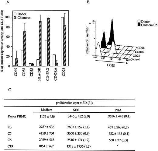
The expression of activation markers on human CD3+ T cells was assessed for the three series of engraftment. Figure 1A shows representative results obtained with seven chimeras engrafted with PBL from donor 1. A mean increase of the percentage of human T cells expressing the early activation marker CD69 (17% v 2% before the graft) was observed, but an important individual variation was obtained among mice, ranging from 0% to 45%. Conversely, the expression of two late activation markers, CD38 and HLA-DR, was strongly and homogeneously induced in all chimeras, on the majority of human T cells (71% ± 18% and 90% ± 4% after engraftment v 39% and 13% before engraftment, respectively). The percentage of human T cells expressing the memory/activation marker CD45RO also increased after the graft (96% ± 2% v 52% before the graft), whereas CD45RA expression concomitantly decreased (10% ± 5% v 23% before the graft). Furthermore, CD25 was not expressed at this stage of engraftment, whether the detection of the interleukin-2 (IL-2) receptor was performed with MoAbs specific to epitope inside (Fig 2A) or outside (data not shown) the binding site of IL-2. In addition, while the percentage of human T cells expressing CD28 molecule remained unchanged upon engraftment (Fig 1A), a decrease in CD28 expression was observed (fluorescence mean 9 in chimera C5 v 58 before the graft) (Fig 1B). This was confirmed for seven other chimeras (mean of fluorescence 17 ± 14 postgraft) (data not shown).
Characterization of spleen human cell chimerism. (A) Expression of several activation markers among CD3+ T cells was analyzed on fresh PBL from donor 1 (□) or engrafted human cells from the spleen of chimeras, killed at days 65, 70, or 77 after the graft (▨). Error bars are indicated. (B) CD28 molecule expression on human CD3+ T cells was compared between fresh donor PBL and splenic cells from chimera C5. The filled peaks correspond to the chimera C5 cells, the open peaks correspond to the fresh donor's cells. Isotypically matched negative IgG1 control antibodies were used for each sample. (C) PBL from donor and splenic cells from four chimeras, killed respectively at day 65 (C3, C5, and C6) or day 70 (C19), were cultured for 4 days in medium alone or in the presence of PHA or SEE at 1 μg/mL. Syngeneic T-cell–depleted irradiated feeder cells were added to cultures of chimeras' cells. Results are expressed as cpm and Stimulation Index (SI) = 3H thymidine incorporation put in brackets, in the presence of PHA or SEE/3H thymidine incorporation in medium alone.
Characterization of spleen human cell chimerism. (A) Expression of several activation markers among CD3+ T cells was analyzed on fresh PBL from donor 1 (□) or engrafted human cells from the spleen of chimeras, killed at days 65, 70, or 77 after the graft (▨). Error bars are indicated. (B) CD28 molecule expression on human CD3+ T cells was compared between fresh donor PBL and splenic cells from chimera C5. The filled peaks correspond to the chimera C5 cells, the open peaks correspond to the fresh donor's cells. Isotypically matched negative IgG1 control antibodies were used for each sample. (C) PBL from donor and splenic cells from four chimeras, killed respectively at day 65 (C3, C5, and C6) or day 70 (C19), were cultured for 4 days in medium alone or in the presence of PHA or SEE at 1 μg/mL. Syngeneic T-cell–depleted irradiated feeder cells were added to cultures of chimeras' cells. Results are expressed as cpm and Stimulation Index (SI) = 3H thymidine incorporation put in brackets, in the presence of PHA or SEE/3H thymidine incorporation in medium alone.
Modifications of the Vβ subset representation in chimeras. (A) The percentage of Vβ+ (BV) CD3+ among total CD3+ T cells from fresh PBL and from the spleen of 9 chimeras killed at times indicated was determined by FACS analysis using anti-CD3 MoAbs and MoAbs specific for several regions of the human TCR. (B and C) The same analysis was performed on human CD4+ (B) and CD8+ (C) T cells from donor and from 3 chimeras.
Modifications of the Vβ subset representation in chimeras. (A) The percentage of Vβ+ (BV) CD3+ among total CD3+ T cells from fresh PBL and from the spleen of 9 chimeras killed at times indicated was determined by FACS analysis using anti-CD3 MoAbs and MoAbs specific for several regions of the human TCR. (B and C) The same analysis was performed on human CD4+ (B) and CD8+ (C) T cells from donor and from 3 chimeras.
Furthermore, we have observed an inability of human T cells to proliferate in response to the bacterial superantigen SEE or to the mitogen PHA, while these cells normally responded to these polyclonal activators before the graft (Fig 1C). This proliferative defect might be due to the in vivo existence of already proliferating lymphocytes. However background 3HTdR incorporation in nonstimulated cultures of chimeras' lymphocytes was only slightly increased (Fig 1C), suggesting that the loss of response to mitogenic stimulation of engrafted lymphocytes was not only due to an excess of ex vivo proliferating lymphocytes. In addition, the SI values upon stimulation with PHA were inferior to one for all chimeras (Fig 1C), indicating that the unresponsiveness was associated with cell death following in vitro stimulation.
Because these features suggested that human T cells were activated in long-term chimeras, it was important to determine whether this cell activation was associated with modifications during the reconstitution of the TCR repertoire.
The human Vβ usage in hu-PBL-SCID chimeras.We studied the human Vβ repertoire of engrafted splenic T cells using a battery of MoAbs specific to variable regions of the β chain of the TCR. Figure 2A summarizes results obtained for nine reconstituted chimeras, engrafted with PBL from donor 1 and killed at day 62, 70, or 77 postgraft. They indicate that, although all the tested human Vβ T-cell subsets were present among splenocytes of reconstituted mice, important modifications of their representation among total CD3+ T cells were observed. Figure 2A shows that five of nine mice exhibited an increase in the representation of one (C3/BV8, C20/BV13S1, C21/BV13S1) or more (C33/BV5S2,3-BV12-BV13-BV17 and C34/BV5S2,3-BV6S7-BV8-BV12-BV13S1-BV17) Vβ subfamilies. Interestingly, the overrepresentation of BV5S2,3, BV12, and BV17 T cells in chimera C34 was associated with the increase of their absolute number (1.68-fold, 2.67-fold, and 1.8-fold, respectively) (data not shown), which strongly suggested a specific expansion of these cells in this chimera. The other four mice (C5, C6, C24, and C35) exhibited a general underrepresentation of most of the Vβ tested, indicating an overrepresentation of some nontested Vβ T-cell subsets. Moreover, the Vβ2 and Vβ5.1 subsets were underrepresented in all chimeras and conversely, no defined β subset was found overrepresented.
However, the modifications of the CD3+ Vβ subsets did not exclude the possibility that they preferentially affected CD4+ or CD8+ T cells. Comparative analysis of the Vβ representation among human CD4+ and CD8+ T cells between chimeras' splenic cells and donors' PBL indicated that the variability in the frequency of the 8 Vβ subsets studied, previously observed in CD3+ T cells, was also detected in CD4+, as well in CD8+ T cells (Fig 2B). In addition, both CD4+ and CD8+ subsets were affected independently. These Vβ representation modifications were also found in absolute number (data not shown). For example, the number of human Vβ17+ CD4+ cells was decreased in the spleen of chimera C34 as 0.14 × 10⁶ Vβ17+ CD4+ T cells were recovered for 0.26 × 10⁶ injected, while the Vβ17+ CD8+ subset was amplified in this chimera as 1.22 × 10⁶ Vβ17+ CD8+ T cells were recovered for 0.48 × 10⁶ injected.
Because available MoAbs did not allow the complete study of the human Vβ repertoire by cytofluorometric approaches, we assessed the presence of the 24 human Vβ subsets by reverse transcriptase (RT)-PCR. Results obtained with this method were generally in agreement with FACS analysis, as a good correlation was found between the intensity of the signal obtained by RT-PCR and the percentage of the corresponding Vβ subset obtained by FACS analysis. Importantly, RT-PCR analyses showed that in some chimeras (two of four), about half of the total Vβ subsets were not detected (data not shown). These modifications of the T-cell repertoire suggest that some positive selection events associated with deletions govern the reconstitution of hu-PBL-SCID mice.
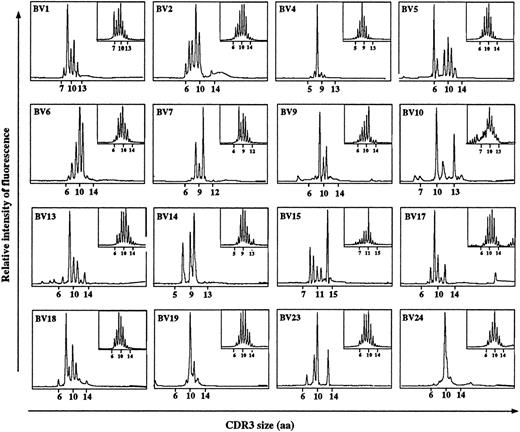
Skewed human Vβ repertoire in hu-PBL SCID chimeras.To further characterize the nature of the modifications affecting human Vβ repertoire in SCID mice, we compared the CDR3 size distribution of 24 Vβ T-cell subsets before and after engraftment of several chimeras. In agreement with previous studies,19 20 the Vβ profiles obtained for donors' PBL were perfectly gaussian (Fig 3). Strikingly, strong modifications of these profiles were induced upon engraftment in SCID mice (representative results are expressed for chimera C5). All types of patterns were found from multipeak profiles (for example BV1, BV2, BV5 subsets) to strongly restricted profiles exhibiting one dominant peak (for example BV4 subset CDR3 = 8, BV19 subset CDR3 = 10, BV24 subset CDR3 = 10). We observed overrepresentation, underrepresentation, and disappearance of some peaks as compared with their representation before the graft, corresponding respectively to the selection (for example BV4, CDR3 = 8 and BV15, CDR3 = 14) or the deletion (for example BV5, CDR3 = 8 and BV23, CDR3 = 11) of T cells with defined CDR3 sizes. All these modifications show that the human T-cell repertoire is submitted to a severe restriction in the SCID environment. It should be mentioned that “new peaks” did not emerge when the pattern of Vβ subsets were compared before and after the graft.
Comparison of CDR3 size distribution of the human Vβ subsets before and after engraftment. RNA extraction from fresh donor's PBL or from spleen cells of chimera C5, killed at day 65 postengraftment, PCR and run off reactions were performed as described in Materials and Methods. Patterns represent the distribution, measured as relative fluorescence intensity, of the aa size of the CDR3 for the PCR reaction products obtained for each Vβ subset studied. Patterns on the bottom left correspond to chimera's profiles and patterns on the top right correspond to fresh donor's profiles. Each peak corresponds to a defined CDR3 size.
Comparison of CDR3 size distribution of the human Vβ subsets before and after engraftment. RNA extraction from fresh donor's PBL or from spleen cells of chimera C5, killed at day 65 postengraftment, PCR and run off reactions were performed as described in Materials and Methods. Patterns represent the distribution, measured as relative fluorescence intensity, of the aa size of the CDR3 for the PCR reaction products obtained for each Vβ subset studied. Patterns on the bottom left correspond to chimera's profiles and patterns on the top right correspond to fresh donor's profiles. Each peak corresponds to a defined CDR3 size.
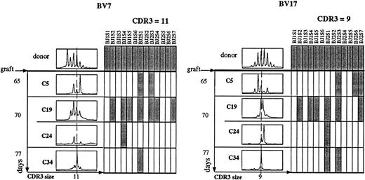
Comparative analysis of the CDR3 size distribution of all Vβ chains was extended to chimeras C19, C24, and C34 and the same kind of restrictions were obtained for these chimeras. Representative analyses of Vβ7+ and Vβ17+ subsets are shown in Fig 4. An important variability in the restriction of CDR3 size pattern was observed among mice either when tested on the same day or in a time course study. This variability was obtained for all the Vβ subsets (data not shown). To further characterize the nature of the Vβ repertoire restriction, we determined whether some Jβ segments were preferentially used. Two CDR3 sizes were chosen: a CDR3 size equal to 11 for the Vβ7+ T cells and a CDR3 size equal to 9 for the Vβ17+ T cells (Fig 4). While all Jβ segments were found associated with the Vβ7 and Vβ17 segments in donors' T cells before the graft, a consistent restriction in the usage of Jβ segments was observed in splenic T cells recovered from hu-PBL-SCID mice. This restriction was variable among engrafted mice and ranged from 6 of 13 Jβ used (C19) to only 1 Jβ used (C24 and C34) for Vβ7 and 7 of 13 Jβ used (C19) to 1 Jβ used (C24) for Vβ17 subset. In mice in which all Vβ subsets could not be detected by RT-PCR, the restriction of CDR3 size distribution and Jβ usage was more pronounced than in mice in which all Vβ subsets were detected. The degree of restriction probably depends on the kinetics of reconstitution. Interestingly, the overrepresentation of some Vβ transcripts with defined CDR3 size was generally associated with the use of only one Jβ segment (eg, Vβ7 subset in chimera C34). In the same way, expansion of some human Vβ T cells observed by FACS analysis was found associated with a severe restriction of their repertoire (for example, in chimera C34, Vβ17 subset is restricted to one CDR3 size associated with only two Jβ segments), indicating that the expansion of these Vβ subsets corresponds to a clonal proliferation. These repertoire analyses argue for the occurrence, in engrafted SCID mice, of clonal specific selection of human T cells. Because it was reported in mice that the in vivo antigenic stimulation induces a similar restriction in the T-cell CDR3 size pattern,14 the skewed TCR repertoire of human cells in chimeras that we report is probably the consequence of an antigenic activation in the SCID environment.
Variability of the human Vβ repertoire restriction between chimera. Fresh donor's PBL and splenic cells from chimeras C5, 19, 24, and 34, killed at day 65, 70, or 77, were treated as described in Fig 2 using specific primers for Vβ7+ and Vβ17+ subsets. Analysis of CDR3 size distribution was performed as described in Fig 2 (left part of the figure). Next, a run off was performed on the same Vβ/Cβ PCR products using fluorescent specific primers to the 13 human Jβ segments. Analysis of the Jβ segment usage by Vβ7+ and Vβ17+ T cells was performed for CDR3 size equal to 11 and 9, respectively (right part of the figure). Hatched boxes correspond to the detection of the corresponding Jβ (annoted BJ) segment associated with the tested Vβ (annoted BV) segment.
Variability of the human Vβ repertoire restriction between chimera. Fresh donor's PBL and splenic cells from chimeras C5, 19, 24, and 34, killed at day 65, 70, or 77, were treated as described in Fig 2 using specific primers for Vβ7+ and Vβ17+ subsets. Analysis of CDR3 size distribution was performed as described in Fig 2 (left part of the figure). Next, a run off was performed on the same Vβ/Cβ PCR products using fluorescent specific primers to the 13 human Jβ segments. Analysis of the Jβ segment usage by Vβ7+ and Vβ17+ T cells was performed for CDR3 size equal to 11 and 9, respectively (right part of the figure). Hatched boxes correspond to the detection of the corresponding Jβ (annoted BJ) segment associated with the tested Vβ (annoted BV) segment.
DISCUSSION
The present report demonstrates the severe restriction of the Vβ T-cell repertoire of human lymphocytes following their long-term engraftment in SCID mice, indicating the in vivo occurrence of antigenic T-cell selection in the SCID environment. This assessment is supported by the activated phenotype of engrafted human T cells. In addition, the chronicity of the antigenic stimulation during the graft is suggested by both the absence of CD25 and the weak expression of CD28 molecules, already reported, respectively, during chronic in vivo21 and in vitro22 T-cell activation. The involvement of exogenous antigens cannot be ruled out, although unlikely, because the reconstituted chimeras were maintained in a sterile environment. The extent of lymphocyte activation together with that of the modifications of the Vβ repertoire argues instead for a major selection process consistent with a xenoreaction. This xenoreaction would promote the reconstitution of chimeras by human cells. Indeed, the xeno-specificity of human CD4+ T cells from chimeras was suggested by a recent study11 showing that human CD4+ T-cell lines derived from long-term chimeras were specific to murine MHC class II molecules. Noteworthy, in this previous study CD8+ T-cell lines derived from the same chimeras were not found xenoreactive.11 However, the present study demonstrates that both CD4+ and CD8+ Vβ repertoire were modified, suggesting that, in addition to CD4+ T cells, the human CD8+ subset was also probably submitted to a xeno-specific selection.
Kinetics of reconstitution of SCID mice intraperitoneally engrafted with PBL from healthy donors probably evolves in two main phases.11,12 During the first phase, most of injected cells remain in the peritoneal cavity and then dramatically disappear, whatever the site screened in the murine host, as a consequence of their nonselection.12 This was confirmed in the present study where half of the Vβ subsets were not detected by RT-PCR in some chimeras (data not shown). During the second phase of reconstitution, xenogeneic selected human T-cell clones proliferate and repopulate several peripheral organs.11 Strikingly, although SCID mice were syngeneic and thus genetically homogeneous, a great variability in the Vβ repertoire of human cells was observed between engrafted mice, confirming previously reported studies.12 Several causes could be proposed to explain this variability. First, the possible clonal heterogeneity of human T cells injected into recipient mice. Indeed, although it is difficult to evaluate the diversity of the human T-cell repertoire, its size was recently estimated to about 2.5 × 10⁹ Vβ specificities.20 Thus, it is unlikely that identical human T-cell clones were injected in each SCID mice. Second, the immunological environment of xenogeneic clones might differ from one recipient to another, making their positive selection variable from one chimera to another. Third, the result of both the massive deletion of engrafted cells and the specific selection and expansion of few clones would induce a very restricted T-cell repertoire established at random.
We have observed an inability of engrafted human T cells to respond in vitro to several polyclonal activators such as superantigen or mitogen, extending previous data obtained upon in vitro stimulation by anti-CD3 MoAbs.11,12 This unresponsiveness was not due to the presence of murine cells in the cultures (data not shown). The refractory state to TCR-mediated activation of engrafted T cells could be the consequence of their in vivo chronic activation, as reported previously 5,11 and shown in the present study. In addition, a significant fraction of these cells could be in a real anergic state, previously characterized by both a defect in Ca2+ flux induction and IL-2 secretion upon in vitro anti-CD3 stimulation.11 This in vitro proliferative defect is reminiscent of the absence of in vivo primary and secondary immune responses in chimeras at this stage of reconstitution.23
Altogether, these results show a strong variability in the repopulation of hu-PBL SCID mice by human cells and point out important modifications in the Vβ repertoire of engrafted T cells. This skewed repertoire probably results from the occurrence in these chimera of a xenoreaction, as previously suggested.12 The fact that such a reaction can promote the reconstitution of the chimeras and its consequences on the engrafted human immune system raises the question of the validity of such a model in studies of human pathologies. For example, the use of hu-PBL-SCID mice for the study of human immunodeficiency virus (HIV)-infection should be done being aware of the limits of the model. Indeed, analysis of some immunological parameters such as Th1/Th2 balance,24 apoptosis25 or cytotoxic T lymphocyte function,26 known to be important in acquired immunodeficiency syndrome pathogenesis, should be carefully performed in the context of the chronic immune activation occurring in this humanized model. However, hu-PBL-SCID mice have been successfully used during the first phase of reconstitution to study in vivo the influence of HIV-infection on the depletion of human CD4 T cells.27 28 In these studies, the activated state of engrafted human T cells could be used to promote the infection of human cells by HIV.
ACKNOWLEDGMENT
The authors particularly acknowledge Drs Madeleine Cochet and Joseph Even for numerous helpful discussions during the course of these studies and for critical review of the manuscript, and Viviane James for her excellent technical support.
Supported by grants from the Agence Nationale de Recherche sur le SIDA (ANRS, Paris) to M.L.G., the Centre National de la Recherche Scientifique (CNRS, Paris), the Pasteur Institute, the Fondation pour la Recherche Médicale (FRM, Paris, France) (Sidaction) to M.L.G., the European Community (EEC) Biomed-1 program (Contract No. BMH1-CT 92-1571). S.G. was supported by the French Research and Space Ministry, relayed by a fellowship from the Fondation pour la Recherche Médicale (FRM) (Sidaction).
Address reprint requests to Marie-Lise Gougeon, PhD, Unité d'Oncologie Virale, Département SIDA et Rétrovirus Institut Pasteur, 28 Rue du Dr. Roux, 75724 Paris Cedex 15, France.


This feature is available to Subscribers Only
Sign In or Create an Account Close Modal