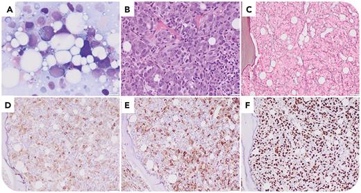
A 71-year-old man presented with pancytopenia (white blood cell count 1.9 × 10⁹/L, hemoglobin 8.0 g/dL, platelets 19 × 109/L). The bone marrow (BM) touch imprint showed 80% blasts with basophilic cytoplasm (panel A; Giemsa stain, original magnification ×1000); BM biopsy showed sheets of immature mononuclear cells (panel B; hematoxylin and eosin stain, original magnification ×400) with extensive reticulin fibrosis (panel C; original magnification ×200). Blasts were positive for CD117 (panel D; original magnification ×200), CD71 (panel E; original magnification ×200), and e-cadherin and negative for myeloperoxidase, supporting erythroid differentiation, with strong overexpression of p53 (panel F; original magnification ×200) by immunohistochemistry. Cytogenetic studies showed a complex karyotype (45,XY,del(5)(q13q32),-21[8]/43,idem,add(2)(q37),-13,-14[6]/46,XY[1]) but intact TP53 by fluorescence in situ hybridization. Next-generation sequencing detected a TP53 c.817C>T, p.R273C mutation with 66% variant allelic frequency, suggesting TP53 copy-neutral loss of heterozygosity. No other pathogenic or likely pathogenic mutations were detected.
This case is a rare form of acute myeloid leukemia (AML) with multihit TP53 alterations and erythroid differentiation with divergent classification in the World Health Organization Classification of Tumours: Haematolymphoid Tumours, 5th Edition (acute erythroid leukemia) and International Consensus Classification (AML with mutated TP53) schemes. Notably, p53 immunohistochemistry is useful for assessing TP53 mutation but is agnostic of TP53 allelic status. Although multihit TP53 alterations are considered a hallmark of acute erythroid leukemia, we believe highlighting erythroid differentiation in the pathology report may have treatment implications, given the B-cell lymphoma-extra large dependency and venetoclax resistance conferred by erythroid differentiation.
For additional images, visit the ASH Image Bank, a reference and teaching tool that is continually updated with new atlas and case study images. For more information, visit https://imagebank.hematology.org.


This feature is available to Subscribers Only
Sign In or Create an Account Close Modal