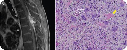
A 25-year-old man with a rare hemoglobin variant (Sallanches), previously confirmed by α-globin gene sequencing demonstrating a homozygous mutation at codon 104 and a transfusion-dependent α-thalassemia phenotype, presented with lower extremity weakness. Pretransfusion hemoglobin levels were typically ∼60 g/L and our approach had been to transfuse only when the patient had symptoms attributable to anemia, which was about 4 times per year. Magnetic resonance imaging (panel A) revealed a cord-compressing mass (17 × 33 × 8.5 mm) at the T6 vertebral body (arrow). There was concern for extramedullary hematopoiesis (EMH), and surgery was undertaken. Pathology (panel B: hematoxylin and eosin stain; original magnification ×40) revealed a giant cell–rich (arrows) lesion, with fibrosis and bone (thick arrow) negative for pancytokeratin, epithelial membrane antigen, glial fibrillary acidic protein, and progesterone receptor, as revealed by immunohistochemistry. The findings were most consistent with a solid-variant aneurysmal bone cyst, a diagnosis confirmed by a fluorescence in situ hydridization stain positive for ubiquitin-specific peptidase 6.
Although patients with α-thalassemia are less likely than those with β-thalassemia to develop EMH, this patient was transfusion-dependent and, as such, at risk for EMH. However, in this case, surgical findings revealed a benign solid-variant aneurysmal bone cyst. The absence of evidence for EMH obviated the need for more aggressive intervention or alteration of our conservative transfusion strategy predicated on preexisting secondary hemochromatosis.
For additional images, visit the ASH Image Bank, a reference and teaching tool that is continually updated with new atlas and case study images. For more information, visit https://imagebank.hematology.org.


This feature is available to Subscribers Only
Sign In or Create an Account Close Modal