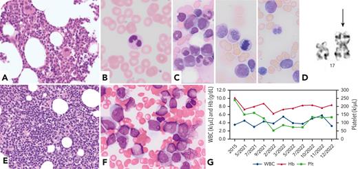
A 71-year-old man presented with persistent anemia. Bone marrow biopsy (BMBx) indicated 40% cellularity with erythroid hypoplasia and no dysplasia (panel A; hematoxylin and eosin [H&E] stain; 40× objective); peripheral blood showed rare dysplasia in circulating neutrophils (panel B; Wright-Giemsa stain; 100× objective); cytogenetic analysis was unremarkable and molecular testing was negative. He was diagnosed with idiopathic cytopenia of undetermined significance (ICUS). His anemia worsened. BMBx at the age of 77 years showed trilineage dysplasia, 6% blasts (panel C; Wright-Giemsa stain; 100× objective), isochromosome 17q (i[17q]) (panel D; karyotype), and mutations in NF1, ASXL2, ETNK1, and SRSF2 genes by next-generation sequencing. Treatment with azacitidine/ipilimumab and a myeloid cell leukemia-1 inhibitor led to temporary blast reductions, but subsequent BMBx at the age of 78 years revealed 7% blasts and persistent i(17q). He developed acute myeloid leukemia (AML) 1 year later (panel E; H&E stain; 40× objective; and panel F; Wright-Giemsa stain; 100× objective). Panel G shows the patient’s peripheral blood trends from the ICUS diagnosis (2015) up until before the development of AML (2022).
Isolated i(17q) is a rare but recurrently identified cytogenetic abnormality in myeloid neoplasms. These neoplasms have a poor clinical outcome and frequently show hybrid myelodysplastic syndrome (MDS)/myeloproliferative neoplasm (MPN) features that do not fulfill World Health Organization criteria for other MDS/MPN entities. This case demonstrates an i(17q) pathway from ICUS to MDS with i(17q) to AML with i(17q).
For additional images, visit the ASH Image Bank, a reference and teaching tool that is continually updated with new atlas and case study images. For more information, visit https://imagebank.hematology.org.


This feature is available to Subscribers Only
Sign In or Create an Account Close Modal