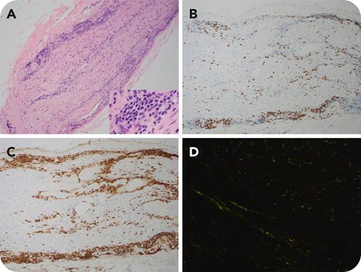
A 53-year-old woman presented with pain in the right upper extremity. Imaging studies suggested the possibility of thoracic outlet syndrome. She underwent decompressive surgery followed by intravenous immunoglobulin and steroids, without improvement. Magnetic resonance imaging (MRI) studies showed no discrete lesions and a brachial plexus nerve root biopsy was performed, which showed infiltration by lymphocytes, plasmacytic lymphocytes, and plasma cells (panel A; ×10 magnification and ×40 magnification [inset], hematoxylin-eosin stain). The lymphocytes were negative for CD3 (panel B; ×10 magnification) but expressed CD20 (panel C; ×10 magnification) and CD138 and were kappa light chain restricted. Congo-red stain revealed amyloid deposition (panel D; ×40 magnification, fluorescence microscopy). Molecular testing revealed a P.LEU265PRO mutation in the MYD88 gene tested using polymerase chain reaction followed by single-nucleotide mutation detection. Immunoglobulin quantification showed normal levels. Serum immunofixation detected a faint immunoglobulin M (IgM)/kappa monoclonal band. Positron emission tomography/computed tomography chest imaging was negative for 18F-fluorodeoxyglucose avid lymphadenopathy; pelvic and spine MRI was negative for lesions. Staging bone marrow biopsy and lumbar puncture showed no evidence of lymphoma.
Lymphoplasmacytic lymphoma (LPL) is a low-grade B-cell lymphoma involving the bone marrow and is often associated with elevated IgM paraproteins. Typically, neurological deficits result from paraproteins causing direct damage to the nerve roots. However, in this case, the pathology was due to brachial plexus infiltration by the LPL and amyloid deposition, with no evidence of bone marrow involvement. Because most neural lymphocytic infiltrates are reactive T cells, conducting additional B-cell markers such as CD19 and CD20 is crucial for prompt diagnosis.
For additional images, visit the ASH Image Bank, a reference and teaching tool that is continually updated with new atlas and case study images. For more information, visit https://imagebank.hematology.org.


This feature is available to Subscribers Only
Sign In or Create an Account Close Modal