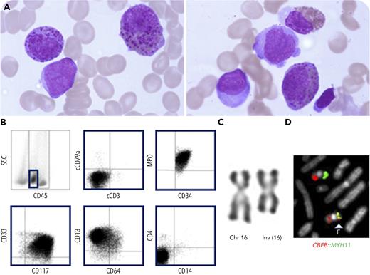
A 56-year-old woman was admitted to our hospital for anemia (hemoglobin, 108 g/L) and leukopenia (1.98 × 10⁹/L) with 10% blasts. Bone marrow aspirate (May-Grünwald-Giemsa stained) revealed 36% blasts and 17% abnormal eosinophils with large coarse basophilic granules (panel A; 100× objective; original magnification ×1000), suggesting an acute myeloid leukemia (AML) with CBFB::MYH11 fusion according to the fifth World Health Organization classification. At the morphologic level, this AML subtype is classically characterized by myelomonocytic differentiation and presence of abnormal eosinophils in bone marrow. In our case, the monocytic population was absent, as confirmed by flow cytometry immunophenotyping, which revealed presence of myeloid blasts (MPO+CD33+CD13+CD34+CD117+) without monocytic contingent (panel B). Karyotype (R banding) showed the characteristic inversion of chromosome 16: inv(16)(p13.1q22) in 19 of 20 metaphases (panel C; 63× objective; original magnification ×630). Fluorescence in situ hybridization with a CBFB::MYH11 double-fusion probe (MetaSystems, Altlussheim, Germany) showed a CBFB::MYH11 fusion signal in 10 of 10 metaphases and highlighted existence of a partial deletion of MYH11 (panel D; 63× objective; original magnification ×630). This fusion was confirmed by reverse transcription polymerase chain reaction.
To our knowledge, cases of nonmonocytic AML with CBFB::MYH11 fusion remain exceptional. Our case highlights the necessity to search for a CBFB::MYH11 fusion in all new AML cases so as not to miss those harboring atypical presentation. Indeed, this AML subtype is associated with a favorable prognosis and can benefit from a tailored therapy.
For additional images, visit the ASH Image Bank, a reference and teaching tool that is continually updated with new atlas and case study images. For more information, visit https://imagebank.hematology.org.


This feature is available to Subscribers Only
Sign In or Create an Account Close Modal