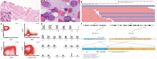
A 67-year-old man presented with anemia (10.4 g/dL), thrombocytopenia (49 000/μL), and circulating blasts. Bone marrow examination revealed hypercellularity, multilineage dysplasia (panel A: hematoxylin and eosin stain, original magnification ×4; inset: micromegakaryocyte, original magnification ×40), and 70% blasts (panel B: Wright-Giemsa stain, original magnification ×100; inset: cytoplasmic blebs and vacuoles in subset, original magnification ×100) with predominantly monocytic differentiation (panel C: flow cytometry, CD34+ blasts coexpress CD56, CD64 [variable], myeloperoxidase (MPO) [partial], CD33), subset (3.6%) with megakaryoblastic differentiation (CD42b), and minimal blast component (<1%) showing T-cell lineage differentiation (cytoplastic-CD3). Karyotype detected t(3;5) and del(20q) (95% of metaphases) (panel D). Targeted next-generation sequencing (NGS) detected somatic variants at high allele frequencies: NRAS p.G12V(c.35G>T), ETV6 p.Y344∗(c.1032C>A), PHF6 p.Y195∗(c.585T>A), RET p.R297C(c.889C>T); copy number loss of ETV6, and CDKN1B on chr12p13. Given the possibility of mixed lineage leukemia, targeted RNA sequencing utilizing a commercially available NGS-based assay (Archer) was performed, identifying an in-frame TBL1XR1::CSF1R fusion (panel E: JBrowse supporting reads; panel F: rearrangement between TBL1XR1 [exon9] and CSF1R [exon11]). This case was best classified as acute myeloid leukemia with a minimal component showing T-cell differentiation. Induction 7 + 3 (cytarabine + daunorubicin) was initiated, followed by intermediate-dose cytarabine reinduction.
TBL1XR1::CSF1R fusion has been reported once in B-lymphoblastic leukemia. This is the first report in a predominantly myeloid acute leukemia. TBL1XR1 is a regulator of hematopoietic stem cell self-renewal and lineage differentiation. We hypothesize that this fusion may lead to overexpression and/or ligand-free activation of CSF1R. An intact tyrosine kinase domain in CSF1R potentially serves as a therapeutic target.
For additional images, visit the ASH Image Bank, a reference and teaching tool that is continually updated with new atlas and case study images. For more information, visit https://imagebank.hematology.org.


This feature is available to Subscribers Only
Sign In or Create an Account Close Modal