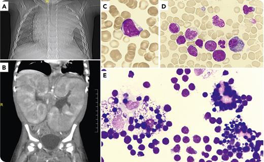
A 2-year-old child presented with diarrhea and abdominal distension. Blood test showed acute renal failure (serum creatinine, 182 μmol/L) and no cytopenias (hemoglobin, 144 g/L; leukocytes, 21.1 × 10⁹/L; and platelets, 313 × 109/L). Imaging test results revealed mediastinal widening with pleural effusions (panel A; thorax radiography) and massive bilateral nephromegaly (panel B; abdominal computed tomography). Peripheral blood smear examination results found 3% of medium-sized blasts with agranular and basophilic cytoplasm, dispersed nuclear chromatin with 2 to 3 nucleoli, and a high nuclear/cytoplasmic ratio (panel C; 60× objective; total magnification ×600; May-Grünwald-Giemsa). Bone marrow (panel D; 60× objective; total magnification ×600) was infiltrated with 52% of blasts with a T-cortical lymphoblastic leukemia immunophenotype (cytoplasmic CD3+, CD7+, CD5+, and CD1a+). In the pleural liquid cytology, numerous histiocytes with different degrees of blast phagocytosis emerged over a uniform lymphoblast background (panel E; 60× objective; total magnification ×600). The patient did not fulfill criteria for hemophagocytic lymphohistiocytosis. Hemophagocytosis was not present in bone marrow. Extensive microbiological testing identified Epstein-Barr virus primoinfection, and chemical parameters in pleural fluid did not suggest infection.
Mediastinal involvement and pleural effusions are frequent in T-lymphoblastic leukemias. Histiocytes are monocytic-derived cells whose phagocytic activity under pathological conditions can target erythrocytes and, less frequently, other cell types. The biological mechanism underlying this finding as well as its clinical impact are still to be elucidated.
For additional images, visit the ASH Image Bank, a reference and teaching tool that is continually updated with new atlas and case study images. For more information, visit http://imagebank.hematology.org.


This feature is available to Subscribers Only
Sign In or Create an Account Close Modal