

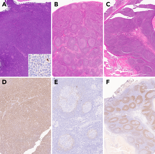
A 53-year-old woman presented with a mass at the base of her tongue. Biopsies of the lesion demonstrated diffuse effacement of the submucosal tissue by intermediate-sized lymphocytes with atypical centrocytic morphology (A). These lymphocytes were CD20+ and CD5+ B cells that were negative for CCND1 (cyclin D1) (A inset). The lymphocytes were diffusely positive for SOX11 (D) and diagnostic of CCND1− mantle cell lymphoma. Fluorescence in situ hybridization confirmed the absence of CCND1 rearrangement. The patient had undergone a right neck lymph node biopsy and bilateral tonsillectomy 9 and 5 years previously, respectively, both with benign diagnoses. These specimens were reevaluated given the patient’s new lymphoma diagnosis. The initial lymph node biopsy demonstrated reactive morphologic features including follicular hyperplasia and dermatopathic changes (B). SOX11+ lymphocytes were present, restricted to the inner mantle zones (E). The subsequent tonsil biopsy demonstrated a subtle expansion of the mantle zones (C) by numerous SOX11+ cells (F).
These specimens illustrate progression of in situ mantle cell neoplasia to a mantle cell lymphoma that first exhibited a mantle zone pattern and later diffuse growth. All were CCND1−, demonstrating that CCND1− mantle cell lymphomas may sometimes be preceded by CCND1− in situ mantle cell neoplasia.
For images A, D, and E, ×40 total magnification; B, C, and F, ×20 total magnification; and A inset, ×400 total magnification.
For additional images, visit the ASH Image Bank, a reference and teaching tool that is continually updated with new atlas and case study images. For more information, visit http://imagebank.hematology.org.

This feature is available to Subscribers Only
Sign In or Create an Account Close Modal