In this issue of Blood, Lou et al1 show that all-trans retinoic acid (ATRA) and different inflammatory signals increase the expression of retinoic acid (RA)-inducible gene I (RIG-I) in bone marrow (BM) mesenchymal stem cells (BMSCs), reducing their reactive oxygen species (ROS) buffering capacity and their hematopoietic supportive function after transplantation.
The impact of cancer treatment on the BM niche and its consequences for hematopoiesis is an emerging research field with therapeutic potential. In their article, Lou et al show that ATRA and other inflammatory signals increase the expression of RIG-I in BMSCs. In turn, RIG-I triggers degradation of the master antioxidant protein nuclear factor erythroid 2–related factor 2 (NRF2), leading to the accumulation of ROS and impaired survival of BMSCs, thereby decreasing their hematopoietic supportive function. These results pinpoint that RIG-I in BMSCs is inadvertently increased by leukemia treatment and may be an actionable target for improving hematopoietic regeneration.
Hematopoietic stem cells (HSCs) need to carefully balance self-renewal and differentiation during regeneration through complex decisions that require a dynamic interplay between HSCs and their niches. Decisions regarding cell fate can be adjusted to organismal demands through master regulators such as the sympathetic nervous system, which influences the activity of HSCs in different BM niches. For instance, chemotherapy or irradiation, which are routinely used for cancer treatment or as conditioning regimens for HSC transplantation, increase the release of cholinergic signals that activate the α7 nicotinic receptor in BMSCs, which in turn preserve HSC quiescence via CXCL12; consequently, not all HSCs become activated (which could cause their exhaustion), but some remain quiescent, thereby retaining their full potential.2
BMSCs are important HSC niche cells in addition to functioning as documented immunomodulators. Co-transplantation of BMSCs with HSCs can accelerate hematopoietic recovery and decrease the risk of graft-versus-host disease.3,4 Intravenously infused BMSCs do not engraft in the BM efficiently or for long. These effects are likely due to the potent immunomodulatory effects of BMSCs, especially when these cells become apoptotic and are engulfed by macrophages.5
HSC transplantation is routinely performed to treat leukemia. However, cumulative evidence indicates that conditioning regimens have bystander niche effects, which may hamper subsequent hematopoietic recovery.6 One example is the semaphorin 3A-neuropilin 1 signaling pathway, which promotes BM vascular regression after myelosuppression and can be blocked to accelerate vascular and hematopoietic regeneration.7 In addition, metabolic exchange and crosstalk between HSCs and their niche might be critical for efficient regeneration. For instance, mitochondrial transfer from HSCs to BMSCs helps to rescue the niche from conditioning-related damage, which subsequently improves hematopoietic regeneration.8 The metabolic adaptation during regeneration can also affect HSC decisions by changing chromatin accessibility. For example, the availability of acetyl-CoA, which is a major regulator of histone acetylation, may shape the epigenetic landscape and ultimately determine whether HSCs undergo self-renewing divisions or differentiate during regenerative hematopoiesis.9 However, the effects of cancer treatment on the marrow niche and the consequences for hematopoiesis have only started to be elucidated.
Lou et al investigated the effects of ATRA on BMSCs. ATRA is used alone or in combination therapies to treat acute promyelocytic leukemia and has been investigated in other hematologic malignancies such as T-cell leukemia and acute myeloid leukemia (AML). Previous studies showed that RIG-I is required for RA antileukemic effects, partly through competitive inhibition of the Src-AKT-mTOR pathway by RIG-I in AML cells.10 However, whether RA and ATRA or other inflammatory signals converging to upregulate RIG-I affect the hematopoietic niche is not clear.
Lou et al found that treatment with ATRA impairs BMSC function (clonogenicity, multilineage differentiation potential, and capacity to support hematopoietic regeneration after transplantation). The reason seems to be ROS accumulation, because treatment with N-acetyl-L-cysteine reverses the effect. To determine the cause of the unbalanced ROS, the authors performed a transcriptomic analysis. The expression of genes related to mitochondrial oxidative phosphorylation (the main source of ROS) is unaffected, but ATRA strongly represses targets of the master antioxidant NRF2. The inhibition seems to be posttranscriptional because ATRA induces proteasomal NRF2 degradation but it does not affect levels of NRF2 messenger RNA (mRNA). Functionally, NRF2 overexpression rescues ROS accumulation, suggesting that ATRA-induced NRF2 suppression causes excessive ROS, which compromise BMSC niche function.
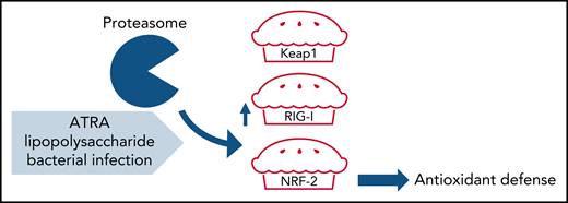
Seeking factors that affect the stability of NRF2, the authors found that ATRA treatment leads to protein (not mRNA) accumulation of Kelch-like ECH-associated protein 1 (Keap1), which facilitates NRF2 ubiquitination and proteasomal degradation. Functionally, Keap1 silencing rescues NRF2 protein levels, so the focus of the Lou et al study moved to ATRA regulation of Keap1. Because both RIG-I and Keap1 are targeted by the ubiquitin ligase Trim25, the authors hypothesized that ATRA-induced RIG-I might outcompete Keap1 in Trim25-dependent ubiquitination, leading to accumulation of Keap1. Competitive immunoprecipitation analyses showed that RIG-I and Keap1 competitively bind to Trim25, and that RIG-I reduces the binding between Trim25 and Keap1 in mice treated with ATRA. Importantly, Keap1 does not accumulate and NRF2 is not reduced by ATRA in RIG-I–deficient mice, which supports the authors’ hypothesis of competition between RIG-I and Keap1 for Trim25-dependent ubiquitination and proteasomal degradation. Consequently, Keap1 accumulation favors NRF2 degradation after treatment with ATRA (see figure).
Model of inflammation-induced RIG-I accumulation and NRF2 degradation compromising antioxidant defense in BMSCs. ATRA, lipopolysaccharide, or bacterial infection favor proteasomal degradation of the master antioxidant protein NRF2, leading to the accumulation of ROS and impaired BMSC survival, thereby decreasing their hematopoietic supportive function.
Model of inflammation-induced RIG-I accumulation and NRF2 degradation compromising antioxidant defense in BMSCs. ATRA, lipopolysaccharide, or bacterial infection favor proteasomal degradation of the master antioxidant protein NRF2, leading to the accumulation of ROS and impaired BMSC survival, thereby decreasing their hematopoietic supportive function.
Supporting the role of RIG-I as gatekeeper, deletion of RIG-I rescues ROS levels, clonogenicity, and osteogenic differentiation of BMSCs, and improves their HSC niche functions after administration of ATRA. NRF2 knockdown similarly reduces BMSCs, their clonogenicity, differentiation, and HSC niche factor expression, and attenuates BMSC protection from ATRA upon RIG-I deletion. Therefore, the authors conclude that NRF2 mediates the effects of ATRA.
Finally, Lou et al extrapolate ATRA effects to other inflammatory signals and stimuli, which similarly increase RIG-I (through STAT1 activation), such as the administration of lipopolysaccharide or bacterial infection. Treatment with ATRA permits the growth of pathogenic bacteria, but this effect is ameliorated in RIG-I–deficient mice because of the preserved BMSC function. This mechanistically rich study highlights the role of the RIG-I–NRF2 axis as a master regulator of antioxidant defense in BMSCs, which is affected by RA and ATRA and several inflammatory insults, thus impairing the BMSC function supporting emergency myelopoiesis and regenerative hematopoiesis.
Conflict-of-interest disclosure: The author declares no competing financial interests.


This feature is available to Subscribers Only
Sign In or Create an Account Close Modal