Abstract
Thrombosis with associated inflammation (thromboinflammation) occurs commonly in a broad range of human disorders. It is well recognized clinically in the context of superficial thrombophlebitis (thrombosis and inflammation of superficial veins); however, it is more dangerous when it develops in the microvasculature of injured tissues and organs. Microvascular thrombosis with associated inflammation is well recognized in the context of sepsis and ischemia-reperfusion injury; however, it also occurs in organ transplant rejection, major trauma, severe burns, the antiphospholipid syndrome, preeclampsia, sickle cell disease, and biomaterial-induced thromboinflammation. Central to thromboinflammation is the loss of the normal antithrombotic and anti-inflammatory functions of endothelial cells, leading to dysregulation of coagulation, complement, platelet activation, and leukocyte recruitment in the microvasculature. α-Thrombin plays a critical role in coordinating thrombotic and inflammatory responses and has long been considered an attractive therapeutic target to reduce thromboinflammatory complications. This review focuses on the role of basic aspects of coagulation and α-thrombin in promoting thromboinflammatory responses and discusses insights gained from clinical trials on the effects of various inhibitors of coagulation on thromboinflammatory disorders. Studies in sepsis patients have been particularly informative because, despite using anticoagulant approaches with different pharmacological profiles, which act at distinct points in the coagulation cascade, bleeding complications continue to undermine clinical benefit. Future advances may require the development of therapeutics with primary anti-inflammatory and cytoprotective properties, which have less impact on hemostasis. This may be possible with the growing recognition that components of blood coagulation and platelets have prothrombotic and proinflammatory functions independent of their hemostatic effects.
Thrombosis and inflammation
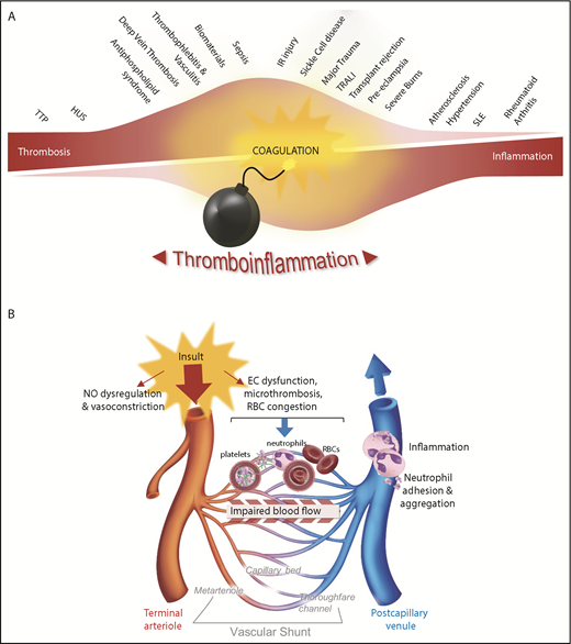
The coordinated activation of inflammatory and hemostatic responses following infection or tissue injury is a phylogenetically conserved defense mechanism that can be traced back to early invertebrates.1 In these primitive organisms, a single cell (the hemocyte) can perform basic inflammatory, immune, and hemostatic functions.1 However, most higher-order mammals have evolved a complex multicellular system encompassing platelets and a variety of leukocyte subsets, including neutrophils, monocytes, and a series of antigen-presenting cells. Similarly, some of the major humoral components of innate immunity have evolved from a rudimentary complement system into a sophisticated series of highly integrated protease cascades, including the complement, coagulation, fibrinolysis, and contact-kinin systems. Despite this close evolutionary development, thrombosis and inflammation have traditionally been viewed as distinct complementary processes. Thrombosis can be best defined as an exaggerated hemostatic response, leading to the formation of an occlusive blood clot obstructing blood flow through the circulatory system. By comparison, inflammation is the term applied to the complex protective immune response to harmful stimuli, such as pathogens, damaged cells, or irritants. However, with the recognition that inflammation stimulates thrombosis, and in turn, thrombosis promotes inflammation, the functional interdependence of these processes has becoming increasingly well defined (Figure 1A).
Thromboinflammation: an important pathogenic process linked to a diverse range of human diseases. (A) A broad spectrum of human disorders is associated with thromboinflammatory complications, many of which have microvascular dysfunction. It is likely that the level of α-thrombin generation is a key determinant of the extent of the thromboinflammatory response. For example, diseases such as sepsis, ischemia reperfusion (IR) injury (the focus of this review), and organ transplant rejection are associated with widespread activation of coagulation throughout the microcirculation, which is accompanied by an intense thrombotic and inflammatory response. At the extremes of this spectrum, the microvascular thrombotic disorder thrombotic thrombocytopenia purpura (TTP) exhibits limited α-thrombin generation and is associated with a limited inflammatory response in the early phase of the disease. At the other end of the spectrum, autoimmune diseases, such as rheumatoid arthritis and systemic lupus erythematosus (SLE), are primarily considered inflammatory disorders, with a limited role for α-thrombin. Nonetheless, other hemostatic components appear to contribute to the pathogenesis of these diseases. (B) Tissue or organ injury by diverse pathogenic mechanisms is commonly associated with microvascular thromboinflammatory responses. Microvascular obstruction can be mediated by all cellular elements in blood, including platelets, neutrophils, and red blood cells (RBCs), with stable occlusion typically linked to the activation of coagulation and fibrin generation. Regardless of the primary insult to tissues or organs (infection, ischemia, or trauma), the ultimate outcome of these disorders is heavily influenced by the extent of the microvascular thrombotic and inflammatory responses. EC, endothelial cell; HUS, hemolytic-uremic syndrome; NO, nitric oxide; TRALI, transfusion-related acute lung injury.
Thromboinflammation: an important pathogenic process linked to a diverse range of human diseases. (A) A broad spectrum of human disorders is associated with thromboinflammatory complications, many of which have microvascular dysfunction. It is likely that the level of α-thrombin generation is a key determinant of the extent of the thromboinflammatory response. For example, diseases such as sepsis, ischemia reperfusion (IR) injury (the focus of this review), and organ transplant rejection are associated with widespread activation of coagulation throughout the microcirculation, which is accompanied by an intense thrombotic and inflammatory response. At the extremes of this spectrum, the microvascular thrombotic disorder thrombotic thrombocytopenia purpura (TTP) exhibits limited α-thrombin generation and is associated with a limited inflammatory response in the early phase of the disease. At the other end of the spectrum, autoimmune diseases, such as rheumatoid arthritis and systemic lupus erythematosus (SLE), are primarily considered inflammatory disorders, with a limited role for α-thrombin. Nonetheless, other hemostatic components appear to contribute to the pathogenesis of these diseases. (B) Tissue or organ injury by diverse pathogenic mechanisms is commonly associated with microvascular thromboinflammatory responses. Microvascular obstruction can be mediated by all cellular elements in blood, including platelets, neutrophils, and red blood cells (RBCs), with stable occlusion typically linked to the activation of coagulation and fibrin generation. Regardless of the primary insult to tissues or organs (infection, ischemia, or trauma), the ultimate outcome of these disorders is heavily influenced by the extent of the microvascular thrombotic and inflammatory responses. EC, endothelial cell; HUS, hemolytic-uremic syndrome; NO, nitric oxide; TRALI, transfusion-related acute lung injury.
A growing body of evidence indicates that some autoimmune disorders, such as rheumatoid arthritis and systemic lupus erythematosus, are regulated by components of the hemostatic system.2 Thrombotic disorders, such as paroxysmal nocturnal hemoglobinuria or atypical hemolytic uremic syndrome, are triggered by humoral components of innate immunity (complement), and many microvascular inflammatory disorders promote platelet activation and coagulation. This is most clearly demonstrated in the acute exaggerated thromboinflammatory responses that accompany sepsis, ischemia-reperfusion (IR) injury, and major trauma. In these latter disorders, the ultimate extent of organ injury is dependent on the primary insult (bacterial invasion, organ ischemia, or traumatic injury), as well as on the extent of the ensuing microvascular thromboinflammatory response (Figure 1B). When severe, thromboinflammation can extend systemically and damage remote organs, particularly the lung (acute lung injury or acute respiratory distress syndrome) and kidneys (acute kidney injury). This is a major issue in critically ill patients and can lead to the development of multiorgan dysfunction syndrome and death. Thus, defining the molecular mechanisms regulating thromboinflammation in specific disease states is of major clinical importance. The primary focus of this review will be on the role of coagulation in promoting acute microvascular thrombosis and inflammation during sepsis and IR injury. These disorders have been most intensively investigated in the context of thromboinflammation, and much has been learned from sepsis and IR injury about the potential benefits and limitations of targeting coagulation processes in the clinic. For more detailed information on the interaction between coagulation and other components of the hemostatic and innate immune system, several excellent review articles have been published.3-7
Endothelium: a critical regulator of thromboinflammation
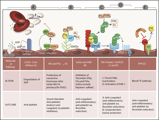
The endothelium lines the lumen of the entire circulatory system, from the chambers of the heart down to the microcapillary beds. However, quantitatively, ∼98% of all endothelial cells reside within the microvasculature, a reflection of the vast surface area of the microcirculatory system in the human body.8,9 Endothelial cells maintain vascular health by exerting antiplatelet, anticoagulant, and anti-inflammatory actions (Figure 2). Quiescent endothelial cells prevent platelet adhesion and activation by producing potent platelet antagonists, such as the adenosine triphosphate/adenosine 5′-diphosphate (ADP) scavenging enzyme CD39/ecto-adenosine diphosphatase,10 prostacyclin (or prostaglandin I2 [PGI2]), and nitric oxide (NO). These molecules also maintain endothelial homeostasis through other mechanisms. NO minimizes leukocyte recruitment to the vessel wall by reducing P-selectin expression on the endothelial surface, decreasing chemokine expression, and reducing transcription of adhesion molecules, such as E-selectin, VCAM-1, and intercellular adhesion molecule-1 (ICAM-1). PGI2 decreases inflammation by reducing leukocyte adhesion, activation, and extravasation. Additionally, the endothelium supports an extensive repertoire of natural anticoagulant and antifibrinolytic pathways, involving glycosaminoglycans (GAGs), thrombomodulin (TM), the activated protein C (APC) pathway, and tissue factor pathway inhibitor (TFPI). The details of these pathways and their roles in regulating microvascular thromboinflammation have been described previously.10,11
Antithrombotic function of endothelial cells. (A) The endothelium presents an antiadhesive phenotype maintained through 3 intrinsic pathways: the CD39/ecto-adenosine diphosphatase (ecto-ADPase) pathway, which depletes the platelet agonist, ADP, and the PGI2 and NO pathways, which inhibit platelet activation. With respect to depletion of ADP, spatial limitations dictate that this primarily occurs for ADP release from platelets adherent to activated endothelium or matrix proteins. (B-E) The endothelium uses several mechanisms to neutralize α-thrombin. (B) First, antithrombin (AT) binds to GAGs on the endothelial cell surface and inactivates α-thrombin (IIa), FXa, and several other coagulation proteases. (C-D) Second, thrombin can be efficiently converted from a procoagulant to anticoagulant protease in the microcirculation by binding the endothelial integral membrane protein TM and the endothelial protein C receptor (EPCR). Once bound to these receptors, thrombin cleaves and activates protein C (PC) to generate APC. (C) APC functions as an anticoagulant by proteolytically inactivating FVa and FVIIIa. (D) If APC remains bound to the EPCR, it induces signaling through PAR1 to elicit cytoprotection and increased endothelial barrier function. (E) Healthy endothelium also expresses high levels of TFPI that inhibit formation of the tissue factor (TF)/FVIIa complex, thereby preventing the initiation of coagulation and subsequent thrombin generation. AMP, ecto-adenosine monophosphate; Pi, inorganic phosphate, PAR1, protease-activated receptor 1; RBC, red blood cell.
Antithrombotic function of endothelial cells. (A) The endothelium presents an antiadhesive phenotype maintained through 3 intrinsic pathways: the CD39/ecto-adenosine diphosphatase (ecto-ADPase) pathway, which depletes the platelet agonist, ADP, and the PGI2 and NO pathways, which inhibit platelet activation. With respect to depletion of ADP, spatial limitations dictate that this primarily occurs for ADP release from platelets adherent to activated endothelium or matrix proteins. (B-E) The endothelium uses several mechanisms to neutralize α-thrombin. (B) First, antithrombin (AT) binds to GAGs on the endothelial cell surface and inactivates α-thrombin (IIa), FXa, and several other coagulation proteases. (C-D) Second, thrombin can be efficiently converted from a procoagulant to anticoagulant protease in the microcirculation by binding the endothelial integral membrane protein TM and the endothelial protein C receptor (EPCR). Once bound to these receptors, thrombin cleaves and activates protein C (PC) to generate APC. (C) APC functions as an anticoagulant by proteolytically inactivating FVa and FVIIIa. (D) If APC remains bound to the EPCR, it induces signaling through PAR1 to elicit cytoprotection and increased endothelial barrier function. (E) Healthy endothelium also expresses high levels of TFPI that inhibit formation of the tissue factor (TF)/FVIIa complex, thereby preventing the initiation of coagulation and subsequent thrombin generation. AMP, ecto-adenosine monophosphate; Pi, inorganic phosphate, PAR1, protease-activated receptor 1; RBC, red blood cell.
Under pathological conditions, such as sepsis, IR injury, and disseminated intravascular coagulation (DIC), humoral mediators perturb the homeostatic function of endothelial cells. In sepsis, components of the bacterial cell wall activate pattern recognition receptors (PRRs) on the endothelial surface, leading to cytokine production. Bacterial endotoxin also potently stimulates tissue factor (TF) expression and increased levels of the plasminogen activation inhibitor 1 (PAI-1), blocking fibrinolysis and subsequently resulting in a procoagulant endothelial surface. In IR injury, production of reactive oxygen species is elevated within the microcirculation, leading to a decrease in NO production and promotion of the adhesive qualities of the endothelial surface. In all cases, endothelial dysfunction is associated with downregulation of key components of the natural anticoagulant system11 (Figure 3).
Proinflammatory and prothrombotic function of inflamed endothelial cells. (A) Perturbation of the endothelium by oxidative stress (reactive oxygen species [ROS]), pathogen infection, or proinflammatory molecules (eg, lipids and cytokines) leads to increased expression of adhesion molecules, such as von Willebrand factor (VWF) and P-selectins, αvβ3 and ICAM-1, leading to the recruitment of platelets and leukocytes. Activated endothelial cells also express TF, leading to activation of FVII, FXa, and, ultimately, thrombin generation. Thrombin cleaves fibrinogen to generate fibrin, as well as multiple protease-activated receptors on the surface of endothelial cells, platelets, and leukocytes, thereby propagating the thromboinflammatory process. (B) Recruitment of platelets and leukocytes to sites of inflamed endothelium: the expression of VWF and P-selectin on the damaged endothelial cell surface supports platelet tethering and rolling. Subsequently, stable platelet adhesion occurs through fibrinogen–integrin αIIbβ3 complexes binding to αvβ3 or ICAM-1 on endothelial cells. Adherent platelets secrete numerous bioactive substances that alter the chemotactic and adhesive properties of endothelial cells. Platelet-derived interleukin-1 (IL-1) induces active TF expression on the endothelium. The expression of P-selectin on adherent activated platelets induces leukocyte tethering followed by their adhesion, mainly through the interaction of macrophage-1 antigen (Mac-1) with GPIb–GPV–GPIX and fibrinogen–αIIbβ3 complexes. PAMPs, pathogen-associated molecular patterns; TNFα, tumor necrosis factor-1.
Proinflammatory and prothrombotic function of inflamed endothelial cells. (A) Perturbation of the endothelium by oxidative stress (reactive oxygen species [ROS]), pathogen infection, or proinflammatory molecules (eg, lipids and cytokines) leads to increased expression of adhesion molecules, such as von Willebrand factor (VWF) and P-selectins, αvβ3 and ICAM-1, leading to the recruitment of platelets and leukocytes. Activated endothelial cells also express TF, leading to activation of FVII, FXa, and, ultimately, thrombin generation. Thrombin cleaves fibrinogen to generate fibrin, as well as multiple protease-activated receptors on the surface of endothelial cells, platelets, and leukocytes, thereby propagating the thromboinflammatory process. (B) Recruitment of platelets and leukocytes to sites of inflamed endothelium: the expression of VWF and P-selectin on the damaged endothelial cell surface supports platelet tethering and rolling. Subsequently, stable platelet adhesion occurs through fibrinogen–integrin αIIbβ3 complexes binding to αvβ3 or ICAM-1 on endothelial cells. Adherent platelets secrete numerous bioactive substances that alter the chemotactic and adhesive properties of endothelial cells. Platelet-derived interleukin-1 (IL-1) induces active TF expression on the endothelium. The expression of P-selectin on adherent activated platelets induces leukocyte tethering followed by their adhesion, mainly through the interaction of macrophage-1 antigen (Mac-1) with GPIb–GPV–GPIX and fibrinogen–αIIbβ3 complexes. PAMPs, pathogen-associated molecular patterns; TNFα, tumor necrosis factor-1.
α-Thrombin: a central mediator of thromboinflammation
TF expression within the vasculature is considered a pivotal step in initiating and sustaining coagulation in a broad range of thromboinflammatory diseases.12 TF is a potent activator of coagulation through its high-affinity binding and activation of factor VII (FVIIa).13 Although primarily produced by cells surrounding the vessel wall, including pericytes and fibroblasts, TF can also be produced intravascularly by endothelial cells, monocytes, and circulating microparticles.14,15 Monocytes can synthesize and express TF and are considered a major source of blood-borne TF,16 with several thromboinflammatory conditions associated with TF expression on circulating monocytes, including chronic inflammation and Gram-negative sepsis.16 Whether neutrophils and platelets can produce pathophysiologically relevant levels of TF remains controversial.17
During sepsis, pathogen-associated molecular patterns, expressed by invading bacteria, are recognized by PRRs present on the surface of endothelial cells, platelets, and leukocytes.18 PRRs transduce signals leading to the release of inflammatory cytokines and chemokines and increased expression of leukocyte-adhesion molecules. They also undermine the natural anticoagulant and fibrinolytic system on endothelial cells19 and increase TF production by monocytes and endothelial cells.20 The importance of TF in promoting thromboinflammation in sepsis has been confirmed using pharmacological TF inhibitors or through the study of mice expressing very low levels of TF, wherein inhibition or low expression of TF in mice exposed to endotoxin attenuates coagulation and inflammation and improves survival.21 Importantly, selective deletion of TF from endothelial cells does not decrease α-thrombin generation or improve survival in a preclinical model of endotoxemia,20 suggesting that nonendothelial sources of TF are the dominant trigger of coagulation in vivo. In humans, TF inhibition alone does not effectively inhibit α-thrombin generation and inflammation during sepsis, suggesting the existence of alternative pathways.22
An additional pathway promoting α-thrombin generation is the contact-activated (intrinsic) pathway of coagulation. In this pathway, coagulation cascade activation is initiated by the exposure of negatively charged surfaces, such as the release of RNA or DNA from damaged or dying cells,23 or secretion of negatively charged inorganic polyphosphates (PolyPs) by platelets.23 This pathway has been shown to be particularly important in IR injury. α-Thrombin is also a potent activator of platelets, facilitating platelet procoagulant function and fibrin generation through the surface expression of phosphatidylserine.24,25 Studies examining α-thrombin generation during cerebral IR injury have identified a crucial role for PolyPs (released by activated platelets) in inducing activation of the contact pathway of coagulation.26 These findings have been corroborated using FXIIa27 and FIXa28 inhibitors, factors involved in the contact pathway of coagulation that appear to reduce thromboinflammation and afford protection from ischemic stroke. Notably, negatively charged molecules on the bacterial cell wall can also promote contact factor activation,29 raising the possibility that this pathway may contribute to activation of the coagulation cascade during sepsis.30
Multifaceted actions of α-thrombin
Given the central importance of α-thrombin in propagating microvascular thrombosis and inflammation, defining the mechanisms of dysregulated α-thrombin generation and a clearer understanding of mechanisms by which this serine protease promotes thromboinflammation are likely to be important for future development of more effective anticoagulant and anti-inflammatory therapies. α-Thrombin can cleave numerous substrates31 that can have pro/anticoagulant or pro-/anti-inflammatory functions (Table 1). Thrombin can enhance its own generation through multiple mechanisms, including the thrombin-FXI feedback activation loop.32 Many of the biological functions of α-thrombin are mediated by G protein–coupled protease-activated receptors (PARs).33 Four PARs have been identified, and they can be activated by a broad range of proteases. PAR1, PAR3, and PAR4 are activated by thrombin and cathepsin G. Additionally, PAR1 and PAR4 can be activated by plasmin and potentially FXa in the case of PAR1.34-36 In contrast, PAR2 is activated by trypsin, tryptase, FVIIa, and FXa.34,35
Major substrates cleaved by thrombin and their functional impact on coagulation, thrombosis or inflammation
| Thrombin substrates . | Action . | Role . |
|---|---|---|
| Fibrinogen | Cleavage of fibrinogen to fibrin | *Prothrombotic: blood clot formation and stability |
| FV | Activation of FV (Fva) | †Procoagulant: enhances thrombin generation through +ve feedback loop |
| FVIII | Activation of FVIII (FVIIIa) | †Procoagulant: increases thrombin generation through +ve feedback loop |
| FXI (cofactor PolyP) | Activation of FXI (FXIa) | †Procoagulant: upregulates thrombin generation through +ve feedback |
| FXIII (cofactor calcium, fibrin) | Activation of FXIII (FXIIIa) | *Prothrombotic: active XIIIa acts on fibrin to cross-link polymers to form insoluble fibrin polymers |
| ADAMTS13 | Inactivation of ADAMTS13 | *Antithrombotic: proteolysis ablates activity against purified VWF |
| GPV (cofactor GPIbα) | Cleavage of GPV subunit of VWF receptor GPIb/V/IX | *Negative regulation of platelet activation |
| PARs | Limited cleavage of protease-activated receptors, initiating subsequent G protein–mediated signaling events | *Prothrombotic: platelet activation via PAR1 and PAR4 |
| ‡Proinflammatory: endothelial PAR1, leading to upregulation of chemokine expression, adhesion molecules (ICAM-1, P-selectin) | ||
| ‡Proinflammatory: monocyte PAR1, increased expression of proinflammatory cytokines (TNF, IL-1, IL-6, MCP-1) | ||
| Protease nexin 1 (PN-1) | Inhibitor of thrombin activity, FXa and FXIa activity132 | †Anticoagulant: expressed in monocytes, platelets and vascular cells. PN-1 is primarily localized at the cell surface as a result of the high affinity of PN-1 for GAGs. Active and heparin-bound PN-1 is secreted during platelet activation and efficiently inhibits thrombin, thereby inhibiting fibrin formation, thrombin-induced platelet activation, and amplification of thrombin generation in vitro. |
| Protein C (in complex with TM and EPCR) | Activation of protein C–APC | †Anticoagulant: inactivation of FVa and FVIIIa |
| ‡Anti-inflammatory/cytoprotective: via PAR1 signaling | ||
| TAFI (and cofactor TM, GAGs) | Activation of TAFI-TAFIa | §Inhibitor of fibrinolysis. TAFIa downregulates fibrinolysis by the removal of C-terminal lysines from fibrin. As a consequence, the binding of plasminogen and t-PA to the fibrin clot is inhibited. |
| ‡Anti-inflammatory: generation of proinflammatory mediators (bradykinin and complement factor C5a) | ||
| Inter-a-inhibitor (IaI) heavy chain 1 (HC1) | Cleavage of IaI HC1–associated hyaluronan (HA) matrices133 | ‡Thrombin cleavage of HC1 dissolves the inflammatory matrix (HA:HC) generated during inflammation as a consequence of covalent modification of HA with the HC of IaI. This leads to a reduction in leukocyte adhesion. |
| C5 (C5 convertase) | Cleavage of C5 (R947), generating the intermediates C5T and C5bT134,135 | ‡Proinflammatory: thrombin partners with C5 convertases during terminal complement pathway activation through the formation of C5bT and possibly C5T, leading to assembly of a functional membrane attack complex responsible for destroying damaged cells and pathogens |
EPCR, endothelial protein C receptor; IL, interleukin; MCP, monocyte chemoattractant protein-1; TAFI, thrombin-activatable fibrinolysis inhibitor; TNF, tumor necrosis factor; VFW, von Willebrand factor.
Pro- vs antithrombotic.
Pro- vs anticoagulant.
Pro- vs anti-inflammation.
Other.
α-Thrombin has numerous effects within the vasculature, cleaving components of the coagulation, complement, and fibrinolytic cascades and through activation of numerous cell types, including endothelial cells, platelets, leukocytes (monocytes, neutrophils, and macrophages), vascular smooth muscle cells, and fibroblasts.37-39 Part of the proinflammatory effects of α-thrombin is mediated through endothelial cell stimulation. α-Thrombin activates endothelial cells predominantly through PAR1 proteolysis, inducing TF expression and Weibel–Palade body mobilization, leading to increased P-selectin expression and von Willebrand factor release.40 α-Thrombin also induces endothelial release of chemokines, cytokines, and growth factors,33 and it upregulates expression of adhesion molecules, including VCAM-1, ICAM-1, and E-selectin.41 Additionally, α-thrombin stimulates platelet procoagulant activity via PAR1 and PAR4 (PAR3 and PAR4 in mice).42 Cleavage of PARs on platelets by α-thrombin triggers the release of granule contents, including ADP, serotonin, P-selectin, CD40 ligand, and α-thrombin itself,43 the generation of thromboxane A2,44 and the release of a diverse array of proinflammatory molecules, including chemokines and growth factors.45 Additionally, α-thrombin is a potent activator of platelet integrin αIIbβ3, promoting rapid platelet aggregation. Although endothelial cells and platelets can be stimulated by α-thrombin, platelets appear to play a critical role in promoting rapid and efficient neutrophil recruitment to sites of localized endothelial injury.46
Anticoagulant therapies for thromboinflammatory diseases: lessons from the clinic
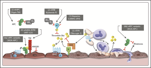
Given the central role of coagulation in promoting microvascular thrombosis and inflammation, it is reasonable to assume that therapies inhibiting coagulation might improve microvascular perfusion, reduce inflammation, and preserve organ function. Most clinical studies examining anticoagulant effects on thromboinflammation have been in the context of sepsis, although specific examples relevant to IR injury will also be highlighted (Figure 4). We will also discuss the evidence for an important role for platelets in thromboinflammation and the possibility that targeting platelets may serve a dual role in reducing microvascular thrombosis and inflammation.
Therapeutic modulation of coagulation processes in sepsis. This illustration highlights some of the therapeutic targets of various anticoagulant approaches trialed in sepsis. (1) Inhibiting the procoagulant and proinflammatory functions of thrombin and FXa was initially trialed through the use of unfractionated heparin (UFH) or low-molecular-weight heparins (LMWHs). (2) Restoring the circulating plasma levels of ATIII has also been trialed via the infusion of recombinant human ATIII. Bolstering the anticoagulant and anti-inflammatory properties of endothelial cells has been attempted through the administration of recombination APC (3), recombinant human TM (rhTM) (4), or TFPI (5). Despite heparins, APC, ATIII, and TFPI having unique pharmacological profiles, acting at distinct points in the coagulation cascade, and possessing additional pharmacological activities independent of their anticoagulant effects, their potential clinical benefits have been undermined by bleeding complications. Improved understanding of the structural regions of APC responsible for anti-inflammatory properties (6a) and cytoprotective properties (6b), independent of anticoagulant effects, has facilitated the generation of recombinant mutants of APC with potentially fewer bleeding side effects (6a-b).
Therapeutic modulation of coagulation processes in sepsis. This illustration highlights some of the therapeutic targets of various anticoagulant approaches trialed in sepsis. (1) Inhibiting the procoagulant and proinflammatory functions of thrombin and FXa was initially trialed through the use of unfractionated heparin (UFH) or low-molecular-weight heparins (LMWHs). (2) Restoring the circulating plasma levels of ATIII has also been trialed via the infusion of recombinant human ATIII. Bolstering the anticoagulant and anti-inflammatory properties of endothelial cells has been attempted through the administration of recombination APC (3), recombinant human TM (rhTM) (4), or TFPI (5). Despite heparins, APC, ATIII, and TFPI having unique pharmacological profiles, acting at distinct points in the coagulation cascade, and possessing additional pharmacological activities independent of their anticoagulant effects, their potential clinical benefits have been undermined by bleeding complications. Improved understanding of the structural regions of APC responsible for anti-inflammatory properties (6a) and cytoprotective properties (6b), independent of anticoagulant effects, has facilitated the generation of recombinant mutants of APC with potentially fewer bleeding side effects (6a-b).
Commonly used anticoagulant agents
Heparins
Unfractionated heparin is a naturally occurring GAG that binds antithrombin III (ATIII), inducing a conformational change that leads to a 1000-fold increase in ATIII’s ability to inhibit thrombin, FXa, and other coagulation serine proteases. Heparin was first trialed for treatment of sepsis in 1966,47 and initial clinical studies were encouraging.48 However, a detailed meta-analysis of 9 trials demonstrated that, in the majority of patients with sepsis, heparin therapy does not reduce mortality, organ injury, or in-hospital stay and is associated with increased bleeding risk. In general, the beneficial effects of heparin on mortality have primarily been observed in patients with sepsis-induced DIC,49 whereas the wider benefit of unfractionated heparin treatment in thromboinflammation remains to be demonstrated.50 Fan et al examined the safety and efficacy of low-molecular-weight heparins (LMWHs) in sepsis patients from 11 trials. This analysis showed that LMWH reduced sepsis severity and decreased 28-day mortality compared with standard treatment but at the expense of increased bleeding.51 However, these studies were primarily performed on Asian patients, so the generalizability of these findings to other ethnicities remains unclear.51 Unfortunately, despite considerable investigation, the overall benefit of LMWH in sepsis remains uncertain.50-52
ATIII
ATIII is a hepatically synthesized serine protease inhibitor and the most abundant anticoagulant circulating in plasma. ATIII inhibits α-thrombin along with FXa, FIXa, FVIIa, FXIa, and FXIIa. In addition to its anticoagulant actions, ATIII may elicit anti-inflammatory effects, by binding endothelial GAGs to enhance PGI2 production, inhibiting lipopolysaccharide (LPS)-mediated signaling in macrophages, and through competition for pathogen binding to endothelial cells.53 ATIII plasma levels decrease precipitously during the early stages of severe sepsis, and ATIII depletion is associated with a poor prognosis. Restoring plasma levels of ATIII through IV infusion has been trialed extensively in patients with sepsis. Although initial small-scale clinical studies were encouraging,54 subsequent large-scale trials55 did not demonstrate any significant reduction in mortality in sepsis patients. However, a retrospective analysis of clinical trial data has suggested that inclusion of patients receiving low-dose heparin may have complicated the interpretation of trial outcomes, because coadministration of heparin and ATIII exacerbates bleeding risk. Patients not receiving prophylactic heparin had a lower 28-day mortality, reaching significance at 90 days, suggesting a potential benefit of ATIII therapy.55 Additional trials on sepsis patients has revealed a 28-day reduction in mortality in patients with DIC, a reduction not observed in sepsis patients without DIC. Thus, ATIII has approval for use in sepsis treatment related to DIC in Japan.56 However, bleeding remains a significant ongoing concern with ATIII infusions, even in the absence of heparin, and a recent phase 3 clinical trial did not demonstrate any benefit for overall mortality.54,56 Consequently, updated International Guidelines for the Management of Sepsis and Septic Shock have made specific recommendations against the use of ATIII52 (Figure 4).
Bolstering the natural anti-inflammatory and antithrombotic properties of the endothelium
The natural anticoagulant and anti-inflammatory properties of endothelial cells are critically important to limit microvascular thrombosis, inflammation and organ injury. Thus, there has been extensive evaluation of the effects of APC, soluble TM, or TFPI in patients with sepsis.
APC
Protein C is a critical physiological anticoagulant activated by the binding of α-thrombin to TM, generating APC. Protein C activation is also enhanced through its binding to the endothelial protein C receptor. Soluble APC’s anticoagulant effects are principally mediated by the cleavage and inactivation of coagulation factors FVa and FVIIIa, whereas APC bound to endothelial protein C receptor has important cytoprotective and anti-inflammatory effects, primarily through cleavage and activation of PAR1, although PAR3 and other APC receptors may also be involved.57 Recombinant human APC has been extensively evaluated in preclinical sepsis models and in sepsis patients. Although several preclinical sepsis models demonstrated a reduction in tissue damage and death with APC treatment, clinical trials have failed to consistently demonstrate a reduction in 28-day all-cause mortality, with most associated with a significantly increased risk for serious bleeding (PROWESS, PROWESS-SHOCK) (Figure 4).58,59 One explanation for the differences in outcome in more recent clinical trials is that sepsis severity in these latter trials was less than in the original PROWESS trial.
Given the biased G protein–coupled receptor signaling properties displayed by APC, studies over the last decade have increasingly focused on the development of APC variants with anti-inflammatory properties and cytoprotective effects, which are specifically engineered to exclude detrimental effects on bleeding.60,61 Preclinical studies utilizing signaling-selective APC “mutants” have provided evidence that modified recombinant APCs with selective anti-inflammatory and cytoprotective actions, but less anticoagulant effects, are a safer treatment option for thromboinflammatory diseases and are currently being trialed in patients with ischemic stroke60 (Figure 4).
Along similar lines, the development of cell-penetrating peptides or “pepducin” technology has also been developed to target selective elements of PAR signaling.62 Although initial studies with a PAR4-selective pepducin (P4pal-10) was able to provide partial protection of liver, lung, and kidney function, it did not improve overall mortality in a murine model of endotoxin-induced systemic inflammation and DIC.63 This, combined with its deleterious effects on the hemostatic response,62 has dampened enthusiasm for this approach. More recently, a distinct class of small molecules that bind selectively to the cytoplasmic face of PAR1 to induce signaling has been developed. Termed parmodulins,64 these molecules have shown promise in LPS-induced thromboinflammatory models in mice. Parmodulins are able to stimulate APC-like cytoprotective signaling through Gq, essentially eliciting an antithrombotic effect at the level of the endothelium, independent of antiplatelet activity. These studies provide proof-of-principle evidence that targeting the cytoplasmic face of G protein–coupled receptors to achieve pathway selective signaling, as opposed to classic orthosteric inhibitors of PAR1,65-67 is an effective and safer strategy to reduce thromboinflammation.
Soluble TM
TM exerts anticoagulant effects in membrane-bound and soluble forms, principally through the activation of PC. Soluble recombinant human TM (rhTM) is currently undergoing clinical evaluation for the treatment of severe sepsis (Figure 4). rhTM has several theoretical advantages over recombinant human APC, including less bleeding68 (due to its reliance on high thrombin levels to exert an anticoagulant effect), and with additional APC-independent actions, including suppression of complement, endotoxin, and HMGB-1 proteins.69 ART-123, an rhTM, has been extensively trialed in Japan, demonstrating improved efficacy and safety in the treatment of DIC relative to heparin.70 In Western countries, phase 2b trials of sepsis patients with suspected DIC have demonstrated lower d-dimer, prothrombin fragment F1.2, and TAT concentrations in patients receiving ART-123.71 Therefore, ART-123 appears to be a safe intervention in critically ill sepsis patients.71
TFPI
TFPI is an endogenous serine protease inhibitor produced by the endothelium that directly inhibits FXa and the FVIIa/TF complex. Preclinical trials have demonstrated that recombinant TFPI (rTFPI) attenuates cytokine responses (tumor necrosis factor-α and interleukin-8 [IL-8]) in a pig peritonitis–induced bacteremia model without improving survival.72 However, rTFPI reduced mortality in a rabbit model of Gram-negative bacterial sepsis73 and in a baboon model of Escherichia coli–induced septic shock. The latter was associated with lower levels of inflammatory and coagulation biomarkers (IL-6 and TAT, respectively).74 rTFPI has been tested and demonstrated to be safe in healthy humans following bolus IV injection of endotoxin. rTFPI attenuated endotoxin-induced α-thrombin generation with complete blockade of coagulation by high-dose rTFPI. Interestingly, rTFPI did not affect endotoxin-induced changes in the fibrinolytic system, leukocyte activation, cytokine and chemokine release, endothelial cell activation, or the acute-phase response.75 rTFPI has also been trialed in phase 2 studies on patients with severe sepsis and it appeared to be safe and effective, with reduction in TAT and IL-6 levels76 and a trend toward reduced mortality. However, results from a randomized double-blind placebo-controlled multicenter phase 3 trial failed to demonstrate an effect of rTFPI (tifacogin) on all-cause mortality (Figure 4). Tifacogin administration was associated with attenuated prothrombin fragment 1.2 and TAT levels, leading to serious bleeding complications.22
Perspectives and future directions
Alternative anticoagulant therapeutics with safer bleeding profiles
The known clinical benefits of anticoagulant therapies in thromboinflammatory states, such as sepsis and stroke, have been largely offset by bleeding complications, and it is highly desirable to develop safer anticoagulants with preserved anti-inflammatory function. This is the basis for development of APC variants. It is also possible that inhibiting the contact pathway of coagulation may prove beneficial.
Inhibition of FXI and FXII
Genetic deletion of FXI or FXII protects against thrombosis in a variety of animal models, including mice, rabbits, and baboons,27,77,78 with minimal bleeding risk.77 This is consistent with observations in humans with congenital FXI deficiency who have a reduced risk for venous thromboembolism or ischemic stroke.79,80 From a safety perspective, FXII-deficient humans do not have a bleeding tendency,78 and FXI deficiency rarely causes spontaneous bleeding,81 although increased bleeding occurs following major hemostatic challenge, such as trauma or surgery.81 The contact activation pathway promotes coagulation and facilitates inflammation by triggering the bradykinin-generating kallikrein-kinin system.82 Several pharmacological inhibitors of FXIIa83-85 have provided protection against sepsis and cerebral IR injury in rats, without increased bleeding risk. Several approaches have been developed to inhibit FXIa, with encouraging results in preclinical studies.86 Among these inhibitors, the 14E11 antibody87 and a natural inhibitor from snake venom (Fasxiator)88 have demonstrated protection in mouse models of sepsis and FeCl3-induced occlusive thrombosis, respectively. Moreover, phase 2 clinical trials using FXI-antisense oligonucleotides have confirmed an important role for FXI in promoting venous thrombosis in patients undergoing knee arthroplasty, with a safer bleeding profile.89
Inhibitors of PolyP
PolyP has emerged over the last several years as a potentially important regulator of coagulation and inflammation and, thus, is a prospective target to reduce thromboinflammation.90 Long-chain PolyP accumulates in infectious microorganisms and is also released by platelets during dense granule exocytosis. PolyP acts at several steps in the coagulation cascade to enhance the rate of α-thrombin generation. PolyP antagonists have been considered for their effectiveness as anticoagulants in vitro, as well as antithrombotic and anti-inflammatory agents in vivo. Recent in vitro coagulation studies and in vivo mouse models of venous and arterial thrombosis, pulmonary thromboembolism, and vascular leakage have demonstrated that PolyP inhibitors have anticoagulant and anti-inflammatory effects,90 while having fewer bleeding side effects than heparin. However, most PolyP inhibitors tested to date, including polycationic substances, such as polyethylenimine, polyamidoamine dendrimers, and polymyxin B, have significant toxicity in vivo. Travers and colleagues91 recently reported on a novel class of polycationic compounds based on universal heparin-reversal agents that exhibit less toxicity, significantly decreasing arterial thrombosis in mice while having less impact on bleeding.
Antiplatelet agents
Experience with aspirin, integrin αIIbβ3, and P2Y12 antagonists
Platelets are the predominant cellular elements promoting microvascular thrombosis, and they play a major role in recruiting leukocytes to sites of endothelial injury and in regulating intravascular leukocyte trafficking.46 Depleting platelets in animal models of sepsis or IR injury markedly reduces leukocyte infiltration into tissues.92 Several preclinical studies have provided evidence that the antiplatelet agents, acetylsalicylic acid or integrin αIIbβ3 antagonists, may reduce sepsis-related mortality.93
Aspirin
More than 50 years ago, aspirin was demonstrated to improve survival of dogs in an E coli–induced canine septic shock model,94 and there is also evidence that aspirin improves survival in a rat septic shock model induced by Salmonella enteriditis endotoxin.95 Although aspirin may have benefit in a subset of patients with pneumonia and in the critically ill,96-99 it appears to have limited impact on the severity of sepsis and does not improve survival.100
GPIIb-IIIa antagonists
GPIIb-IIIa antagonists block fibrinogen binding to integrin αIIbβ3 and inhibit platelet aggregation. These inhibitors have shown benefit in animal models of sepsis. Abciximab reduced vascular leakage and subsequent tissue edema in a rat LPS–induced sepsis model,101 and blockade of integrin αIIbβ3 delayed thrombocytopenia, preserved red and white blood cell counts, and reduced renal damage in a baboon sepsis model.102 Integrin αIIbβ3 antagonists have also been shown to reduce endothelial damage and mortality in mouse models of sepsis103,104 ; however, these inhibitors have not been trialed in humans with sepsis.
P2Y12 receptor antagonists
P2Y12 receptor antagonists are widely used antiplatelet agents that have been demonstrated to reduce platelet–leukocyte interactions and alter inflammatory biomarkers, which are associated with improved lung function in mouse models of pneumonia.93,105 Similarly, P2Y12 receptor antagonists have recently been demonstrated to improve lung function in humans with pneumonia, although larger-scale clinical trials are required to support these preliminary findings.100,106
The case for antiplatelet therapy in the context of experimental IR injury is more compelling, because platelet depletion or inhibitors of platelet adhesion or activation markedly improve microvascular perfusion, reduce inflammation, and preserve organ function.106,107 These findings have been uniformly demonstrated in various animal models of cardiac, renal, cerebral, gut, and liver IR injury.108-111 Clinically, antiplatelet agents (aspirin, P2Y12 antagonists, integrin αIIbβ3 inhibitors) have been demonstrated to improve microvascular perfusion in the heart and brain during IR injury; however, in both organs these benefits may be partially offset by an increased risk for hemorrhagic transformation.112,113
ITAM-bearing receptors
Progress in defining new pathways promoting thromboinflammation suggests that platelet receptors belonging to the immunoreceptor tyrosine-based activation motif (ITAM) receptors may offer alternative therapeutic targets. Platelets contain the ITAM-bearing receptor C-type lectin-like-2 (CLEC-2), which binds podoplanin, and the collagen receptor, glycoprotein VI (GPVI). These receptors are potentially attractive therapeutic targets, because deficiency of either receptor does not produce a major bleeding disorder. GPVI mimetics, such as revacept, decrease cerebral infarct size and edema and improved outcome after ischemic stroke.114 In phase 1 clinical trials, Revacept was also able to specifically and efficiently inhibit collagen-induced platelet aggregation without increasing bleeding.114,115 Administration of anti-GPVI antibodies in mice has also been demonstrated to provide protection against stroke,116 as well as pneumonia-derived sepsis, again without increasing bleeding.117 Further to this, specific deletion of CLEC-2 (but not GPVI) reduces thrombosis in the liver in a mouse model of systemic Salmonella typhimurium infection.118 However, the enthusiasm for CLEC-2 as a therapeutic target may be tempered by the finding that CLEC-2 deficiency leads to enhanced systemic inflammation and accelerated organ injury in mouse sepsis models.119
TLT-1
The importance of platelets in promoting septic complications is further underscored through the study of the human triggering receptor expressed in myeloid cells (TREM) gene cluster, which encodes for several TREM proteins. Interestingly, TREM-like transcript (TLT-1) has been demonstrated to be specific to platelets and megakaryocytes, where it is sequestered within platelet α-granules and translocated to the cell surface following activation with LPS or thrombin.120 Although platelet membrane-bound TLT-1 has been shown to facilitate platelet aggregation through the binding of fibrinogen, a soluble form of TLT-1 (sTLT-1) has also been identified as a potent endogenous regulator of sepsis-associated inflammation,121 with studies in trem−/− mice demonstrating that TLT-1 promotes survival during sepsis.122 With elevated levels of sTLT-1 identified in the blood of patients diagnosed with sepsis, and elevated sTLT-1 strongly correlated to DIC score and with high levels of d-dimer,123 it has been suggested that platelet-derived TLT-1 contributes to the progression of acute lung injury and acute respiratory distress syndrome.123
Targeting the physical interactions between platelets and neutrophils
Platelets and neutrophils are the predominant cell types promoting acute thromboinflammatory responses. Moreover, the formation of neutrophil–platelet aggregates contributes to microvascular obstruction and inflammation in various thromboinflammatory disorders, including acute lung injury, the acute coronary syndromes, and ischemic stroke.124-126 Preclinical studies have confirmed that targeting adhesion molecules on platelets (P-selectin, GPIb, αIIbβ3) and neutrophils (PSGL-1, Mac-1) inhibits neutrophil–platelet aggregates and improves microvascular dysfunction and inflammation. A phase 1 trial has confirmed the safety and dosing of inclacumab, a monoclonal antibody against P-selectin, and established that it does not extend bleeding time or impact on platelet aggregation.127
Conclusions
Reducing the deleterious impact of microvascular thrombosis and inflammation in the context of sepsis and ischemia-reperfusion injury continues to represent a major therapeutic challenge. Part of the difficulty reflects the complex and variable nature of the innate immune and hemostatic responses that drive the various stages of the thromboinflammatory process. Accordingly, it is unlikely that a single “magic therapeutic bullet” is going to be highly effective at reducing thromboinflammatory complications. Understanding the pathogenic heterogeneity of different thromboinflammatory disorders, the diverse genetic backgrounds of patients, and the appropriate timing of therapeutic interventions will ultimately be critical for optimized treatment. This is evident in the management of the coagulopathy of sepsis; certain patient groups may respond to specific therapies if administered at appropriate stages of the disease process.
Clinical trials over the last few decades have consistently revealed that bleeding risk is a major limitation in the use of antithrombotic approaches in the setting of sepsis and IR injury. Although the heparins, APC, ATIII, and TFPI have unique pharmacological profiles, act at distinct points in the coagulation cascade, and possess activity independent of their anticoagulant effects, their potential clinical benefit is diminished by a substantial increase in the risk of serious bleeding. This is perhaps not surprising, because microvascular dysfunction is often associated with microvascular thrombosis and bleeding,128 with the extent of microvascular bleeding correlating with organ dysfunction and patient outcome.129 It remains to be seen whether emerging strategies that primarily have anti-inflammatory and cytoprotective properties, with less impact on coagulation, will be effective, because previous anti-inflammatory approaches that do not affect coagulation and tissue perfusion have not successfully reduced organ injury or improved survival.130 Moreover, although there is much anticipation around novel antithrombotic therapeutics that have less impact on bleeding, it remains unclear whether targeting only 1 major thromboinflammatory pathway will be sufficient to limit microvascular dysfunction, inflammation, and bleeding. Experience with the coronary no-reflow phenomenon suggests that a cocktail of therapeutic agents (including vasodilators and platelet and potentially leukocyte inhibitors) may be necessary to improve microvascular function in specific patients.131 Thus, a formidable challenge remains in identifying combination antithrombotic and anti-inflammatory approaches that are effective and can be safely used in the clinic.
Acknowledgments
The authors thank Emily McCarthy for assistance with preparing the manuscript.
This project was supported by the Australian National Health and Medical Research Council of Australia Project grant APP1127267 (S.P.J.). S.P.J. is the recipient of a National Health and Medical Research Council of Australia Senior Principal Research Fellowship.
Authorship
Contribution: S.P.J., R.D., and S.M.S. cowrote and reviewed the manuscript, and S.M.S. prepared the figures.
Conflict-of-interest disclosure: The authors declare no competing financial interests.
Correspondence: Shaun P. Jackson, Heart Research Institute and Charles Perkins Centre, Level 3, D17, University of Sydney, Orphan School Creek Rd, Camperdown, NSW 2006, Australia; e-mail: shaun.jackson@sydney.edu.au.
REFERENCES
Author notes
S.P.J., R.D., and S.M.S. contributed equally to this review.


![Figure 3. Proinflammatory and prothrombotic function of inflamed endothelial cells. (A) Perturbation of the endothelium by oxidative stress (reactive oxygen species [ROS]), pathogen infection, or proinflammatory molecules (eg, lipids and cytokines) leads to increased expression of adhesion molecules, such as von Willebrand factor (VWF) and P-selectins, αvβ3 and ICAM-1, leading to the recruitment of platelets and leukocytes. Activated endothelial cells also express TF, leading to activation of FVII, FXa, and, ultimately, thrombin generation. Thrombin cleaves fibrinogen to generate fibrin, as well as multiple protease-activated receptors on the surface of endothelial cells, platelets, and leukocytes, thereby propagating the thromboinflammatory process. (B) Recruitment of platelets and leukocytes to sites of inflamed endothelium: the expression of VWF and P-selectin on the damaged endothelial cell surface supports platelet tethering and rolling. Subsequently, stable platelet adhesion occurs through fibrinogen–integrin αIIbβ3 complexes binding to αvβ3 or ICAM-1 on endothelial cells. Adherent platelets secrete numerous bioactive substances that alter the chemotactic and adhesive properties of endothelial cells. Platelet-derived interleukin-1 (IL-1) induces active TF expression on the endothelium. The expression of P-selectin on adherent activated platelets induces leukocyte tethering followed by their adhesion, mainly through the interaction of macrophage-1 antigen (Mac-1) with GPIb–GPV–GPIX and fibrinogen–αIIbβ3 complexes. PAMPs, pathogen-associated molecular patterns; TNFα, tumor necrosis factor-1.](https://ash.silverchair-cdn.com/ash/content_public/journal/blood/133/9/10.1182_blood-2018-11-882993/4/m_blood882993f3.png?Expires=1769095712&Signature=EebcfCuISYIxmCzvwf0RmO9zLwdmI8RqQGfEPEFTxtHmGOwaobIRrwM1tAXx3tBG9HSM1s2~6s0HXF5NuFzXxOia3SYxCmmvtFffKHH1UUc83eX3OBzqxJ2DFGLs33TpeSSglQmVk0BjOwiH2s39c56VBfQwOJJsHbnOkKbQcYr3XAps2f2g8lMRJO4TOL0jBaZ3880s0fkQq7xx~~M-Lkc7Uefy8vsOWQQFG-7zL0pWH30w0HC5Oqn~gOR0~st17QVdLioBgcTgZUJ6w7bCBc9Tw2gz8hssjGFOkMYbH3csCNeyhLsn8xqLWrTn4h0xo2ShC2L3S~nS-~P3pdQlpQ__&Key-Pair-Id=APKAIE5G5CRDK6RD3PGA)

This feature is available to Subscribers Only
Sign In or Create an Account Close Modal