

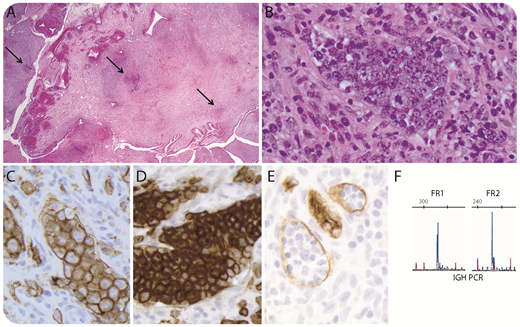
A 28-year-old, otherwise asymptomatic, woman with dysfunctional uterine bleeding was evaluated with hysteroscopic biopsy of a polyp that showed mild stromal chronic inflammation and scattered foci of atypical lymphocytes (panel A: hematoxylin and eosin stain, original magnification ×40; arrows show lymphoid infiltrate). These foci comprised vascular channels filled with large, cohesive pleomorphic, nucleolated lymphocytes (panel B: hematoxylin and eosin stain, original magnification ×400). Immunostains confirmed an intravascular large B-cell lymphoma with high Ki67 index and CD30 expression (panel C: CD20 stain; panel D: CD30 stain; panel E: CD34 stain; all original magnification ×400). CD5, Epstein Barr virus, and HHV8 stains were negative. Immunoglobulin heavy chain by polymerase chain reaction was clonal in FR1/FR2 polymerase chain reaction reactions (panel F); fluorescence in situ hybridization showed no BCL2, BCL6, or MYC rearrangements.
Intravascular large B-cell lymphoma represents an uncommon variant with preferential growth of neoplastic cells within blood vessels, with a median presenting age of 67 years. Two clinical presentations are described: a classical form presenting incidentally or with symptoms related to dysfunction of the affected organ, and a hemophagocytic syndrome-associated form. This case is unusual in its presentation as a benign-appearing endometrial polyp in a young woman. Awareness of this rare, yet aggressive, entity is important because subtle intravascular atypical cell infiltrates may be overlooked at sites, such as the endometrium, where chronic inflammation is common.
For additional images, visit the ASH Image Bank, a reference and teaching tool that is continually updated with new atlas and case study images. For more information, visit http://imagebank.hematology.org.

This feature is available to Subscribers Only
Sign In or Create an Account Close Modal