

An 83-year-old man presented with hematuria and bone pain. Computed tomography–scan imaging demonstrated multiple osteolytic lesions and vertebral collapse. A complete blood count showed anemia with hemoglobin of 9.1 g/dL and normal white blood count and platelets, 4.95 × 10⁹/L and 198 × 109/L, respectively. Total protein was at 105 g/L, with electrophoresis revealing a monoclonal band, identified as immunoglobulin G ĸ at 50 g/L. Bone marrow smear examination found plasma cells comprising 38% of total nucleated cells (TNCs) and an unexpected infiltration (3%) of abnormal mast cells (MCs) with a spindle-shaped morphology (panels A-B; May-Grünwald Giemsa staining, original magnification ×1000). Serum tryptase was elevated at 161 µg/L, and next-generation sequencing revealed D816Y mutation at the exon 17 of the kit gene, found in the MCs, confirming a diagnosis of systemic mastocytosis with an associated hematological neoplasm (SM-AHN). Flow cytometry analysis showed aberrant expression of CD2 and CD25 on the MCs.
After 3 months of lenalidomide and dexamethasone, bone marrow plasma cells were at 2% of TNCs, but the infiltrate of abnormal MCs remained unchanged, and serum tryptase was still elevated at 182 µg/L. This case of SM-AHN involving D816Y mutation and myeloma demonstrates the benefit of microscopic examination as relatively common diseases can hide even the rarest hematologic malignancies.
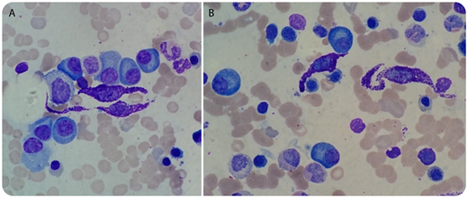
For additional images, visit the ASH Image Bank, a reference and teaching tool that is continually updated with new atlas and case study images. For more information, visit http://imagebank.hematology.org.

This feature is available to Subscribers Only
Sign In or Create an Account Close Modal