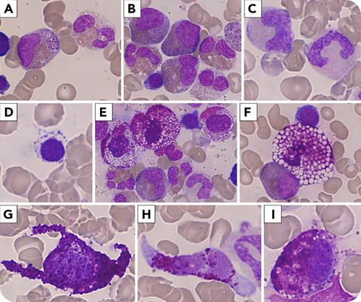
A 50-year-old man presented with flushes, fever, intestinal disorders, urticaria pigmentosa, and splenomegaly. Anemia and thrombocytopenia associated with hypereosinophilia (16.5 g/L) were found. Cytomorphological examination of a bone marrow aspiration revealed dystrophic eosinophilic precursors, erythroid and granular myeloid-associated dysplasia (panels A-D; in all panels, May-Grünwald-Giemsa stain; original magnification ×1000) related to a myelodysplastic syndrome with multilineage dysplasia (MDS-MD). It associated with a high infiltration (8%) of abnormal mast cells (MCs). Most MCs were degranulated, displaying a very atypical foamy aspect with large optically empty vacuoles, and were often grouped within islet-like cell clusters (panels E-F). Flow cytometry analysis showed aberrant expression of CD2 and CD25 on KIT+/FceRI+ MCs. The serum tryptase level was at 773 ng/mL. Trephine biopsy confirmed the diagnosis of aggressive systemic mastocytosis (ASM) with associated hematological neoplasm (AHN). Molecular biology found neither D816V KIT mutation nor any mutation on panexonic sequencing of the KIT gene. Screening for BCR-ABL1 and FIP1L1-PDGFRA was negative. The disease evolved 9 months later into MC leukemia refractory to many lines of treatment, and the patient died 2 years after the diagnosis.
This is a very unique case of an ASM-AHN (MDS-MD) with foamy MCs, contrasting with classical cytomorphology of spindle-shaped (panels G-H) or hypogranulated MCs (panel I) also present to a lesser extent.
For additional images, visit the ASH Image Bank, a reference and teaching tool that is continually updated with new atlas and case study images. For more information, visit http://imagebank.hematology.org.


This feature is available to Subscribers Only
Sign In or Create an Account Close Modal