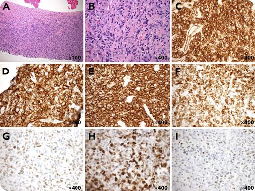
A 49-year-old man presented with an 8.8 × 6.5 cm, left infraclavicular subpectoral mass. A biopsy showed predominantly spindle-ish cells (panels A-B, hematoxylin and eosin stain; original magnifications shown on pictures) and was therefore worked up for soft tissue tumors initially. The spindle-ish cells were strongly positive for CD31 with membranous and cytoplasmic staining (panel C) but were negative for CD34, AE1/AE3, smooth muscle actin, desmin, and S100 (not shown). Further workup revealed positive stains for CD45 (panel D), CD20 (panel E), Bcl-2 (panel F), Mum-1 (panel G), and C-myc (panel H); partially positive stains for Bcl-6 (panel I); weakly positive stains for Pax-5 (not shown); and high Ki-67 proliferation index (90%, not shown). The cells were negative for CD3, CD10, LMP1, P53, CD5, and Bcl-1 (not shown). The overall findings are most consistent with a diffuse large B-cell lymphoma (DLBCL), activated B-cell subtype, with spindle-ish cell morphology and strong aberrant expression of CD31.
A few previous reports indicated that CD31 was detected in small cell lymphomas (by flow cytometry), plasmablastic lymphoma (weak stain), and cutaneous spindle cell B-cell lymphoma (focal weak stain). We believe this is the first documented case of DLBCL with strong diffuse expression of CD31. The spindle-ish cell morphology and strong CD31 expression create diagnostic difficulties.
For additional images, visit the ASH Image Bank, a reference and teaching tool that is continually updated with new atlas and case study images. For more information, visit http://imagebank.hematology.org.


This feature is available to Subscribers Only
Sign In or Create an Account Close Modal