Key Points
SLAMF7-CAR T cells are effective against proteasome inhibitor/immunomodulatory drug–refractory myeloma.
SLAMF7-CAR T cells confer fratricide of SLAMF7+/high normal lymphocytes.
Abstract
SLAMF7 is under intense investigation as a target for immunotherapy in multiple myeloma. In this study, we redirected the specificity of T cells to SLAMF7 through expression of a chimeric antigen receptor (CAR) derived from the huLuc63 antibody (elotuzumab) and demonstrate that SLAMF7-CAR T cells prepared from patients and healthy donors confer potent antimyeloma reactivity. We confirmed uniform, high-level expression of SLAMF7 on malignant plasma cells in previously untreated and in relapsed/refractory (R/R) myeloma patients who had received previous treatment with proteasome inhibitors and immunomodulatory drugs. Consequently, SLAMF7-CAR T cells conferred rapid cytolysis of previously untreated and R/R primary myeloma cells in vitro. In addition, a single administration of SLAMF7-CAR T cells led to resolution of medullary and extramedullary myeloma manifestations in a murine xenograft model in vivo. SLAMF7 is expressed on a fraction of normal lymphocytes, including subsets of natural killer (NK) cells, T cells, and B cells. After modification with the SLAMF7-CAR, both CD8+ and CD4+ T cells rapidly acquired and maintained a SLAMF7– phenotype and could be readily expanded to therapeutically relevant cell doses. We analyzed the recognition of normal lymphocytes by SLAMF7-CAR T cells and show that they induce selective fratricide of SLAMF7+/high NK cells, CD4+ and CD8+ T cells, and B cells. Importantly, however, the fratricide conferred by SLAMF7-CAR T cells spares the SLAMF7–/low fraction in each cell subset and preserves functional lymphocytes, including virus-specific T cells. In aggregate, our data illustrate the potential use of SLAMF7-CAR T-cell therapy as an effective treatment against multiple myeloma and provide novel insights into the consequences of targeting SLAMF7 for the normal lymphocyte compartment.
Introduction
The development of chimeric antigen receptors (CARs) specific for antigens expressed in multiple myeloma is pursued aggressively because of the documented potential of CAR-modified T cells to induce durable complete remissions in advanced hematologic malignancies1-3 and the lack of a curative treatment for the majority of myeloma patients. Several candidate antigens for CAR T cells are under investigation in myeloma, including CD19, which is only infrequently expressed on malignant plasma cells, and B-cell maturation antigen (BCMA). Recent data from a phase I clinical trial with BCMA-CAR T cells showed partial and complete responses in a fraction of patients, but also several limitations, including low- or nonuniform expression of BCMA on myeloma cells, which precluded treatment with the BCMA-CAR T cells, and relapse with BCMA–/low myeloma cells after BCMA-CAR T-cell therapy.4 These data highlight the still unmet need to identify and validate a uniformly applicable CAR target in this disease.
SLAMF7 (aliases: CD319, CRACC, and CS-1) is a member of the signaling lymphocytic activation molecule (SLAM) family of transmembrane receptors that modulate the function of immune cells through immune-receptor tyrosine-based switch motifs and intracellular adaptor proteins. SLAMF7 expression was first documented in natural killer (NK) cells and a proportion of CD8+ and, to a lesser extent, CD4+ T cells and B cells, where it mediates activating or inhibitory functions through the EAT-2 adaptor protein.5-7 SLAMF7 is also present on subsets of monocytes, macrophages, and dendritic cells.8,9 In the life cycle of normal B cells, SLAMF7 is highly expressed in pro–B cells and plasma cells, and high-level expression is retained in malignant plasma cells in multiple myeloma and its antecedent, “premalignant” stages: monoclonal gammopathy of undetermined significance and smoldering myeloma.10-12 The functional role of SLAMF7 in myeloma pathogenesis is incompletely understood, but several lines of evidence suggest a role in myeloma cell survival and interaction with stroma cells in the bone marrow niche.13
Previous work has shown that SLAMF7 is uniformly expressed on malignant plasma cells in newly diagnosed (ND) myeloma and retained in relapsed myeloma after intensive chemoradiotherapy (total therapy).10 Intriguingly, there is no known expression of SLAMF7 in any other normal human tissue, making it a candidate antigen for CAR T cells in myeloma.
There is experience with targeting SLAMF7 on myeloma from the use of the anti-SLAMF7 antibody huLuc63 (elotuzumab) that has been postulated to exert its antimyeloma effect through antibody-dependent cellular cytotoxicity, activation of NK cells, and blockade of SLAMF7 binding to its ligands in the bone marrow niche.10,14,15 Elotuzumab has only minimal clinical efficacy as a single agent, but is effective in combination with lenalidomide and dexamethasone and has been approved for the treatment of relapsed myeloma after previous therapy with proteasome inhibitors and immunomodulatory drugs (IMiDs).16 In this study, we investigated the manufacture and phenotype of T cells that we engineered to express a SLAMF7-specific CAR with a targeting domain derived from huLuc63 and assessed the recognition of myeloma cells as well as SLAMF7+ normal lymphocytes in preclinical models.
Materials and methods
Construction of SLAMF7-CAR–encoding lentiviral vector
A codon-optimized, single-chain variable fragment comprising the variable heavy and variable light chains of the anti-SLAMF7 monoclonal antibody (mAb) huLuc63,13 separated by a (G4S)3 linker, was synthesized (GeneArt, ThermoFisher, Regensburg, Germany) and cloned into the epHIV7 lentiviral vector, where it was fused to an immunoglobulin G4 (IgG4)-Fc hinge-CH2-CH3 spacer, a CD28-CD3ζ signaling module in cis with a T2A element and truncated epidermal growth factor receptor (EGFRt).17,18 For in vivo experiments, the IgG4-Fc spacer was modified to prevent binding of Fc receptors (4/2NQ modification).19 A CD19-CAR has been described in previous work.19,20
Analysis of CAR T-cell effector function in vitro
Cytolytic activity was analyzed in a bioluminescence-based assay using firefly luciferase (ffluc)-transduced target cells.21 Interferon-γ (IFN-γ) and interleukin-2 (IL-2) were measured by enzyme-linked immunosorbent assay (BioLegend, Koblenz, Germany) in supernatants obtained after a 20-hour coculture of T cells with K562 (effector-to-target [E:T] ratio = 4:1) and myeloma target cells (E:T ratio = 2:1). Proliferation of live (7-aminoactinomycin D [7-AAD]–) carboxyfluorescein diacetate succinimidyl ester-labeled CD4+ and CD8+ T cells was analyzed after a 72-hour coculture with target cells by flow cytometry.
Experiments in a xenograft myeloma model in vivo
The University of Würzburg Institutional Animal Care and Use Committee approved all mouse experiments. Six- to eight-week-old female NOD-scid IL2rγnull (NSG) mice were obtained from Charles River Laboratories and inoculated by tail vein injection with 2 × 10⁶ MM.1S/ffluc_eGFP. On day 14, the development of systemic myeloma was documented by bioluminescence imaging, and groups of 3 to 5 mice were treated with 5 × 10⁶ T cells (ie, 2.5 × 10⁶ CD4+ and 2.5 × 10⁶ CD8+) expressing either the SLAMF7-CAR, a control CD19-CAR, or phosphate-buffered saline by tail vein injection. Bioluminescence imaging was done on an IVIS Lumina system (PerkinElmer, Waltham, MA) after intraperitoneal injection of d-luciferin (0.3 mg/g body weight; Biosynth, Staad, Switzerland), and data was analyzed using LivingImage software (PerkinElmer).
Flow cytometry–based cytotoxicity assay
Target cells were stained with eFluor670 (eBioscience, Frankfurt, Germany) for detection by flow cytometry after coculture with CAR-modified or untransduced T cells. The percentage of live target cells was determined using 7-AAD or fixable viability stain 450 (BD Biosciences, Heidelberg, Germany) and quantified using 123count eBeads (eBioscience). Specific lysis was calculated using the formula: 1 –  . IFN-γ production was analyzed 4 hours after stimulation with 50 ng/mL phorbol 12-myristate 13-acetate (PMA) and 1 µg/mL ionomycin (both Sigma-Aldrich, Munich, Germany) by intracellular staining using the Cytofix/Cytoperm Plus kit (BD Biosciences).
Results
Expression of a SLAMF7-CAR in T cells of myeloma patients and healthy donors
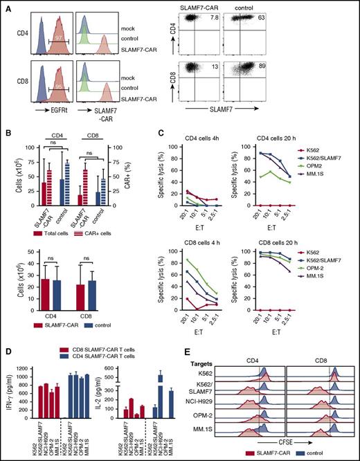
We prepared SLAMF7-CAR–modified CD4+ and CD8+ T cells from myeloma patients (n = 7) and healthy donors (n = 4) (supplemental Figure 1A, available on the Blood Web site). Following anti-CD3/anti-CD28 bead stimulation and lentiviral transduction, we enriched transgene-positive T cells to >90% purity using the EGFRt transduction marker,18 amplified T cells through antigen-dependent expansion with SLAMF7+ feeder cells, and confirmed uniform surface expression of the SLAMF7-CAR through labeling with recombinant SLAMF7 protein (Figure 1A). We detected SLAMF7 protein by flow cytometry on a fraction of input T cells before lentiviral transduction and observed rapid loss of SLAMF7 surface expression in CD8+ and CD4+ T cells after modification with the SLAMF7-CAR. At the end of the manufacturing process, SLAMF7-CAR T cells were SLAMF7–/low (Figure 1A). We performed additional flow cytometric analyses during antigen-dependent expansion and detected SLAMF7 on SLAMF7-CAR T cells early after stimulation, but not at later time points during the expansion cycle (supplemental Figure 2A). Stimulation with PMA/ionomycin rapidly induced re-expression of SLAMF7 protein on the surface of SLAMF7-CAR T cells, although the expression level as assessed by mean fluorescence intensity (MFI) was lower than in control CD19-CAR T cells (supplemental Figure 2B). Importantly, CD8+ and CD4+ SLAMF7-CAR T cells could be expanded to therapeutic doses during primary expansion with anti-CD3/anti-CD28 beads, as well as the subsequent 9-day antigen-dependent expansion cycle, which yielded >2 × 10⁷ from 1 × 10⁶ input EGFRt+ T cells, and similar cell counts like parallel cultures of CD19-CAR T cells (>20-fold expansion) (Figure 1B). Before functional testing, SLAMF7-CAR T cells had an effector phenotype and expressed higher levels of PD-1, TIM-3, and LAG-3 than CD19-CAR–modified control T cells (supplemental Figure 1B-C). In aggregate, these data show that T cells acquire a SLAMF7–/low phenotype after modification with our SLAMF7-CAR and can be rapidly expanded to clinically relevant doses using conventional protocols.
Phenotype and antimyeloma function of SLAMF7-CAR T cells in vitro. (A) Staining with anti–epidermal growth factor receptor antibody to detect the EGFRt transduction marker on CD8+ and CD4+ SLAMF7-CAR T cells (red). Untransduced T cells were used as controls (blue, left histograms). Staining with SLAMF7 protein to detect the SLAMF7-CAR on SLAMF7-CAR T cells. CD19-CAR T cells (control) and untransduced T cells (mock) were used as references (right histograms). Expression of SLAMF7 on SLAMF7-CAR and CD19-CAR (control) T cells (dot plots). (B) Expansion of CD4+ and CD8+ T cells after anti-CD3/anti-CD28 bead stimulation and lentiviral transduction with SLAMF7-CAR and CD19-CAR as control (upper bar diagram). The left y-axis shows the absolute number of T cells, and the right y-axis shows the percentage of CAR-transduced (ie, EGFRt+) T cells obtained from 0.5 × 10⁶ input T cells after 9 days of culture. The subsequent antigen-dependent expansion of SLAMF7-CAR and CD19-CAR (control) T cells after stimulation with irradiated feeder cells. The diagram shows the absolute number of T cells obtained from 1 × 10⁶ input (EGFRt-enriched) T cells within 9 days of culture (lower bar diagram). The same SLAMF7+CD19+EBV-LCL feeder cells were used to stimulate SLAMF7-CAR and CD19-CAR (control) T cells. (C) Cytotoxic/cytolytic activity of CD4+ and CD8+ SLAMF7-CAR T cells within 4 and 20 hours of coculture, respectively, with myeloma cell lines. SLAMF7– native K562 cells were used as a negative control. K562 cells that had been transduced with SLAMF7 were used as a positive control (K562/SLAMF7). (D) Secretion of IFN-γ and IL-2 analyzed by enzyme-linked immunosorbent assay in supernatants obtained after a 20-hour coculture of effector and target cells. Assays were performed with 50 000 effector T cells and 10 000 target cells in 96-well round bottom plates with a volume of 200 μl. CD19-CAR–transduced CD4+ and CD8+ T cells were used as controls, did not produce cytokines above background, and are therefore not shown in the diagram. (E) Proliferation assessed by carboxyfluorescein diacetate succinimidyl ester dilution after a 72-hour coculture of effector and target cells. Assays were performed with 50 000 effector T cells and 10 000 irradiated target cells in 96-well plates with a volume of 200 μl without the addition of exogenous cytokines. Data in panels A-E are representative of the results obtained in independent experiments with CAR T cells prepared from 4 healthy donors. ns, not significant.
Phenotype and antimyeloma function of SLAMF7-CAR T cells in vitro. (A) Staining with anti–epidermal growth factor receptor antibody to detect the EGFRt transduction marker on CD8+ and CD4+ SLAMF7-CAR T cells (red). Untransduced T cells were used as controls (blue, left histograms). Staining with SLAMF7 protein to detect the SLAMF7-CAR on SLAMF7-CAR T cells. CD19-CAR T cells (control) and untransduced T cells (mock) were used as references (right histograms). Expression of SLAMF7 on SLAMF7-CAR and CD19-CAR (control) T cells (dot plots). (B) Expansion of CD4+ and CD8+ T cells after anti-CD3/anti-CD28 bead stimulation and lentiviral transduction with SLAMF7-CAR and CD19-CAR as control (upper bar diagram). The left y-axis shows the absolute number of T cells, and the right y-axis shows the percentage of CAR-transduced (ie, EGFRt+) T cells obtained from 0.5 × 10⁶ input T cells after 9 days of culture. The subsequent antigen-dependent expansion of SLAMF7-CAR and CD19-CAR (control) T cells after stimulation with irradiated feeder cells. The diagram shows the absolute number of T cells obtained from 1 × 10⁶ input (EGFRt-enriched) T cells within 9 days of culture (lower bar diagram). The same SLAMF7+CD19+EBV-LCL feeder cells were used to stimulate SLAMF7-CAR and CD19-CAR (control) T cells. (C) Cytotoxic/cytolytic activity of CD4+ and CD8+ SLAMF7-CAR T cells within 4 and 20 hours of coculture, respectively, with myeloma cell lines. SLAMF7– native K562 cells were used as a negative control. K562 cells that had been transduced with SLAMF7 were used as a positive control (K562/SLAMF7). (D) Secretion of IFN-γ and IL-2 analyzed by enzyme-linked immunosorbent assay in supernatants obtained after a 20-hour coculture of effector and target cells. Assays were performed with 50 000 effector T cells and 10 000 target cells in 96-well round bottom plates with a volume of 200 μl. CD19-CAR–transduced CD4+ and CD8+ T cells were used as controls, did not produce cytokines above background, and are therefore not shown in the diagram. (E) Proliferation assessed by carboxyfluorescein diacetate succinimidyl ester dilution after a 72-hour coculture of effector and target cells. Assays were performed with 50 000 effector T cells and 10 000 irradiated target cells in 96-well plates with a volume of 200 μl without the addition of exogenous cytokines. Data in panels A-E are representative of the results obtained in independent experiments with CAR T cells prepared from 4 healthy donors. ns, not significant.
SLAMF7-CAR T cells confer potent antimyeloma effector functions in vitro
To assess the effector function of SLAMF7-CAR T cells, we used the SLAMF7+ myeloma cell lines MM.1S, OPM2, and NCI-H929 as well as K562 cells that we had transduced with SLAMF7 (supplemental Figure 3A-B). CD8+ SLAMF7-CAR T cells exerted rapid, specific, and high-level cytolysis of all SLAMF7+ target cells (Figure 1C). With MM.1S as target cells, we observed >80% specific lysis after 4 hours and >90% specific lysis after 20 hours of coculture with CD8+ SLAMF7-CAR T cells at a 20:1 E:T ratio. CD4+ SLAMF7-CAR T cells also exerted a direct cytotoxic effect against each of the myeloma cell lines (Figure 1C). The analysis of supernatants obtained after a 20-hour coculture of CD8+ and CD4+ SLAMF7-CAR T cells with myeloma cell lines confirmed specific secretion of IFN-γ and IL-2 (Figure 1D). In addition, SLAMF7-CAR T cells underwent specific and productive proliferation after stimulation with each of the myeloma cell lines, with up to 4 cell divisions within the 72-hour assay period (Figure 1E). Overall, SLAMF7-CAR T cells prepared from healthy donors and myeloma patients displayed similarly potent effector functions (Figure 1C-E; supplemental Figure 4A-C). In contrast, CD19-CAR–modified and untransduced T cells prepared from the same healthy donors and patients did not exert any discernable specific antimyeloma reactivity.
SLAMF7-CAR T cells exert rapid and complete cytolysis of primary myeloma cells
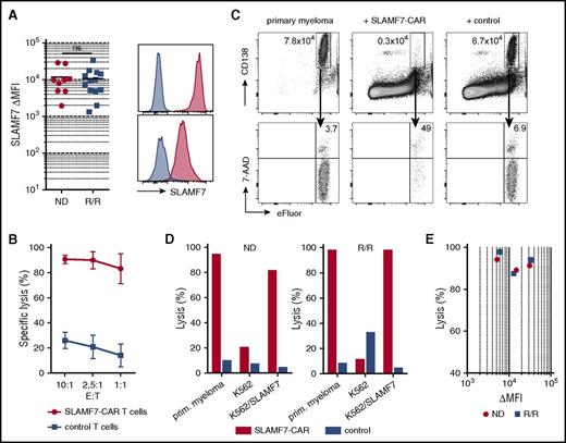
In the next set of experiments, we analyzed primary CD38+ CD138+ malignant plasma cells that we isolated from the bone marrow of patients with ND (n = 9) or relapsed/refractory (R/R) myeloma (n = 13). In the latter cohort, patients had received previous treatment with chemotherapy regimens containing the proteasome-inhibitor bortezomib (n = 5), the IMiDs lenalidomide and/or pomalidomide (n = 7), and had undergone high-dose chemotherapy (melphalan, >100mg/m2) and subsequent autologous hematopoietic stem cell transplantation (n = 9). We confirmed that uniform high-level SLAMF7 expression was present on malignant plasma cells in each of the patients (22 of 22, 100%) (Figure 2A). The antigen density of SLAMF7 on malignant plasma cells as assessed by MFI varied between patients, but was similarly high and within a similar range in ND and R/R myeloma cases (Figure 2A). We did not detect a SLAMF7– myeloma cell subpopulation by flow cytometry in any of the patients.
Expression of SLAMF7 on malignant plasma cells and recognition of primary myeloma cells by SLAMF7-CAR T cells in vitro. (A) Expression of SLAMF7 on CD38+CD138+ primary myeloma cells obtained from patients with ND or R/R multiple myeloma. Data are presented as differences in MFI (ΔMFI) obtained by staining with anti-SLAMF7 mAb 162.1 and isotype control. Histograms show SLAMF7 expression on primary myeloma cells obtained from the patient with the highest (upper) and the lowest ΔMFI (lower histogram) in the R/R myeloma group, respectively. Staining with anti-SLAMF7 mAb (red) and isotype control (blue). (B) Primary myeloma cells were labeled with eFluor670 fluorescent dye and cocultured with autologous SLAMF7-CAR or CD19-CAR (control) CD8 T cells (10 000 myeloma target cells, E:T ratio = 10:1 to 1:1). After 4 hours of incubation, live (7-AAD–) CD138+eFluor+ myeloma cells were quantified by flow cytometry using counting beads and specific lysis calculated using untreated myeloma cells as a comparator. (C) Exemplary dot plots obtained in the flow cytometry–based cytotoxicity assay (E:T ratio = 10:1). (D) Specific lysis of primary myeloma obtained from a patient with ND (left) and a patient with R/R multiple myeloma (right bar diagram) by autologous SLAMF7-CAR or CD19-CAR (control) T cells (E:T ratio = 10:1). Native SLAMF7– K562 cells and K562/SLAMF7 cells are included as a negative and positive control, respectively. (E) Aggregate data on specific lysis of primary myeloma cells obtained from 3 ND and 4 R/R multiple myeloma patients (E:T ratio = 10:1). The x-axis shows SLAMF7 expression on primary myeloma cells before the assay as ΔMFI, and the y-axis shows the specific lysis obtained with SLAMF7-CAR T cells.
Expression of SLAMF7 on malignant plasma cells and recognition of primary myeloma cells by SLAMF7-CAR T cells in vitro. (A) Expression of SLAMF7 on CD38+CD138+ primary myeloma cells obtained from patients with ND or R/R multiple myeloma. Data are presented as differences in MFI (ΔMFI) obtained by staining with anti-SLAMF7 mAb 162.1 and isotype control. Histograms show SLAMF7 expression on primary myeloma cells obtained from the patient with the highest (upper) and the lowest ΔMFI (lower histogram) in the R/R myeloma group, respectively. Staining with anti-SLAMF7 mAb (red) and isotype control (blue). (B) Primary myeloma cells were labeled with eFluor670 fluorescent dye and cocultured with autologous SLAMF7-CAR or CD19-CAR (control) CD8 T cells (10 000 myeloma target cells, E:T ratio = 10:1 to 1:1). After 4 hours of incubation, live (7-AAD–) CD138+eFluor+ myeloma cells were quantified by flow cytometry using counting beads and specific lysis calculated using untreated myeloma cells as a comparator. (C) Exemplary dot plots obtained in the flow cytometry–based cytotoxicity assay (E:T ratio = 10:1). (D) Specific lysis of primary myeloma obtained from a patient with ND (left) and a patient with R/R multiple myeloma (right bar diagram) by autologous SLAMF7-CAR or CD19-CAR (control) T cells (E:T ratio = 10:1). Native SLAMF7– K562 cells and K562/SLAMF7 cells are included as a negative and positive control, respectively. (E) Aggregate data on specific lysis of primary myeloma cells obtained from 3 ND and 4 R/R multiple myeloma patients (E:T ratio = 10:1). The x-axis shows SLAMF7 expression on primary myeloma cells before the assay as ΔMFI, and the y-axis shows the specific lysis obtained with SLAMF7-CAR T cells.
We then evaluated the ability of SLAMF7-CAR T cells to eliminate primary myeloma cells and overcome resistance to proteasome-inhibitor and IMiD therapy in R/R patients. We set up flow cytometry–based cytotoxicity assays and confirmed SLAMF7-CAR T cells were highly effective at all tested E:T ratios, ranging from 10:1 to 1:1 (Figure 2B). We performed the cytotoxicity assay with myeloma cells obtained from ND (n = 3) and R/R patients (n = 4) and enumerated residual live target cells after coculture with CD8+ SLAMF7-CAR–modified or control CD19-CAR T cells (E:T ratio = 10:1) (Figure 2C). SLAMF7-CAR T cells conferred near-complete cytolysis of primary myeloma cells within 4 hours in all patients (n = 7; percent specific lysis range, 87.5%-97.9%; mean, 92.5%), irrespective of the SLAMF7 expression level in a given patient. There was no difference in cytolytic activity against myeloma cells from ND and R/R patients (Figure 2D-E).
SLAMF7-CAR T cells eradicate systemic myeloma in a xenograft model
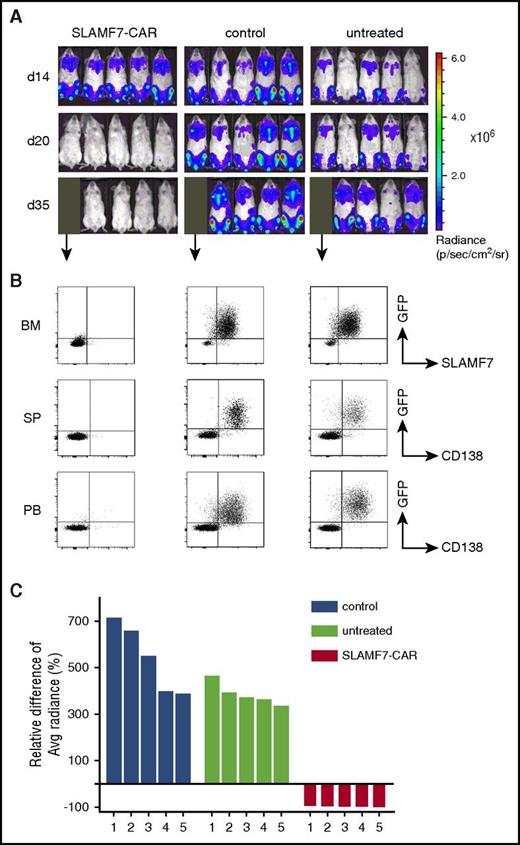
We proceeded to experiments in a xenograft model in which we injected MM.1S/ffluc into immunodeficient NSG mice and monitored myeloma burden and distribution by bioluminescence imaging and flow cytometry (Figure 3A-B). Within 14 days of MM.1S/ffluc inoculation, all mice developed systemic myeloma. Mice were then treated with a single dose of SLAMF7-CAR T cells or control CD19-CAR T cells (1:1 ratio of CD4+ and CD8+ T cells; total dose, 5 × 10⁶) or were left untreated. At 7 days after T-cell transfer, we observed a reduction in bioluminescence signal in all mice that had been treated with SLAMF7-CAR T cells (Figure 3A,C). The antimyeloma response was sustained until the end of the observation period for each mouse in this treatment cohort (Figure 3A; supplemental Figure 5A). In contrast, all of the mice that received CD19-CAR T cells or were left untreated presented with rapidly increasing bioluminescence signal (>2 log within 14 days), indicating disease progression. We obtained peripheral blood, bone marrow, and spleens from mice in each treatment group on day 35 after myeloma inoculation (day 21 after T-cell transfer) and performed flow cytometric analyses that revealed complete elimination of MM.1S from the hematopoietic system after treatment with SLAMF7-CAR T cells (Figure 3B). Accordingly, Kaplan-Meier analyses showed complete survival of all mice that had been treated with SLAMF7-CAR T cells at the end of the 8-week observation period, whereas all mice that had received control CD19-CAR T cells or were left untreated had to be euthanized prematurely due to progressive disease (supplemental Figure 5B). Also, in vivo, we observed similarly potent antimyeloma efficacy with SLAMF7-CAR T cell lines prepared from healthy donors and myeloma patients (Figure 3A-C; supplemental Figure 6A-D). Of note, after clearance of MM.1S from its medullary and extramedullary manifestations, we detected a subsequent increase in bioluminescence signal in all mice from the SLAMF7-CAR treatment cohort due to the emergence of MM.1S cells in anatomical sanctuaries, especially the peritoneum, which had initially not been affected. These observations are consistent with previous reports of late relapses after CAR T-cell therapy in murine xenograft models.22,23
Antimyeloma efficacy of SLAMF7-CAR T cells in vivo. NSG mice were inoculated with CD38+CD138+SLAMF7+ ffluc_eGFP–transduced MM.1S myeloma cells (IV) and, 14 days later, treated with a single dose of SLAMF7-CAR T cells or CD19-CAR (control) T cells (both: IV, 2.5 × 10⁶ CD4+ and 2.5 × 10⁶ CD8+; total dose: 5 × 10⁶) or remained untreated (n = 5 mice per group). (A) Serial bioluminescence imaging to assess myeloma progression/regression. Radiance was measured in regions of interest that encompassed the entire body of mice. Labels on the left state the day after inoculation with MM.1S myeloma cells. (B) Flow cytometric analysis of peripheral blood (PB), bone marrow (BM), and spleens (SP) to detect residual MM.1S myeloma cells in exemplary mice that were euthanized in each treatment group on day 35 after tumor inoculation (ie, day 21 after treatment). (C) Waterfall plot shows the relative increase/decrease in bioluminescence signal between day 14 (before treatment) and day 20 (6 days after treatment) in individual mice. (A-C) The data shown are representative of 3 independent experiments with SLAMF7-CAR and control CD19-CAR T cells prepared from 3 healthy donors.
Antimyeloma efficacy of SLAMF7-CAR T cells in vivo. NSG mice were inoculated with CD38+CD138+SLAMF7+ ffluc_eGFP–transduced MM.1S myeloma cells (IV) and, 14 days later, treated with a single dose of SLAMF7-CAR T cells or CD19-CAR (control) T cells (both: IV, 2.5 × 10⁶ CD4+ and 2.5 × 10⁶ CD8+; total dose: 5 × 10⁶) or remained untreated (n = 5 mice per group). (A) Serial bioluminescence imaging to assess myeloma progression/regression. Radiance was measured in regions of interest that encompassed the entire body of mice. Labels on the left state the day after inoculation with MM.1S myeloma cells. (B) Flow cytometric analysis of peripheral blood (PB), bone marrow (BM), and spleens (SP) to detect residual MM.1S myeloma cells in exemplary mice that were euthanized in each treatment group on day 35 after tumor inoculation (ie, day 21 after treatment). (C) Waterfall plot shows the relative increase/decrease in bioluminescence signal between day 14 (before treatment) and day 20 (6 days after treatment) in individual mice. (A-C) The data shown are representative of 3 independent experiments with SLAMF7-CAR and control CD19-CAR T cells prepared from 3 healthy donors.
In aggregate, these data demonstrate the potential to redirect the specificity of CD8+ and CD4+ T cells against SLAMF7 on malignant plasma cells through the expression of a SLAMF7-specific CAR. The acquisition of a SLAMF7–/low phenotype in CD8+ and CD4+ SLAMF7-CAR T cells is compatible with potent T-cell effector functions and significant antimyeloma reactivity in vitro and in vivo.
SLAMF7-CAR T cells confer selective fratricide of SLAMF7+ normal lymphocytes
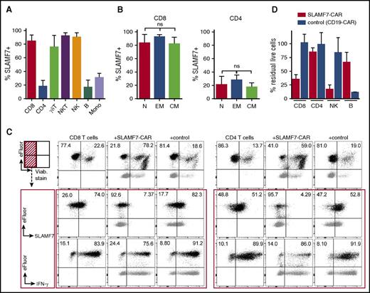
SLAMF7 is a receptor with an immunoregulatory function in normal lymphocytes, and accordingly, we detected surface expression of SLAMF7 protein on a fraction of naive and memory CD4+ and CD8+ T cells, NK and NKT cells, γδ T cells, monocytes, and B cells that we obtained from peripheral blood of myeloma patients (Figure 4A-B). We sought to determine the consequences of targeting SLAMF7 on myeloma for the normal lymphocyte compartment and performed flow cytometry–based cytotoxicity assays with coculture of patient-derived SLAMF7-CAR T cells and autologous (native) CD4+ and CD8+ T cells, NK cells, and B cells. We found that CD8+ SLAMF7-CAR T cells induced specific fratricide of SLAMF7+/high target cells in each of the lymphocyte subsets (Figure 4C-D; supplemental Figure 7A). However, in each lymphocyte subset, the SLAMF7–/low fraction was spared from fratricide and remained viable and functional as evidenced by IFN-γ secretion that could be elicited in CD8+ and CD4+ T cells after stimulation with PMA/ionomycin immediately at the end of the fratricide assay (Figure 4C). During the fratricide assay, SLAMF7-CAR T cells produced IFN-γ, as demonstrated by intracellular cytokine staining (supplemental Figure 7B). Cocultures with CD19-CAR T cells as effector cells were used as the control and induced complete elimination of B cells, as expected, but did not cause perturbations in any of the other lymphocyte subsets. For comparison, SLAMF7-CAR T cells mediated only partial B-cell depletion and preserved a fraction of viable SLAMF7–/low B cells. Experiments with SLAMF7-CAR T-cell lines from several myeloma patients and healthy donors affirmed our observations and showed that, on average, 17.3% of NK cells, 35.4% of CD8+ T cells, 66.5% of B cells, and 85% of CD4+ T cells were viable at the end of the fratricide assay with SLAMF7-CAR T cells (Figure 4D).
SLAMF7-CAR T cells exert selective fratricide of SLAMF7+normal lymphocytes. (A) Expression of SLAMF7 on normal lymphocyte subsets obtained from peripheral blood of myeloma patients (n = 10) analyzed by flow cytometry using the anti-SLAMF7 mAb 162.1. The diagram shows the mean percentage of SLAMF7+/high CD8 T cells (CD3+CD4–CD8+), CD4 T cells (CD3+CD4+CD8–), γδ T cells (Vγ9δ2 TCR+), NKT cells (CD3+CD56+), and NK cells (CD3–CD56+), B cells (B) (CD3–CD19+), and monocytes (CD3–CD14+). (B) Expression of SLAMF7 on naive (N) (CD45RA+CD45RO–CD62L+), effector memory (EM) (CD45RA–CD45RO+CD62L–), and central memory (CM) (CD45RA–CD45RO+CD62L+) CD4+ and CD8+ T cells obtained from peripheral blood of myeloma patients (n = 10). (C) CD8+ and CD4+ T cells were isolated from peripheral blood of myeloma patients, labeled with eFluor670, and used as target cells in 12-hour coculture assays with autologous CD8+ SLAMF7-CAR and control CD19-CAR T cells (non–eFluor labeled; E:T ratio = 4:1). The percentage of viable eFluor+ target cells before and after coculture was determined by staining with viability dye (top row of histograms); expression of SLAMF7 on viable target cells before and after coculture was determined by staining with anti-SLAMF7 mAb 162.1 (middle row); and the ability of viable target cells to produce IFN-γ in response to stimulation with PMA/ionomycin before and after coculture with SLAMF7-CAR and control CD19-CAR T cells was determined by intracellular cytokine staining (bottom row). The dot plots show overlays of eFluor+ target (black) and eFluor– effector (gray) cells. The numbers in the upper quadrants provide percentages of eFluor+ cells. (D) The diagram shows the mean percentage of residual live (7-AAD–) cells in each of the normal lymphocyte subsets after coculture with SLAMF7-CAR or control CD19-CAR T cells. (C-D) Data shown are representative for 4 independent experiments with SLAMF7-CAR and control CD19-CAR T cells from different donors.
SLAMF7-CAR T cells exert selective fratricide of SLAMF7+normal lymphocytes. (A) Expression of SLAMF7 on normal lymphocyte subsets obtained from peripheral blood of myeloma patients (n = 10) analyzed by flow cytometry using the anti-SLAMF7 mAb 162.1. The diagram shows the mean percentage of SLAMF7+/high CD8 T cells (CD3+CD4–CD8+), CD4 T cells (CD3+CD4+CD8–), γδ T cells (Vγ9δ2 TCR+), NKT cells (CD3+CD56+), and NK cells (CD3–CD56+), B cells (B) (CD3–CD19+), and monocytes (CD3–CD14+). (B) Expression of SLAMF7 on naive (N) (CD45RA+CD45RO–CD62L+), effector memory (EM) (CD45RA–CD45RO+CD62L–), and central memory (CM) (CD45RA–CD45RO+CD62L+) CD4+ and CD8+ T cells obtained from peripheral blood of myeloma patients (n = 10). (C) CD8+ and CD4+ T cells were isolated from peripheral blood of myeloma patients, labeled with eFluor670, and used as target cells in 12-hour coculture assays with autologous CD8+ SLAMF7-CAR and control CD19-CAR T cells (non–eFluor labeled; E:T ratio = 4:1). The percentage of viable eFluor+ target cells before and after coculture was determined by staining with viability dye (top row of histograms); expression of SLAMF7 on viable target cells before and after coculture was determined by staining with anti-SLAMF7 mAb 162.1 (middle row); and the ability of viable target cells to produce IFN-γ in response to stimulation with PMA/ionomycin before and after coculture with SLAMF7-CAR and control CD19-CAR T cells was determined by intracellular cytokine staining (bottom row). The dot plots show overlays of eFluor+ target (black) and eFluor– effector (gray) cells. The numbers in the upper quadrants provide percentages of eFluor+ cells. (D) The diagram shows the mean percentage of residual live (7-AAD–) cells in each of the normal lymphocyte subsets after coculture with SLAMF7-CAR or control CD19-CAR T cells. (C-D) Data shown are representative for 4 independent experiments with SLAMF7-CAR and control CD19-CAR T cells from different donors.
Targeting SLAMF7 preserves functional lymphocytes, including virus-specific T cells
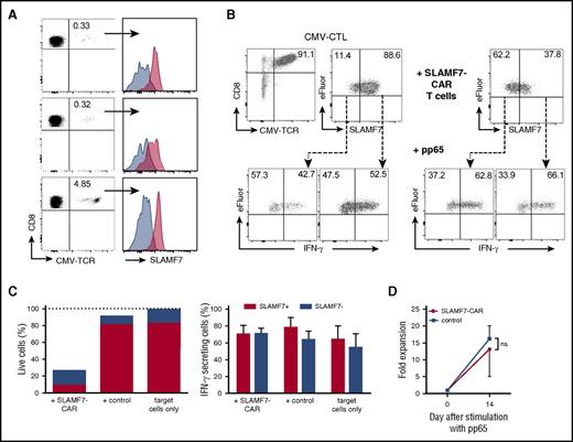
We were interested in determining whether SLAMF7–/low T cells were able to respond to a specific antigen and confer protection against common pathogens like cytomegalovirus (CMV). We detected expression of SLAMF7 on a fraction of CMV-specific and Epstein-Barr virus (EBV)-specific CD8+ memory T cells in the peripheral repertoire of HLA-A*02+ CMV/EBV-seropositive donors (n = 3, respectively) (Figure 5A; supplemental Figure 8). Next, we established CD8+ T-cell lines that recognize the CMV pp65 A2/NLV epitope through their endogenous T-cell receptor (CMV-CTL) and ascertained that they contained a SLAMF7+/high and SLAMF7–/low fraction, similar to the distribution observed with freshly isolated endogenous CMV-specific CD8+ T cells (Figure 5A-B). SLAMF7+/high and SLAMF7–/low CMV-CTL responded equally well to stimulation with pp65 peptide as assessed by IFN-γ secretion. Then, we cocultured CMV-CTL with autologous CD8+ SLAMF7-CAR–modified T cells, which led to the depletion of the SLAMF7+/high CTL fraction and relative enrichment of SLAMF7–/low CTL (Figure 5C). Restimulation of SLAMF7–/low CMV-CTL induced rapid high-level IFN-γ production, comparable with the response that had been observed before the fratricide assay (Figure 5B-C). Finally, we transduced T-cell lines that contained a proportion of CMV-CTL with the SLAMF7-CAR to exert continuous pressure against SLAMF7 expression and confirmed their ability to numerically expand after stimulation with pp65 peptide-pulsed antigen-presenting cells at a similar rate as control CMV-CTL, which had been modified with a CD19-CAR for comparison (Figure 5D).
SLAMF7-mediated fratricide spares a fraction of functional CMV-specific CD8+T cells. (A) SLAMF7 expression on primary CD8+CD45RA–CD45RO+ memory T cells specific for the CMV pp65 A2/NLV epitope in 3 HLA-A*02+ healthy donors. Overlay histograms show staining with anti-SLAMF7 mAb 162.1 (red) and isotype control (blue). (B) CMV-specific CD8+ T-cell lines (CMV-CTL) were prepared from CMV-specific memory T cells and the expression of SLAMF7 was analyzed (2 top left dot plots). CMV-CTL was labeled with eFluor670 and cocultured for 4 hours with autologous SLAMF7-CAR or control CD19-CAR T cells. The expression of SLAMF7 on residual live (ie, 7-ADD–) CMV-CTL was reanalyzed at the end of the coculture (top right dot plot). Residual live CMV-CTL was then stimulated with pp65 NLV peptide-loaded K562/HLA-A2 cells, and IFN-γ production in the SLAMF7+/high and SLAMF7–/low fraction was analyzed by intracellular cytokine staining (2 bottom right dot plots). IFN-γ production in SLAMF7+/high and SLAMF7–/low CMV-CTL before the fratricide assay was analyzed for comparison (2 bottom left dot plots). (C) Bar diagrams show the mean percentage of live (7-AAD–) (left) and IFN-γ–secreting (right diagram) SLAMF7+/high and SLAMF7–/low CMV-CTL after coculture with SLAMF7-CAR and control CD19-CAR T cells. CMV-CTL alone is provided for comparison (target cells only). (D) CMV-CTL were transduced with the SLAMF7-CAR (or CD19-CAR for comparison), and CAR-transduced T cells were enriched using the EGFRt marker. The CAR-transduced CMV-CTL was then stimulated with pp65 NLV peptide–loaded K562/HLA-A2 cells. The diagram shows the fold expansion of CMV-CTL within 14 days of culture. (C-D) The data shown are representative of the results obtained in 2 independent experiments with CAR-transduced CMV-CTL from different donors.
SLAMF7-mediated fratricide spares a fraction of functional CMV-specific CD8+T cells. (A) SLAMF7 expression on primary CD8+CD45RA–CD45RO+ memory T cells specific for the CMV pp65 A2/NLV epitope in 3 HLA-A*02+ healthy donors. Overlay histograms show staining with anti-SLAMF7 mAb 162.1 (red) and isotype control (blue). (B) CMV-specific CD8+ T-cell lines (CMV-CTL) were prepared from CMV-specific memory T cells and the expression of SLAMF7 was analyzed (2 top left dot plots). CMV-CTL was labeled with eFluor670 and cocultured for 4 hours with autologous SLAMF7-CAR or control CD19-CAR T cells. The expression of SLAMF7 on residual live (ie, 7-ADD–) CMV-CTL was reanalyzed at the end of the coculture (top right dot plot). Residual live CMV-CTL was then stimulated with pp65 NLV peptide-loaded K562/HLA-A2 cells, and IFN-γ production in the SLAMF7+/high and SLAMF7–/low fraction was analyzed by intracellular cytokine staining (2 bottom right dot plots). IFN-γ production in SLAMF7+/high and SLAMF7–/low CMV-CTL before the fratricide assay was analyzed for comparison (2 bottom left dot plots). (C) Bar diagrams show the mean percentage of live (7-AAD–) (left) and IFN-γ–secreting (right diagram) SLAMF7+/high and SLAMF7–/low CMV-CTL after coculture with SLAMF7-CAR and control CD19-CAR T cells. CMV-CTL alone is provided for comparison (target cells only). (D) CMV-CTL were transduced with the SLAMF7-CAR (or CD19-CAR for comparison), and CAR-transduced T cells were enriched using the EGFRt marker. The CAR-transduced CMV-CTL was then stimulated with pp65 NLV peptide–loaded K562/HLA-A2 cells. The diagram shows the fold expansion of CMV-CTL within 14 days of culture. (C-D) The data shown are representative of the results obtained in 2 independent experiments with CAR-transduced CMV-CTL from different donors.
Collectively, these experiments reveal that SLAMF7-CAR T cells confer selective fratricide of SLAMF7+/high normal lymphocytes, whereas SLAMF7–/low normal lymphocytes are not eliminated. Our data suggest that adoptive transfer of SLAMF7-CAR T cells in a clinical setting will deplete SLAMF7+/high normal lymphocytes, but preserve a reduced T-cell compartment that can confer protective immunity to pathogens like CMV.
Discussion
SLAMF7 has attracted attention as a target for immunotherapy in myeloma because of previous work that demonstrated uniform high-level expression on primary myeloma cells and the majority of myeloma cell lines and absence of expression on normal solid tissues.10,13 However, SLAMF7 is known to be expressed on normal lymphocytes, including NK cells, a fraction of CD8+ and CD4+ T cells, and B cells, and, as we show for the first time, is also present on a fraction of γδ T cells.7,11,24 Therefore, the effort of deriving SLAMF7-specific, CAR-modified T cells is essentially puzzled by a conundrum: can T cells be redirected to a target on myeloma cells that is also present on T cells themselves? We show that expression of the SLAMF7-CAR leads to a rapid reduction in SLAMF7 protein expression on the T-cell surface and acquisition of a SLAMF7–/low phenotype in both CD8+ and CD4+ SLAMF7-CAR T cells. We provide evidence that several mechanisms contribute to this conversion, including deletion (or more specifically, fratricide) of SLAMF7+/high T cells during culture and outgrowth of SLAMF7-CAR–modified T cells that have internalized SLAMF7 protein and maintain the SLAMF7–/low phenotype. An additional mechanism that may accentuate the SLAMF7–/low phenotype is the sequestration of SLAMF7 protein through the SLAMF7-CAR in the cytoplasm of T cells.
Our longitudinal analysis of SLAMF7 expression in SLAMF7-CAR–modified and control T cells during in vitro culture demonstrated that SLAMF7 expression is dynamic and not static and revealed that SLAMF7-CAR T cells retain expression of SLAMF7. Indeed, SLAMF7 protein reappeared on SLAMF7-CAR T cells at high levels for a short period of time after activation during antigen-dependent expansion and after supraphysiologic stimulation with PMA/ionomycin. The functional role of SLAMF7 in T cells and other lymphocyte subsets is incompletely understood, and depends on the SLAMF7 isoform, adaptor protein, and specific cell subtype.5,6,24,25 It has been shown in previous work that a deficiency in SLAMF7 reduces the cytolytic activity of NK cells, whereas the presence of SLAMF7 on antigen-presenting cells decreased antigen-induced CD4+ T-cell proliferation and cytokine secretion.24,26 In addition, it has recently been shown that SLAMF7 negatively regulates survival and proliferation of innate-like CD8+ and NKT cells.27 In our experiments, SLAMF7-CAR T cells exerted specific cytolytic/cytotoxic activity, cytokine secretion, and proliferation in vitro and in vivo, suggesting that high-level SLAMF7 expression on the T-cell surface is dispensable for exerting the cardinal T-cell effector functions and for expanding and surviving in short-term culture. The long-term consequences of reduced or lost SLAMF7 expression in T cells remain unknown and may vary between killer and helper, naive and memory T-cell subsets.10,11,24
We demonstrate that SLAMF7-CAR T cells confer specific fratricide of native SLAMF7+/high normal lymphocytes, which has implications for the clinical translation of SLAMF7-CAR T-cell therapy. A conceivable side effect of the administration of SLAMF7-CAR T cells is depletion of SLAMF7+/high lymphocytes, a projection that is supported by clinical experience with the anti-SLAMF7 mAb huLuc63 (elotuzumab), which induces a significant reduction in lymphocyte counts.16,28 Importantly, however, lymphocyte counts return to normal within a few weeks after cessation of elotuzumab treatment.16,29 The depth of lymphoreduction may be more pronounced with SLAMF7-CAR T cells compared with elotuzumab, because the antigen density on target cells required for CAR recognition is lower than for antibodies.30 However, it is also conceivable that patients reconstitute normal counts of SLAMF7–/low lymphocytes, similar to the phenotype of SLAMF7-CAR T cells that we observed in our in vitro culture. Nevertheless, it would be prudent to equip SLAMF7-CAR T cells with a safety switch to terminate fratricide of normal lymphocytes if needed. The transgene cassette of our SLAMF7-CAR contains an EGFRt depletion marker that we have shown can be used to deplete CAR T cells in vivo through administration of the anti-epidermal growth factor receptor mAb cetuximab.31 However, because several lymphocyte subsets that mediate antibody-dependent cellular cytotoxicity are likely diminished by SLAMF7-CAR T cells, inducible caspase 9 or herpes simplex virus thymidine kinase may be preferable safety switches in this particular context.32,33
A concern associated with profound and persistent lymphoreduction are infectious complications. We show that fratricide induced by SLAMF7-CAR T cells eliminates a substantial proportion of normal lymphocytes in vitro, but preserves a fraction of viable T cells, B cells, and NK cells. These include virus-specific T cells that we show are capable of responding to and expanding after stimulation with CMV antigen, suggesting that patients receiving SLAMF7-CAR T cells will retain some degree of immunocompetence against common viral pathogens. Our data are in line with the clinical elotuzumab experience, which did not disclose an increase in viral infections.16,29 Further, we have shown that CMV-specific T cells have significant proliferative potential, and even the lowest numbers can mount an effective immune response.34
Strategies for the clinical implementation of SLAMF7-CAR T-cell therapy include their use in the context of autologous or allogeneic hematopoietic stem cell transplantation either pretransplant to achieve minimal residual disease negativity or posttransplant to consolidate the antimyeloma effect. Because the extent and long-term consequences of lymphocytic fratricide are uncertain, the use of SLAMF7-CAR T cells before transplant for a defined therapeutic window of time, followed by CAR T-cell depletion through a suicide gene and reconstitution of patient or donor hematopoiesis through an auto- or allograft would be attractive for initial clinical evaluation. Most crucially, SLAMF7 is not expressed on hematopoietic stem cells.10 The use of a lymphodepleting conditioning regimen before systemic administration of CAR T cells is well established from clinical trials with CD19-CARs1,35 and will be instrumental to prevent severe cytokine release syndrome after infusion of SLAMF7-CAR T cells that may result from the recognition of endogenous SLAMF7+ lymphocytes. Another measure to prevent excessive toxicity is the formulation of SLAMF7-CAR T-cell products that contain defined proportions of CD8+ and CD4+ T cells, which we have shown translates into more consistent and predictable pharmacokinetics of CAR T cells in vivo and allows for lowering the absolute number of adoptively transferred CAR T cells.36
We have equipped our SLAMF7-CAR with a targeting domain comprising the variable heavy and variable light chains of the huLuc63 mAb (elotuzumab). Encouragingly, there is now a significant body of clinical experience with huLuc63 that has disclosed no unexpected toxicity and no off-target reactivity. Although we did not perform a direct side-by-side analysis, our data showed superior antimyeloma efficacy of SLAMF7-CAR T cells compared with elotuzumab in the NSG/MM.1S myeloma xenograft model in vivo.10,13 Recently, engraftment of primary human myeloma cells has been accomplished in a sophisticated murine model that may facilitate future analyses of CAR T-cell efficacy in vivo.37 The SLAMF7-CAR used in our studies contained a CD28 costimulatory moiety. In our in vivo model, we were not readily able to detect CAR T cells in the course of the experiment in peripheral blood and in bone marrow/spleens at the end of the observation period by flow cytometry. It is unclear whether the SLAMF7–/low phenotype affects the long-term engraftment and persistence of SLAMF7-CAR T cells, and we are currently generating SLAMF7-CARs with 4-1BB costimulation, which may translate into superior engraftment and persistence.38 In previous work, we have demonstrated that target epitope selection and CAR extracellular domain design affect tumor recognition and CAR T-cell function and applied these insights to the design of our SLAMF7-CAR.39 We tailored our SLAMF7-CAR to contain a IgG4-Fc Hinge-CH2-CH3 extracellular spacer domain and removed Fc motifs to prevent CAR T-cell elimination by Fc receptor–expressing cells.19 We also evaluated other anti-SLAMF7–targeting domains (eg, Luc9013 ) that proved to be less effective than huLuc63 (data not shown). The huLuc63 targeting domain has been humanized to reduce the likelihood of acute or chronic CAR T-cell rejection.40,41
Several alternative antigens, including CD19, BCMA, CD44v6, and CD38, are under investigation as targets for CAR T cells in myeloma.4,42,43 In a recently reported clinical trial, BCMA-CAR T cells induced transient partial and complete responses in a proportion of patients, but were neutralized in 1 patient through the loss of BCMA expression on myeloma cells.4 Loss of SLAMF7 expression has, to our knowledge, not been reported with elotuzumab and may not readily be compensated by myeloma cells given the role of SLAMF7 in myeloma pathogenesis and survival in the bone marrow niche.13 Other investigators have attempted in previous work to prepare SLAMF7-specific T cells from the endogenous T-cell repertoire and through genetic engineering and focused on analyzing antimyeloma efficacy,44,45 but did not consider the phenotypic and functional consequences of targeting SLAMF7 for the (SLAMF7-specific) effector T cells and the normal lymphocyte compartment.
In summary, we believe SLAMF7-CAR T cells have significant therapeutic potential and, with sufficient care in defining the appropriate clinical setting, monitoring, and supportive therapy, can be implemented as a safe and effective treatment against multiple myeloma.
The online version of this article contains a data supplement.
The publication costs of this article were defrayed in part by page charge payment. Therefore, and solely to indicate this fact, this article is hereby marked “advertisement” in accordance with 18 USC section 1734.
Acknowledgments
The authors thank Ina Hellmann, Roger R. Beerli, and Ulf Grawunder (NBE-Therapeutics Ltd, Basel, Switzerland) for providing the SLAMF7 protein and huLuc63 mAb. The authors also thank Silvia Koch for technical assistance and Silke Frenz and Elke Spirk for support in performing experiments in the preclinical in vivo models.
This work was supported by the European Union’s Horizon 2020 research and innovation program under grant agreement 733297. This work was also supported by German Cancer Aid (Max Eder Program Award 110313 [M.H.]), the Myeloma Crowd Research Initiative (MCRI Award 2015 [M.H.]), and IZKF Würzburg (Interdisziplinäres Zentrum für Klinische Forschung, Projekt D-244 and Z-4/109 [M.H.]). In addition, this work was made possible by philanthropic support from cdw Stiftungsverbund and the patient advocacy group, Hilfe im Kampf Gegen Krebs e.V. (Würzburg, Germany). S.D. was supported by Interdisziplinäres Zentrum für Klinische Forschung (IZKF) Würzburg and is a fellow of the Clinician Scientist Program of the Else-Kröner Forschungskolleg. M.H. was supported by the Young Scholar Program of the Bavarian Academy of Sciences and is an Extraordinary Member of the Bavarian Academy of Sciences.
Authorship
Contribution: T.G. and S.D. designed and performed the research, analyzed the data, and wrote the manuscript; J.R., S.P., and C.B. performed the research and analyzed the data; M.S. provided biological material and analyzed the data; H.E. designed the research, analyzed the data, wrote the manuscript, and supervised the project; and M.H. conceived the project, designed the research, analyzed the data, wrote the manuscript, and supervised the project.
Conflict-of-interest disclosure: M.H. is coinventor on a patent application (PCT/US2013/055862) related to CAR technologies that has been filed by the Fred Hutchinson Cancer Research Center (Seattle, WA) and licensed by JUNO Therapeutics, Inc. In addition, M.H. is coinventor on patent applications related to CAR technologies that have been filed by the University of Würzburg. The remaining authors declare no competing financial interests.
Correspondence: Michael Hudecek, Medizinische Klinik und Poliklinik II, Universitätsklinikum Würzburg, Oberdürrbacher Strasse 6, 97080 Würzburg, Germany; e-mail: hudecek_m@ukw.de.
References
Author notes
T.G. and S.D. contributed equally to this work.

This feature is available to Subscribers Only
Sign In or Create an Account Close Modal