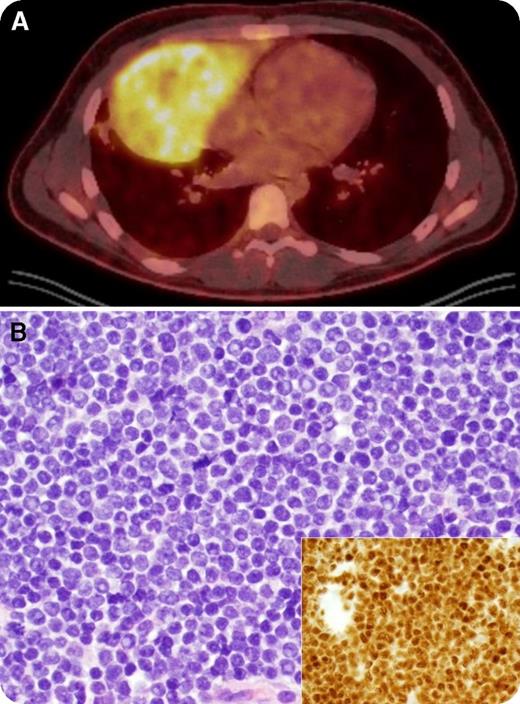
An 18-year-old man presented with a 2-month history of unintentional 23-kg weight loss, night sweats, and left-sided neck swelling. Physical examination revealed left supraclavicular lymphadenopathy. A complete blood count was within normal limits (white blood cells, 6.6 × 10⁹/L) and a peripheral blood smear only identified a rare immature cell. Positron emission tomography showed a 16- × 9-cm hypermetabolic mediastinal mass mirroring the heart in the right hemithorax (panel A, “double-heart” sign) and extending into the neck. Biopsy of the mediastinal mass revealed a diffuse infiltrate of intermediate-sized T lymphoblasts with high nuclear-to-cytoplasmic ratio, fine chromatin, prominent nucleoli, and frequent mitotic figures (panel B; original magnification ×400; main panel, hematoxylin and eosin stain; inset, terminal deoxynucleotidyl transferase stain). The bone marrow was 50% involved, establishing a diagnosis of T-cell acute lymphoblastic leukemia (T-ALL). Cervical lymphadenopathy resolved completely on physical examination within 2 days of starting steroids as a component of AALL1231 and the patient achieved a complete remission after 1 cycle.
The differential diagnosis of a mediastinal mass in a young adult includes thymoma, T-ALL, large B-cell lymphoma, Hodgkin lymphoma, and germ cell tumors. T-ALL masses are often exquisitely sensitive to steroids; hence, empiric steroid treatment before biopsy may result in rapid resolution of the mass and inadequate or nondiagnostic biopsy specimens.
For additional images, visit the ASH IMAGE BANK, a reference and teaching tool that is continually updated with new atlas and case study images. For more information visit http://imagebank.hematology.org.


This feature is available to Subscribers Only
Sign In or Create an Account Close Modal