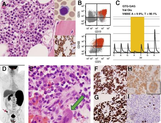
A 44-year-old man presented with splenomegaly, pancytopenia, and occasional circulating hairy cells (panel A upper inset; original magnification ×1000, Wright-Giemsa stain). A B-cell infiltrate was detected in the bone marrow (panel A, original magnification ×400, hematoxylin and eosin stain; lower inset is CD20 immunohistochemistry, original magnification ×400); by flow cytometry, B cells were positive for CD11c, CD19, CD20, CD25, and CD103 (panel B). Pyrosequencing confirmed a BRAF c.1799T>A p.V600E mutation (panel C). A diagnosis of hairy cell leukemia was established. The patient was observed and presented 9 months later with leg pain. 18-Fluorodeoxyglucose positron emission tomography demonstrated splenomegaly, abdominal lymphadenopathy, and a hypermetabolic right iliac bone lesion (panel D, arrow). Biopsy of the bone lesion showed sheets of medium-sized cells with frequent nuclear grooves, dispersed chromatin, and abundant cytoplasm (panel E; original magnification ×400, hematoxylin and eosin stain); occasional multinucleated giant cells (panel E, green arrow) exhibiting similar nuclear features were also identified. These cells were positive for CD1a (panel F; original magnification ×400), langerin (panel G; original magnification ×400), and BRAF V600E protein (panel H; original magnification ×400; inset is a BRAF V600E+ giant cell), and were negative for CD19, CD20 (panel I; original magnification ×400), and PAX5, confirming the diagnosis of Langerhans cell histiocytosis. We performed polymerase chain reaction followed by capillary electrophoresis using DNA and showed monoclonal peaks of identical size in both neoplasms.
This case exhibits the concurrent presence of 2 distinct neoplasms arising from divergent differentiation of a BRAF mutated common progenitor cell.
For additional images, visit the ASH IMAGE BANK, a reference and teaching tool that is continually updated with new atlas and case study images. For more information visit http://imagebank.hematology.org.


This feature is available to Subscribers Only
Sign In or Create an Account Close Modal