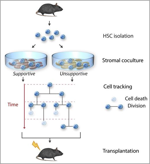
In this issue of Blood, Kokkaliaris et al use a sophisticated imaging approach to track the behavior of individual murine hematopoietic stem cells (HSCs) cultured on stromal cells. The authors identify cell death as a critical factor limiting the in vitro culture of HSCs, which can be mitigated by the matrix protein dermatopontin (DPT). These studies lay the groundwork for the in vitro manipulation of HSCs for therapeutic purposes.1
Experimental approach for the analysis of the behavior over time of individual primary murine HSCs using sophisticated methods to track all cell-death and cell-division events. HSCs were cocultured with stromal cell lines that differ in their ability to maintain long-term HSC function to identify molecules that mediate that stromal support. Professional illustration by Somersault18:24.
Experimental approach for the analysis of the behavior over time of individual primary murine HSCs using sophisticated methods to track all cell-death and cell-division events. HSCs were cocultured with stromal cell lines that differ in their ability to maintain long-term HSC function to identify molecules that mediate that stromal support. Professional illustration by Somersault18:24.
HSCs serve as the foundation of the late fetal and adult hematopoietic systems and their transplantation provides a curative approach to multiple hematologic, immunologic, and genetic disorders. When cultured in vitro, HSCs rapidly differentiate and lose transplantable stem cell activity.2 The study of cells at the population level can obscure the unique properties and behaviors of individual cells within that population. Here, the authors track the biological behavior, including every cell-division or cell-death event, of individual adult murine HSCs cultured ex vivo for up to 2 weeks (see figure). These HSCs were cocultured with stromal lines known to differ in their capacity to support the ex vivo persistence of HSCs. The AFT024 stromal line, established by the authors nearly 2 decades ago, can maintain high levels of transplantable multilineage stem cell activity for extended periods of time in culture.3
The authors find that most HSCs placed in culture rapidly die. However, the supportive AFT024 stromal cells support the survival of HSCs and this survival effect requires direct contact of HSCs with the stromal cells. The differential behavior of HSCs on supportive and nonsupportive stromata was confirmed by elegant experiments with intermixed stromal cultures, monitoring the differential behavior of HSCs in contact with the differing stromal cells within the same dish. To identify potential factors mediating the survival benefit provided by AFT024 cells, previously identified genes differentially expressed between the supportive and nonsupportive stromata were tested using both loss- and gain-of-function approaches. Knockdown of 3 genes, delta-like 1 homolog, fibroblast activation protein, and DPT, each reduced the capacity of the modified AFT024 cells to support the proliferation of cocultured HSCs and also impacted the ability of the cocultured HSCs to maintain long-term engraftment of transplanted mice. Of these 3 genes, DPT appears to play the most significant role in the capacity of the AFT024 cell line to support HSC function. Even exogenously added DPT enhanced the survival and proliferation ex vivo–cultured HSCs.
This study raises many questions. How does DPT, as an extracellular protein, mediate its effect on HSC survival? DPT is expressed by osteoblasts, but not significantly by “bone marrow” cells (http://biogps.org/#goto=genereport&id=56429). Do cells in the bone marrow express DPT and what is its spatial distribution in the marrow? Is DPT yet another component of the ever more complex HSC niche in the bone marrow?4 Because AFT024 cells can also support human HSCs, do the findings presented here translate to human stem cells?
The AFT024 cell line was established from the fetal liver of the mouse.3 Unlike the adult bone marrow where most, if not all, long-term HSCs are quiescent, the HSC population in the fetal liver rapidly expands in number before transitioning to the bone marrow. This rapid increase is likely related not only to increased cell cycling of fetal HSCs,5 but also to the progressive expansion of the remodeling vascular niche,6 and to the influx and maturation of a wave of pre-HSCs.7 It would be of interest to determine whether DPT is expressed in the fetal liver and if it facilitates fetal HSC expansion. Given the rare number of HSCs normally present during fetal and adult life, their enhanced ex vivo survival and maintenance mediated by DPT represents a first step toward the holy grail of in vitro HSC expansion to facilitate the provision of adequate numbers of HSCs from nonadult sources and the engineering of “designer” HSCs for research and therapeutic purposes.
Conflict-of-interest disclosure: The author declares no competing financial interests.

