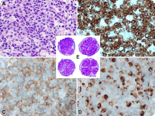
A 78-year-old white American man presented with fever, jaundice, and respiratory distress. Laboratory tests showed the following: white blood cells 4.2 (4-11) × 109/L, hemoglobin 11.7 (13.1-17.2) g/dL, platelets 21 (140-440) × 109/L, bilirubin 9.2 (0.2-1.2) mg/dL, aspartate aminotransferase 1345 (10-37) U/L, alanine aminotransferase 599 (5-40) U/L, alkaline phosphatase 313 (40-125) U/L, lactate dehydrogenase 2102 (98-192) U/L, international normalized ratio 2.4 (0.89-1.29), activated partial thromboplastin time 149 (22-36) seconds, haptoglobin <20 (30-200) mg/dL, and fibrinogen 56 (200-425) mg/dL. A chest–computed tomography scan revealed pulmonary infiltrates and mediastinal lymphadenopathy. Subcarinal lymph node biopsy showed numerous small-to-medium neoplastic cells (panel A) that expressed CD2, cCD3 (panel B), CD7, CD30, CD56 (panel C), TIA1, granzyme B, perforin, and Epstein-Barr virus (EBV; panel D) and were negative for CD4, CD5, and CD8. The Ki67 proliferation index on this specimen showed 90% nuclear staining. Peripheral blood flow cytometry showed 17% abnormal natural killer cells (panel E) expressing CD2, CD7dim, TIA1, and CD56dim and was negative for sCD3 and terminal deoxynucleotidyl transferase. A diagnosis of EBV-associated aggressive natural killer cell leukemia (ANKL) was established. The patient deteriorated rapidly and died.
ANKL is a rare, aggressive, EBV-associated leukemia primarily affecting young Asians. B symptoms, organomegaly, hemophagocytosis, and disseminated intravascular coagulation are common. Complex cytogenetic abnormalities are relatively frequent, and clonal T-cell receptor gene rearrangements are absent. ANKL has been reported in fewer than 20 patients of white Western European descent.
For additional images, visit the ASH IMAGE BANK, a reference and teaching tool that is continually updated with new atlas and case study images. For more information visit http://imagebank.hematology.org.


This feature is available to Subscribers Only
Sign In or Create an Account Close Modal