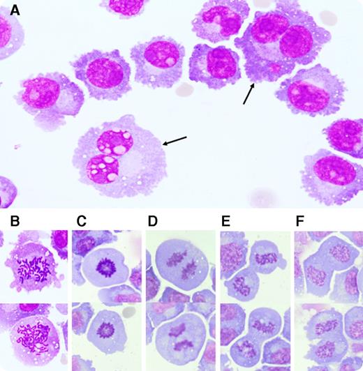
A 72-year-old man presented in an acutely confused state. He had been diagnosed with immunoglobulin A κ, 17p-deleted multiple myeloma (MM) 3 years prior. He had since received numerous lines of treatment, including several immunomodulatory agents (thalidomide, lenalidomide, pomalidomide) and proteasome inhibitors (bortezomib, carfilzomib). Because his initial workup was noncontributory to diagnosis, a lumbar puncture was performed. The cerebrospinal fluid (CSF) showed an elevated protein level and low glucose levels. Microscopic examination of the CSF revealed an infiltration by MM cells (panel A), confirmed by flow cytometry, including numerous binuclear (panel A, arrows) and dividing cells at all phases of mitosis; (panel B) prophase-chromatin condensation into chromosomes; (panel C) metaphase-chromosomes coiling in the middle of the cell; (panel D) anaphase-chromosomes splitting, and movement of sister chromatids to opposite poles; and (panel E) telophase-sister chromatids reaching opposite poles. Karyokinesis (nucleus division) was also observed, without completion of cellular division (panel F), a finding that might explain the generation of binucleated cells.
The incidence of extramedullary MM seems to be rising. Whether this reflects increased selective pressure by novel therapies or is simply a consequence of longer survival has yet to be determined. Our observation suggests that MM cells do not merely survive but also proliferate within the CSF, even in the absence of bone marrow microenvironmental support.
For additional images, visit the ASH IMAGE BANK, a reference and teaching tool that is continually updated with new atlas and case study images. For more information visit http://imagebank.hematology.org.


This feature is available to Subscribers Only
Sign In or Create an Account Close Modal