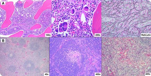
A 14-year-old white young man was transferred from an outside hospital with a 4-week history of fever, 30-lb weight gain, and diarrhea. He was noted to have anasarca, elevated C-reactive protein (CRP), elevated creatinine, anemia, and thrombocytopenia. A detailed clinical work-up for infectious and autoimmune etiologies was negative. Bone marrow biopsy showed a hypercellular marrow with megakaryocytic hyperplasia, clustering, and reticulin fibrosis (panel A, left to right). Ultrasound revealed enlarged lymph nodes that were fluorodeoxyglucose-avid by positron emission tomography–computed tomography scan. A right inguinal lymph node excisional biopsy (panel B, left to right) showed regressed germinal centers, expanded concentric mantle zones penetrated by hyalinized vessels, and vascular interfollicular areas that were consistent with idiopathic multicentric Castleman disease (iMCD). The overall clinicopathological picture was compatible with TAFRO syndrome (thrombocytopenia, anasarca, myelofibrosis/fever, renal dysfunction/reticulin fibrosis, and organomegaly) or Castleman-Kojima disease, a recently described variant of iMCD. In contrast to typical cases of iMCD, gamma globulin levels are normal and thrombocytopenia is a consistent feature.
The etiology of iMCD is unclear and is hypothesized to be a systemic inflammatory disorder mediated by redundant cytokines. The patient responded well to corticosteroids and siltuximab with prompt resolution of thrombocytopenia, ascites, and renal dysfunction and decrease in CRP and interleukin-6 levels.
For additional images, visit the ASH IMAGE BANK, a reference and teaching tool that is continually updated with new atlas and case study images. For more information visit http://imagebank.hematology.org.


This feature is available to Subscribers Only
Sign In or Create an Account Close Modal