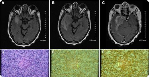
A 65-year-old woman initially presented with chronic extreme headaches. Initial magnetic resonance imaging (MRI) scans (panel A) revealed abnormal signaling possibly representing dural inflammation or a meningioma. A complete blood count at that time was within normal limits. Four weeks later, her symptoms included progressively worsening, right temporal throbbing headaches with associated right eye visual disturbance and right facial paresthesias. Repeat MRI (panels B and C) revealed enlargement of the dural mass now extending into the right cavernous sinus and right optic canal. Initial cerebrospinal fluid studies were negative for cytology and infection. A biopsy revealed a myeloid sarcoma (panels D-F). A peripheral smear revealed 10% circulating blasts, and a subsequent bone marrow biopsy revealed acute myeloid leukemia (AML) with 8:21 translocation.
In patients with AML, less than 1% present initially with prominent extramedullary disease, including myeloid sarcoma. Myeloid sarcoma may present simultaneously with bone marrow disease, precede bone marrow disease, or be seen in disease relapse. The presence of a myeloid sarcoma is diagnostic of AML independent of the blast count. Myeloid sarcoma may be found in many isolated sites, including the dura, periosteum, and bone. Myeloid sarcoma must be considered in the differential diagnosis in the presence of a rapidly progressing dural mass.
For additional images, visit the ASH IMAGE BANK, a reference and teaching tool that is continually updated with new atlas and case study images. For more information visit http://imagebank.hematology.org.


This feature is available to Subscribers Only
Sign In or Create an Account Close Modal