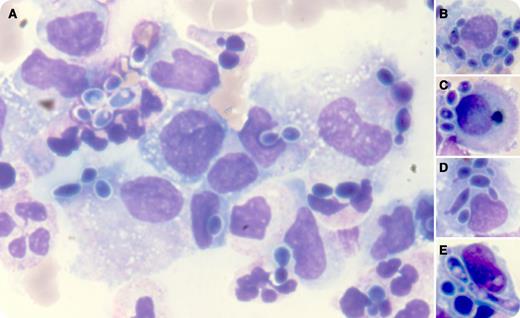
A 42-year-old man was diagnosed with a severe acute respiratory distress syndrome following inhalation of his stomach contents. The previous history revealed liver cirrhosis due to alcoholic liver disease. Seven days later, he presented with a high fever and a painful abdominal distension due to ascites. The blood cell count was as follows: hemoglobin, 10.9 g/dL; white blood cells, 12.230 × 10⁹/L (neutrophils, 9.124 × 10⁹/L); and platelets, 124 × 109/L. The ascitic fluid analysis showed total nucleated cells of 0.242 × 10⁹/L (neutrophils, 29%; lymphocytes, 9%; monocytes, 53%; peritoneal cells, 9%). Microscopic examination showed a mix of isolated, clumped, and budding “pearls” inside or outside the macrophagic cells (panel A). The yeasts had been phagocytized by neutrophils, macrophages, and peritoneal cells and sometimes were arranged as a ring in their cytoplasm (panels B-E). They were identified as Candida albicans, and similar yeasts were identified in the tracheal aspirate. In conjunction with the management of the whole disorder, the patient received fluconazole through an intravenous catheter and recovered after weeks.
Candida sp. infections are well documented in immunocompromised patients. In such a context, the infection is not always accompanied by increased neutrophils in the blood or in the ascitic fluid. Peritoneal cells are capable of phagocytizing microorganisms as neutrophils and monocytes/macrophages do.
For additional images, visit the ASH IMAGE BANK, a reference and teaching tool that is continually updated with new atlas and case study images. For more information visit http://imagebank.hematology.org.


This feature is available to Subscribers Only
Sign In or Create an Account Close Modal