Key Points
XmAb5574 is an Fc-engineered CD19 monoclonal antibody that is well tolerated as a single agent in patients with relapsed or refractory CLL.
XmAb5574 has preliminary efficacy as a single agent in CLL and is of interest for further study in this disease.
Abstract
CD19 is ubiquitously expressed on chronic lymphocytic leukemia (CLL) cells and is therefore an attractive candidate for antibody targeting. XmAb5574 (aka MOR00208) is a novel humanized CD19 monoclonal antibody with an engineered Fc region to enhance Fcγ receptor binding affinity. Here we report results of a first in human phase 1 trial of XmAb5574 in patients with relapsed or refractory CLL. Twenty-seven patients were enrolled to 6 escalating dose levels, with expansion at the highest dose level of 12 mg/kg. Nine doses of XmAb5574 were infused over 8 weeks. No maximal tolerated dose was reached, and the drug was generally well tolerated, with infusion reactions of grades 1 and 2 being the most common toxicities. Grade 3 and 4 toxicities occurred in 5 patients and included neutropenia, thrombocytopenia, increased aspartate aminotransferase, febrile neutropenia, and tumor lysis syndrome. XmAb5574 showed preliminary efficacy, with 18 patients (66.7%) responding by physical examination criteria and laboratory studies, and 8 patients (29.6%) responding by computed tomography criteria. Pharmacokinetics showed a half-life of 14 days with clearance that was not dose-dependent. In conclusion, this phase 1 trial demonstrates safety and preliminary efficacy of a novel Fc-engineered CD19 monoclonal antibody XmAb5574 and justifies movement into the phase 2 setting. This trial was registered at www.clinicaltrials.gov as #NCT01161511.
Introduction
Chronic lymphocytic leukemia (CLL) is the most prevalent form of adult leukemia and is currently incurable outside of allogeneic stem cell transplantation. Although many patients do well with initial therapy, those who relapse have a relatively short overall survival. Unfortunately, these patients with advanced disease are also more refractory to conventional therapy.
The treatment of CLL has progressed significantly over the past 2 decades. Soon after the introduction of purine nucleoside analogs, which were shown to be superior to chlorambucil,1 the chimeric CD20 monoclonal antibody rituximab was introduced. At high doses2 or with dose-intensive treatment,3 single-agent rituximab has demonstrated efficacy; however, complete responses and extended remissions are rare. The efficacy of rituximab has been improved by combining it with traditional cytotoxic agents such as fludarabine4,5 or fludarabine and cyclophosphamide,6 which have produced high complete response rates and extended progression-free survival (PFS) compared with historical controls. As well, the addition of rituximab to fludarabine and cyclophosphamide has been shown to improve survival over chemotherapy alone in patients with untreated CLL.7
The efficacy of rituximab has shown the potential of antibody therapy in CLL and has paved the way for other monoclonal antibodies in this disease. CD20 has been the most common target, with ofatumumab, a fully humanized monoclonal antibody showing efficacy as a single agent in relapsed disease,8 and the humanized type II glycoengineered antibody obinutuzumab in combination with chlorambucil improving survival over chlorambucil alone in patients with treatment-naive CLL.29 Other targets have been effective as well, including CD52 with alemtuzumab, which is effective but limited by significant infectious toxicity.9
One obvious antibody target in CLL is CD19, which is a 95-kDa transmembrane glycoprotein of the immunoglobulin superfamily containing 2 extracellular immunoglobulin-like domains and an extensive cytoplasmic tail. The protein is a pan-B lymphocyte surface receptor and is ubiquitously expressed from the earliest stages of pre–B-cell development onwards until it is downregulated during terminal differentiation into plasma cells. It is B lymphocyte lineage-specific and not expressed on hematopoietic stem cells and other immune cells, except some follicular dendritic cells.10,11 CD19 functions as a positive regulator of B-cell receptor signaling and is important for B-cell activation and proliferation and in the development of humoral immune responses.12 It acts as a costimulatory molecule in conjunction with CD21 and CD81 and is critical for B-cell responses to T-cell–dependent antigens.13 Upon ligand binding, the cytoplasmic tail of CD19 is physically associated with a family of tyrosine kinases that trigger downstream signaling pathways via the Src family of protein tyrosine kinases.
CD19 is an attractive target for lymphoid malignancies because of ubiquitous expression.11,14-17 The clinical development of CD19-directed antibodies had previously been limited by the internalization of the CD19 antigen; however, improved antibody modification technology has restored this potential therapeutic target.
XmAb5574 (aka MOR00208) is an Fc-engineered humanized monoclonal antibody (mAb) that binds CD19. XmAb5574 has been optimized using Xencor’s proprietary XmAb technology,18 which applies a novel method of humanization that maximizes the human sequence content, enhances affinity for antigen, and engineers the Fc region to increase binding affinity for various Fcγ receptors (FcγR). In particular, binding to the human V158 polymorphic variant of FcγRIIIa has been increased 47-fold and binding to the human F158 polymorphic variant of FcγRIIIa has been increased by 136-fold relative to the nonengineered immunoglobulin G1 analog of XmAb5574.19 The increase in binding of XmAb5574 Fc to FcγR, resulting from XmAb engineered mutations, significantly enhances in vitro antibody-dependent cell-mediated cytotoxicity (ADCC), antibody-dependent cell-mediated phagocytosis, and cytotoxicity relative to the unmodified antibody.20 XmAb5574 has not been shown to mediate complement-dependent cytotoxicity.20 In this report, we will describe a first in human phase 1 study of XmAb5574 in patients with relapsed or refractory CLL, including detailed analysis of toxicity, pharmacokinetics, and preliminary efficacy.
Patients and methods
Patient selection
Patients were eligible if they were >18 years of age, met the diagnostic criteria for CLL or small lymphocytic lymphoma (SLL) according to International Workshop on Chronic Lymphocytic Leukemia (IWCLL) 2008 guidelines,21 had active disease requiring therapy, and had relapsed or refractory disease after at least 1 purine analog–containing regimen (or alternate regimen if there was a relative contraindication to purine analog therapy). Patients were required to have adequate kidney and liver function. Platelet count could not be <50 000/mm3 and absolute neutrophil count (ANC) was required to be ≥1000/mm3 if white blood cell count (WBC) was <50 000/mm3. There was no limit for ANC in patients with WBC ≥50 000/mm3. Patients previously treated with CD19 antibody therapeutics were excluded.
Study design
Initial enrollment was in an accelerated manner to limit the number of patients potentially exposed to a subtherapeutic dose. During the accelerated dose escalation, 1 patient was enrolled to each cohort and dose escalation could occur after that patient completed cycle 1 if there was no dose-limiting toxicity (DLT) or grade 2 treatment-related toxicities. If a DLT or grade 2 toxicity was reached, or beginning at the 3 mg/kg dose level, the dose escalation strategy would revert to standard 3 × 3 design. In this design, 3 patients were initially enrolled to each cohort, and if 0 patients had a DLT, escalation would occur. If 1 patient experienced a DLT, expansion to 6 patients would occur, and if no patient experienced a DLT, dose escalation would occur. If 2 patients in a cohort experienced a DLT, the next lower dose would be expanded and considered as the maximum tolerated dose or recommended phase 2 dose.
Premedication for infusion reactions included acetaminophen 650 mg, diphenhydramine 25 to 50 mg IV, and dexamethasone 20 mg IV for the first infusion. Diphenhydramine and dexamethasone doses could be lowered for doses 2 and 3, and premedication was optional after that time in patients who did not experience infusion reactions.
Patients received 9 total infusions of XmAb5574: days 1, 4, 8, 15, and 22 of cycle 1 and days 1, 8, 15, and 22 of cycle 2.
Once the first 5 patients had been treated in the maximal planned cohort, additional patients enrolled at this dose level who had at least stable disease after 2 cycles were given the option of continuing to receive XmAb5574 every 28 days for an additional 4 infusions.
Correlative laboratory studies
All patients enrolled to this study had fluorescence in situ hybridization (FISH) and immunoglobulin heavy chain variable region mutational status performed at baseline as previously described.22,23
Flow cytometry was performed at baseline and designated time points. After viability assessment, samples were stained with panels of directly conjugated monoclonal antibodies. After 30 minutes of incubation at room temperature in the dark, the red cells were lysed using the TQ-Prep instrument and ImmunoPrep reagent (both from Beckman Coulter). Samples were analyzed on a FC500 flow cytometer (Beckman-Coulter). Multiparametric analysis was performed with gating strategy based on CD45 staining and light side-scatter characteristics that allow separation of lymphocyte, monocyte, and myeloid cell populations. Detailed immunophenotypic characterization of the lymphocyte gate was performed using the Prism plot algorithm (Beckman Coulter). Enumeration of B cells was performed in the context of CD24 antigen expression as an alternative pan B-cell marker in CLL because administration of XmAb5574 renders CD19 undetectable.
Serum samples were assayed for XmAb5574 by Prevalere Life Sciences, a division of ICON Development Solutions, LLC (Whitesboro, NY), using a validated method. Prevalere executed pharmacokinetic (PK) testing using a validated enzyme-linked immunosorbent assay method for quantitation of XmAb5574 in human serum. The lower limit of detection was 0.2 ng/mL.
PK parameters including maximum concentration (Cmax), time of Cmax, terminal phase half-life, area under the serum concentration–time curve from time 0 to infinity (AUC∞), clearance, and volume of distribution were estimated using either noncompartmental or compartmental methods, whichever best described the observed data. All PK parameters were computed using actual elapsed time to PK sampling event and to dose event start and stop, calculated relative to the first dose start of infusion. The dose used to compute PK parameters was the actual dose delivered during the infusion duration. Dose proportionality across dose levels was characterized by plotting Cmax and AUC∞ vs dose. Similarly, the kinetic parameters’ terminal half-life, time of Cmax, clearance, and volume of distribution across dose levels were to be characterized by plots of these parameters vs dose. Pharmacokinetic parameters were derived by fitting a 2-compartment IV infusion model to the time concentration profiles for each patient using PK model 10 in the WinNonlin Phoenix software.
Analysis of serum human anti-human antibody (HAHA) was performed by Prevalere Life Sciences. Testing for HAHA was performed at cycle 1, days 1 and 15; cycle 2, days 1, 15, 22, and 28; and 4 weeks and 12 weeks after last dose. For patients in the maintenance cohort, HAHA analysis was also performed on cycle 3, day 15; cycles 4, 5, and 6, day 1; and cycle 7, days 1 and 28. Antibodies against XmAb5574 were measured in human serum using an electrochemiluminescent immunoassay method using Meso Scale Discovery (MSD) technology with ruthenylated (Sulfo-tagged) XmAb5574 and biotinylated XmAb5574. The signal produced is proportional to the amount of anti-XmAb5574 antibody present. Study samples with a response at or above the assay cut point were considered potentially positive. Study samples with a response below the assay cut point were considered negative. Immunocompetition with spiked XmAb5574 was used to confirm results in potentially positive samples.
Toxicity and response assessment
Safety assessments were performed weekly while the patients were receiving therapy, and then every 4 weeks for an additional 12 weeks. Hematologic toxicity was graded according to the IWCLL 2008 criteria21 and nonhematologic toxicity was graded by the National Cancer Center Institute Common Terminology Criteria for Adverse Events version 4.0. DLT was assessed during the first cycle of therapy and was defined as any 1 of the following adverse events with a possible, probable, or unknown relationship to therapy: >grade 3 tumor lysis syndrome or grade 3 tumor lysis syndrome requiring dialysis, ≥grade 4 fatigue lasting for ≥7 days, any other ≥grade 3 nonhematologic toxicity (excluding nausea, vomiting, electrolyte abnormality, or liver function abnormality in the absence of 3 days of maximal antiemetic/electrolyte replacement therapy), grade 4 neutropenia (ANC < 0.5 × 10⁹) lasting for ≥7 days in patients with pretreatment ANC > 1 × 10⁹, or any other grade ≥3 hematologic toxicities lasting for greater than 3 days excluding lymphocytopenia.
Responses were determined according to IWCLL 2008 guidelines,21 which incorporate physical examination and clinical laboratory data as well as computed tomography (CT) scan data for CLL, and per the 2007 International Working Group Response Criteria for SLL.24 Responses were evaluated on cycle 2, day 1; end of cycle 2; and 4, 8, and 12 weeks after the end of cycle 2. PFS was measured from cycle 1, day 1, to the first date that recurrent or progressive disease or death from any cause was documented. Patients were censored at the last follow-up date if they were lost to follow-up or chose not to provide future information.
Statistical considerations
The study is multicenter, open-label, single-arm phase 1 dose escalation study. The primary objective was to define the maximal tolerated dose/recommended dose of XmAb5574 for further study, to characterize the safety and tolerability of IV dosing of XmAb5574, and to characterize pharmacokinetics, pharmacodynamics, and immunogenicity of XmAb5574. The secondary objective was to describe preliminary antitumor activity in this patient population. Given the descriptive nature of the objectives, formal statistical analyses of efficacy were not performed.
Differences in absolute cell counts from preinfusion on cycle 1, day 1, to postinfusion were evaluated graphically by box plots as well as by using Wilcoxon signed-rank tests for comparison. The trend for B-cell, T-cell, and natural killer (NK) cell absolute counts during the treatment period was illustrated using mean and standard error bar graph. Linear mixed models were fitted to assess whether there was a significant general trend in these markers over the first 2 cycles.
Results
Patient demographics
After providing written informed consent, 27 patients with relapsed or refractory CLL/SLL were enrolled to this Institutional Review Board–approved study conducted in accordance with the principles of the Declaration of Helsinki between November 30, 2010, and April 17, 2012. Each of these patients received at least 1 dose of therapy. Patient demographics are outlined in Table 1. The patients were generally high risk, with 14 (52%) having high-risk disease by Rai stage and 24 (89%) having immunoglobulin heavy chain variable unmutated disease. On FISH analysis, 8 (30%) had del(11q22.3) and 10 (37%) had del(17p13.1). Patients had a median of 4 prior therapies, with a range of 1 to 13.
Demographics of patients treated with XmAb5574
| Characteristic . | No. . |
|---|---|
| Total patients | 27 |
| Median age | 66 (range 40-84) |
| Gender | |
| Male | 18 (67%) |
| Female | 9 (33%) |
| Rai stage | |
| Low risk (0) | 1 (4%) |
| Intermediate risk (1-2) | 9 (33%) |
| High risk (3-4) | 15 (56%) |
| Unknown | 2 (7%) |
| ECOG performance status | |
| 0 | 11 (41%) |
| 1 | 15 (56%) |
| 2 | 1 (4%) |
| Median number of prior therapies | 4 (range 1-13) |
| Prior fludarabine | 20 (74%) |
| Prior chemoimmunotherapy | 25 (93%) |
| Prior CD20 antibody | 27 (100%) |
| IgVH unmutated | 24 (89%) |
| Median baseline WBC | 17.5 × 10⁹/L |
| Median baseline hemoglobin | 11.5 g/dL |
| Median baseline platelets | 115 × 109/L |
| Hierarchical FISH | |
| Del(13q14.3) | 5 (19%) |
| Trisomy 12 | 2 (7%) |
| Del(11q22.3) | 7 (26%) |
| Del(17p13.1) | 10 (37%) |
| Median β2 microglobulin | 3.6 (range 1.6-9.3 mg/L) |
| Palpable splenomegaly | 16 (59%) |
ECOG, Eastern Cooperative Oncology Group; IgVH, immunoglobulin heavy chain variable region.
Treatment administered and follow-up
One patient was accrued each to the 0.3 mg/kg and 1 mg/kg dose cohorts. Three patients each were accrued to the 3 mg/kg, 6 mg/kg, and 9 mg/kg dose cohorts. Sixteen patients, inclusive of an expansion cohort, were accrued to the maximum dose evaluated, 12 mg/kg. All 27 patients enrolled received at least 1 dose of XmAb5574, with 22 patients receiving all 9 of the initial planned doses of therapy. Of the 5 patients who did not receive all 9 doses, 2 experienced disease progression, 1 experienced unacceptable adverse events (DLT of grade 4 neutropenia), 1 was removed from the study by the treating physician, and 1 completed the study but missed 1 dose because of an adverse event (grade 3 thrombocytopenia). No patients had dose reductions during the trial. Five patients had at least 1 dose delayed for an adverse event. Eighteen patients had the infusion paused at least once for infusion reactions.
Eight patients participated in the maintenance cohort to assess the safety of this for future investigation. One patient received only 3 additional infusions; the other 7 received all 4 planned additional infusions.
Adverse events
XmAb5574 was generally well tolerated, with only 1 patient discontinuing therapy because of toxicity. All treatment-related adverse events are outlined in Table 2. All treatment emergent toxicities regardless of attribution can be found in supplemental Table 1 on the Blood Web site. One case of DLT of grade 4 neutropenia (lasting ≥7 days) was seen at the 12 mg/kg dose. Five patients experienced grade 3 or 4 treatment-related adverse events, which included neutropenia (3 patients), thrombocytopenia (2 patients), increased aspartate aminotransferase (AST; 1 patient), febrile neutropenia (1 patient), and tumor lysis syndrome (1 patient).
Adverse events at least possibly attributable to XmAb5574
| Toxicity . | 0.3 mg/kg (N = 1) . | 1 mg/kg (N = 1) . | 3 mg/kg (N = 3) . | 6 mg/kg (N = 3) . | 9 mg/kg (N = 3) . | 12 mg/kg (N = 16) . | Total (%) . |
|---|---|---|---|---|---|---|---|
| Any event | 1 | 1 | 3 | 3 | 2 | 14 | 24 (88.9) |
| DLTs | |||||||
| Grade 4 neutropenia lasting ≥7 d with febrile neutropenia | 1 | 1 (3.7) | |||||
| Other grade 3/4 toxicities | |||||||
| Neutropenia | 1 | 1 | 2(7.4) | ||||
| Thrombocytopenia | 2 | 2(7.4) | |||||
| Tumor lysis syndrome | 1 | 1(3.7) | |||||
| Increased AST | 1 | 1(3.7) | |||||
| Grade 1/2 toxicities occurring in >1 patient | |||||||
| Infusion reaction | 1 | 1 | 1 | 2 | 2 | 11 | 18(66.7) |
| Increased AST | 1 | 2 | 1 | 4(14.8) | |||
| Increased alanine aminotransferase | 2 | 1 | 2 | 5(18.5) | |||
| Neutropenia | 1 | 1 | 2(7.4) | ||||
| Thrombocytopenia | 1 | 2 | 3(11.1) | ||||
| Fever | 1 | 1 | 1 | 1 | 4(14.8) | ||
| Chills | 1 | 1 | 1 | 3(11.1) | |||
| Peripheral sensory neuropathy | 1 | 2 | 3(11.1) | ||||
| Diarrhea | 1 | 1 | 2(7.4) | ||||
| Flushing | 1 | 1 | 2(7.4) | ||||
| Hyperuricemia | 1 | 1 | 2(7.4) | ||||
| Hypocalcemia | 1 | 1 | 2(7.4) | ||||
| Increased lipase | 1 | 1 | 2(7.4) |
Grade 1 and 2 toxicities assessed as possibly related to XmAb5574 that occurred in more than 10% of patients included infusion reactions, increased AST, increased alanine aminotransferase, neutropenia, thrombocytopenia, fever, chills, and peripheral neuropathy. Infusion reactions were the most common toxicity and occurred in 67% of patients; however, no grade 3 or 4 infusion reactions were seen. In general, this reaction occurred early in the infusion, with the majority occurring within the first 15 minutes, and quickly responded to slowing of infusion rate or pausing of dose and administering additional acetaminophen, diphenhydramine, and dexamethasone as clinically indicated. All infusion reactions occurred during the first infusion and responded to treatment. All patients completed the day 1 infusion and only 1 patient had recurrence of infusion symptoms during day 1. No infusion reactions occurred during subsequent infusions for any of the patients.
Three patients were treated with filgrastim for neutropenia either before or during study treatment. No patient required erythropoietin or thrombopoietin receptor agonists. Hemoglobin and platelet counts remained relatively stable throughout the treatment period (supplemental Figure 1). Besides premedication, steroids were given to 2 patients with autoimmune hemolytic anemia at study entry, 2 received steroids for respiratory symptoms associated with pneumonia, and 1 received steroid premedication for CT scanning and also for a bug bite reaction.
Response to therapy
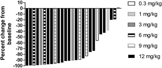
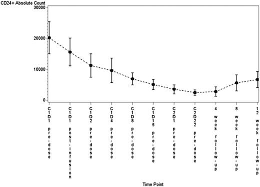
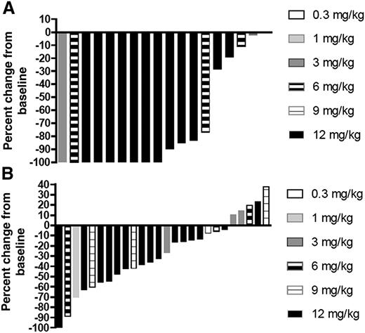
Twenty-seven patients were evaluable for response. Blood disease cleared in most patients, with a median reduction in absolute lymphocyte count from baseline of 90.8% (Figure 1) and a decrease in CLL cell count (Figure 2). On the basis of physical examination and laboratory studies alone, 18 patients (66.7%) achieved a partial response (PR), and the remaining 9 patients (33.3%) achieved stable disease. Best lymph node reduction for all patients is shown in Figure 3. Using CT criteria as well as examination and laboratory data, 8 patients (29.6%) achieved a PR, with an additional 16 patients (59.3%) achieving stable disease. Two patients had progressive disease by CT criteria. No patient dosed below 3 mg/kg had an objective response. Evaluating only the 16 patients at the 12 mg/kg dose level, which is the recommended phase 2 dose, 12 patients (75%) had a PR by physical examination criteria and 6 patients (37.5%) had a PR by CT criteria. Patients who responded by physical examination criteria tended to do so quickly, with 14/18 patients achieving a PR at the first response evaluation (cycle 2, day 1). CT responses lagged somewhat, with 3/8 patients achieving a PR at cycle 2, day 28; 3 at the 4-week follow-up time point; and 2 patients in the expansion cohort achieving a PR at cycle 5 and cycle 7, respectively.
Change in lymphocyte count from baseline. The lowest lymphocyte count for each patient during the observation period was compared with baseline.
Change in lymphocyte count from baseline. The lowest lymphocyte count for each patient during the observation period was compared with baseline.
Change in B-cell counts during therapy. B cells were characterized using CD5, CD19, CD24, CD43, and CD79b. After XmAb5574, CD19 was not detectable, so CD24 was primarily used to identify B cells. No patient had >1% normal B cells, so all CD24+ cells were determined to be CLL cells. Over the course of therapy, CLL cell count decreased significantly (P = .01), with lowest level seen at the end of therapy.
Change in B-cell counts during therapy. B cells were characterized using CD5, CD19, CD24, CD43, and CD79b. After XmAb5574, CD19 was not detectable, so CD24 was primarily used to identify B cells. No patient had >1% normal B cells, so all CD24+ cells were determined to be CLL cells. Over the course of therapy, CLL cell count decreased significantly (P = .01), with lowest level seen at the end of therapy.
Best change in sum of product of lymph nodes. Physical examination (A) or CT (B). Physical examination, including lymph node measurement, was performed at the time of each infusion and then at 4, 8, and 12 weeks postinfusion. CT scans were obtained on cycle 2, day 28.
Best change in sum of product of lymph nodes. Physical examination (A) or CT (B). Physical examination, including lymph node measurement, was performed at the time of each infusion and then at 4, 8, and 12 weeks postinfusion. CT scans were obtained on cycle 2, day 28.
When looking at baseline characteristics related to response by physical examination or CT criteria, baseline lymph node size did appear to be associated with response; patients with the largest lymph node ≤5 cm (n = 18) had a 77.8% PR rate by examination criteria and 38.9% by CT criteria and patients with the largest lymph node >5 cm (n = 9) had a 44.4% PR rate by examination and 11.1% by CT. Cytogenetic abnormalities by FISH, including del(17p13.1), did not appear to be associated with response, with 60% of patients with del(17p13.1) (6 of 10 patients) achieving a PR by examination criteria and 30% achieving a PR by CT criteria.
At evaluation 12 weeks after cycle 2, day 28, of XmAb5574, 5 patients (18.5%) had progressed by CT criteria and 8 (29.6%) by physical examination criteria. No patient died during the observation period. PFS was defined as the time of first dose to the time of progression or death, whichever came first. PFS for all patients, including those in the extended treatment cohort, was 199 days (Figure 4A; 95% confidence interval: 168-299 days). For all patients on all dose levels who received 9 doses or less, PFS was 189 days (Figure 4B), and for patients on the extended treatment cohort alone, PFS was 420 days (Figure 4C; 95% confidence interval: 168 days, not reached).
PFS. PFS by Kaplan-Meier method is shown for all patients (A), those who received up to 9 doses on all dose levels (B), and those who were included in the extended dosing cohort (C).
PFS. PFS by Kaplan-Meier method is shown for all patients (A), those who received up to 9 doses on all dose levels (B), and those who were included in the extended dosing cohort (C).
Effects on T cells, NK cells, and serum immunoglobulins
Flow cytometry was performed for absolute T (CD3+) and NK (CD56+/CD16+) cell counts as well as T-cell activation (CD3+/CD16−/CD56−/CD69+ cells) and NK cell degranulation (CD3−/CD16+/CD56+/CD63+ or CD3−/CD16+/CD56+/CD69+). Samples were collected on cycle 1, day 1, predose and end of infusion; cycle 1, days 2, 4, 8, and 15 predose; cycle 2, days 1 and 22 predose; and 4, 8, and 12 weeks after the infusions were complete. During this period, there was no significant change in T- or NK cell counts (supplemental Figure 2A-B; P = .45, P = .23, respectively); however, from cycle 1, day 1, predose to end of infusion, there was a significant decrease in both T-cell and NK cell numbers (supplemental Figure 2C-D; P < .0001, P < .0001, respectively). There was no change in T- or NK cell activation during the infusion period (data not shown).
Quantitative immunoglobulins were measured in all patients at baseline, end of cycle 2, and at 12-week follow up. There were no significant differences in immunoglobulin levels during this period (supplemental Figure 3; for baseline to 12-week follow-up: IgA, P = .10; IgG, P = .20; IgM, P = .09).
HAHA
Eight total patients of 27 evaluated tested positive for HAHA antibodies pretreatment. No patient had HAHA titer increase from the predose level, suggesting that this drug is not immunogenic. Individual patient titers can be found in supplemental Table 2. PK data from 1 patient on dose level 1 with positive HAHA showed an accelerated decline in the terminal portion of the concentration-time curve, suggesting that there may have been antibody-mediated drug clearance; however, this phenomenon was not seen in any other patients. Patients with HAHA did not appear to have increased likelihood of infusion reactions or a different response to therapy than patients without HAHA.
Pharmacokinetics
Of the 27 patients, PK parameters for 25 of these patients fit a 2-compartment model. Neither the patient enrolled at 0.3 or 1 mg/kg fit the expected PK model, and all PK data presented are from the 3 mg/kg cohort and above. Key PK parameters are summarized by cohort in Table 3. Clearance and volume of distribution are noted to be similar to other full-length monoclonal antibodies, and distribution was limited to the systemic circulation as evidenced by an estimate of volume of distribution. Cmax increased in a slightly less than dose-proportional manner, and AUC increased in a dose-proportional manner. Clearance and half-life showed no dose dependence. A trend of accumulation in concentration was observed with each infusion, and the serum concentration of XmAb5574 reached a plateau suggestive of steady-state at or before infusion 9. Across the dose range from 3 to 12 mg/kg, half-life averaged 13.5 days, supporting dosing intervals of 1 to 3 weeks.
Key pharmacokinetic parameters
| . | . | Cmax (ng/mL) . | AUC∞ (day × ng/mL) . | Clearance (mL/day/kg) . | V1 (mL/kg) . | V2 (mL/kg) . | Vss (mL/kg) . | α Half-life (day) . | β Half-life (day) . | K10 Half-life (day) . |
|---|---|---|---|---|---|---|---|---|---|---|
| Cohort 3 (3.0 mg/kg) | N | 3 | 3 | 3 | 3 | 3 | 3 | 3 | 3 | 3 |
| Mean | 43 309 | 465 218 | 6.542 | 67.25 | 67.18 | 134.4 | 0.6514 | 15.00 | 7.266 | |
| SD | 2,978 | 70 169 | 0.9330 | 3.552 | 46.10 | 42.55 | 0.4147 | 3.637 | 1.498 | |
| CV% | 6.9 | 15.1 | 14.3 | 5.3 | 68.6 | 31.7 | 63.7 | 24.2 | 20.6 | |
| Cohort 4 (6.0 mg/kg) | N | 3 | 3 | 3 | 3 | 3 | 3 | 3 | 3 | 3 |
| Mean | 102 363 | 880 104 | 6.99 | 56.45 | 30.74 | 87.19 | 0.7944 | 9.496 | 5.791 | |
| SD | 9,668 | 173 357 | 1.323 | 7.714 | 7.721 | 8.557 | 0.6252 | 3.169 | 1.595 | |
| CV% | 9.4 | 19.7 | 18.9 | 13.7 | 25.1 | 9.8 | 78.7 | 33.4 | 27.5 | |
| Cohort 5 (9.0 mg/kg) | N | 3 | 3 | 3 | 3 | 3 | 3 | 3 | 3 | 3 |
| Mean | 132 687 | 1 462 480 | 6.758 | 68.80 | 31.57 | 100.4 | 1.981 | 12.37 | 7.340 | |
| SD | 23 721 | 541 763 | 2.173 | 13.01 | 6.192 | 8.758 | 1.756 | 2.461 | 1.419 | |
| CV% | 17.9 | 37.0 | 32.2 | 18.9 | 19.6 | 8.7 | 88.6 | 19.9 | 19.3 | |
| Cohort 6 (12.0 mg/kg) | N | 16 | 16 | 16 | 16 | 16 | 16 | 16 | 16 | 16 |
| Mean | 169 279 | 1 791 460 | 7.192 | 71.43 | 58.56 | 130.0 | 0.9119 | 14.12 | 7.237 | |
| SD | 35.891 | 480.648 | 2.088 | 16.50 | 20.09 | 33.34 | 0.4420 | 4.691 | 2.134 | |
| CV% | 21.2 | 26.8 | 29.0 | 23.1 | 34.3 | 25.6 | 48.5 | 33.2 | 29.5 |
CV%, coefficient of variation percent; K10, first order elimination rate constant; V1, volume of compartment 1; V2, volume of compartment 2; Vss, volume of distribution at steady state.
Cmax and AUC∞ are model-predicted values following a single dose at the given dose level and are not observed Cmax and dose interval AUC.
Discussion
This trial represents the first human experience with the Fc-engineered CD19 monoclonal antibody XmAb5574. No maximum tolerated dose was reached in the dose levels examined, and the drug was well tolerated. Although infusion reactions were common, they were manageable in all cases with supportive care and generally did not recur during subsequent doses. Grade 3 and 4 toxicities were primarily hematologic and in most cases did not require discontinuation of therapy. Infectious toxicities were notably low, with febrile neutropenia occurring in only 1 patient. One patient did develop tumor lysis syndrome, which required rasburicase and intravenous fluids; however, subsequent infusions were tolerated without incident. It is notable that this patient had received no prior chemotherapy and had been treated only with single-agent CD20 antibody therapy before receiving XmAb5574. We also saw no significant decrease in immunoglobulin levels during the course of therapy; however, this will need to be further addressed in studies with a longer duration of exposure. Importantly, there is no evidence of immunogenicity with this antibody.
Although not the primary end point of this phase 1 trial, efficacy with this antibody is encouraging, with 67% of patients achieving a PR by clinical criteria and 30% by using IWCLL 2008 criteria. These response rates are impressive for a single-agent antibody and compare favorably with results for rituximab given on a weekly schedule25,26 as well as ofatumumab8 and obinutuzumab in the relapsed setting.27 Besides CD20, another target being explored is CD37, in which 1 monoclonal antibody (BI 836826) and 1 therapeutic protein (Tru-016) are under investigation; Tru-016 has shown an overall response rate of 20% in a phase 1 study.28 As well, several other CD19 antibodies are in clinical trials with varying modifications to enhance antibody activity. Responses to XmAb5574 were fairly durable as well, especially given the limited duration of exposure to antibody in most patients. Response duration was prolonged in those receiving maintenance therapy, which suggests that a longer duration of antibody administration is reasonable in future trials.
Although the preliminary efficacy of this antibody is encouraging, it is expected that the clinical utility of this antibody will not lie in its use as a single agent but rather in combination approaches. This has been validated using CD20 antibodies, where the addition of the CD20 antibodies rituximab and obinutuzumab has improved overall survival over fludarabine+cyclophosphamide7 or chlorambucil,29 respectively. Given the ubiquitous nature of CD19 and its strong expression, it is expected that this antibody will similarly improve outcomes when combined with active agents. Currently, the nature of CLL therapy appears to be evolving from chemotherapy-based combinations and toward combinations of targeted therapies with antibodies. Because of the dependence of XmAb5574 on ADCC, it is not expected that this agent would combine synergistically with ibrutinib,30 but it may be an appropriate combination partner with a Bruton’s tyrosine kinase inhibitor without inducible T-cell kinase inhibition or a phosphatidylinositol 3-kinase inhibitor. As well, in vitro studies suggest that the combination of XmAb5574 with lenalidomide may enhance NK cell ADCC; this combination is under clinical investigation.
In conclusion, this phase 1 trial demonstrates safety and preliminary efficacy of the novel Fc-engineered CD19 monoclonal antibody XmAb5574. These data justify movement into the phase 2 setting both alone and in combination, and validate a new target for CLL and other B-cell malignancies.
The online version of this article contains a data supplement.
The publication costs of this article were defrayed in part by page charge payment. Therefore, and solely to indicate this fact, this article is hereby marked “advertisement” in accordance with 18 USC section 1734.
Acknowledgments
This work was funded by Xencor, the Leukemia and Lymphoma Society, the D Warren Brown Foundation, the Conquer Cancer Foundation, and the National Cancer Institute (K23 CA178183, 5 P50-CA140158, and P01 CA95426).
Authorship
Contribution: J.A.W. and J.C.B. designed and performed research, analyzed data, and wrote the article; F.A., I.W.F., J.G.B., and G.L. designed and performed research; E.W. and S.M. performed research; Y.H. analyzed data; and P.A.F. designed and performed research and analyzed data.
Conflict-of-interest disclosure: P.A.F. is an employee of Xencor, Inc. The remaining authors declare no competing financial interests.
Correspondence: Jennifer A. Woyach, 455A OSU CCC, 410 West 12th Ave, Columbus, OH 43210; e-mail: jennifer.woyach@osumc.edu.


