Abstract
Acute promyelocytic leukemia (APL) is an AML subtype that is characterized by aberrant expansion of immature myeloid progenitors and precursors that are arrested at the promyelocyte stage. Almost all APL cases are characterized by the t(15;17)(q22;q11.2) translocation that creates the PML-RARA fusion oncogene. Human APL cells are known to have a canonical expression signature and a specific methylation phenotype that is unique to this form of AML. Our laboratory previously created a mouse model of APL by expressing a human PML-RARA cDNA from the mouse Cathepsin G (Ctsg) locus (Ctsg-PML-RARA), which activates human PML- RARA expression in early myeloid progenitor cells, with peak expression in promyelocytes. After a long latent period (6-12 months), ~60% of these mice develop a clonal, APL-like myeloid malignancy. The long latent period is probably due to the requirement for cooperating mutations that synergize with PML-RARA to accelerate the disease.
Human APL samples have a unique gene expression signature that distinguishes them from all other subtypes of AML. We evaluated RNA-Seq data derived from Poly A+ enriched cDNAs obtained from purified promyelocytes derived from 3 young (6 week old) WT and 3 Ctsg-PML-RARA mice. We identified 779 annotated genes that are significantly dysregulated in murine promyelocytes expressing PML-RARA with a log2 fold change >= 2 and P<0.05. Some of these genes included Spib/Pu.1, Pou2af1, Jak2, Runx1, and many others. We also identified a set of 24,018 RNAs in promyelocytes that were defined as novel transcripts. This set contains 7,413 lncRNAs with an FPKM value of >= 2. Differential expression analysis yielded 56 dysregulated lncRNA regions in PML-RARA expressing promyelocytes.
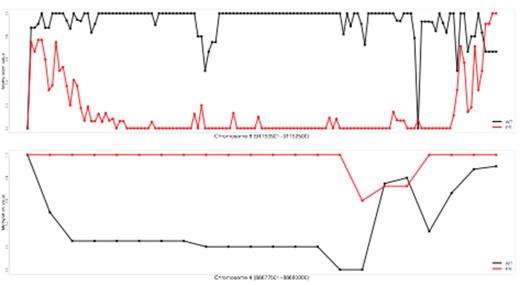
To explore the association between gene dysregulation and DNA methylation in promyelocytes, we carried out whole-genome bisulfite sequencing using DNA derived from the purified promyelocytes of a 6 week old Ctsg-PML-RARA mouse, and a WT littermate. We generated a total of approximately 800 million sequencing reads, of which 78% mapped uniquely to the reference genome (mm9); we were able to map ~19 million CpGs with at least 10x coverage. Differential methylation analysis performed on ~4.5 million 1 Kb windows spanning the entire genome identified 17,633 differentially methylated regions with a mean difference of >= 25% and a q-value of < 0.01, the vast majority of which (17,264, 98%) were hypomethylated in the Ctsg-PML-RARA promyelocytes. These windows overlap several known genes, including Runx1, Jak2, Dnmt3a, Gata2, and the Hoxa and Hoxb gene clusters. Using more strict criteria (> 50% mean methylation difference), we identified 87 differentially methylated regions of at least 2 Kb in size. Of these 87 distinct regions, 74 (85%) were hypomethylated in PML-RARA promyelocytes, and 13 were hypermethylated; examples of both as shown in Figure 1. These data strongly suggest that PML-RARA has at least two distinct mechanisms by which it can modify DNA methylation. In regions where CpGs are hypomethylated, PML-RARA may be blocking the normal methylation of CpGs by the de novo DNA methyltransferases Dnmt3a and/or Dnmt3b. In contrast, PML-RARA may be directing de novo methyltransferases to act on the hypermethylated regions. Regardless, these data, when coupled with comprehensive chromatin accessibility mapping and complete RNA sequencing data, should provide new insights into the mechanisms used by PML-RARA to alter gene expression and initiate APL.
Examples of differentially methylated regions. Black=WT cells. Red=PML-RARA expressing cells. Each CpG in the region is represented as a dot. Scale is 0-100% methylated at each position. Top panel: a region on chromosome 8 that is hypomethylated in PML-RARA expressing promyelocytes. Bottom panel: a region on chromosome 4 that is hypermethylated in PML-RARA expressing promyelocytes.
Examples of differentially methylated regions. Black=WT cells. Red=PML-RARA expressing cells. Each CpG in the region is represented as a dot. Scale is 0-100% methylated at each position. Top panel: a region on chromosome 8 that is hypomethylated in PML-RARA expressing promyelocytes. Bottom panel: a region on chromosome 4 that is hypermethylated in PML-RARA expressing promyelocytes.
No relevant conflicts of interest to declare.
Author notes
Asterisk with author names denotes non-ASH members.


This feature is available to Subscribers Only
Sign In or Create an Account Close Modal