Abstract
Daratumumab is a human antibody that binds to CD38 on the cell surface and induces cell killing by multiple mechanisms including complement mediated cytotoxicity (CDC), antibody-dependent cellular cytotoxicity (ADCC), antibody-dependent cell phagocytosis (ADCP) and apoptosis. In pre-clinical and clinical studies, daratumumab has been shown to effectively kill multiple myeloma (MM) cells and to enhance the potency of other treatments against MM. The purpose of the study was to investigate in vitro and in vivo efficacy of daratumumab against 9 acute myeloid leukemia (AML) cell lines and patient-derived samples.
First, we evaluated the expression of CD38, complement inhibitory proteins (CIP) CD46, CD55, CD59, and FcgR1 (CD64) on AML cell lines (n=9), AML patient cells (n=10) and healthy donor bone marrow using flow cytometry. CD38 enumeration showed a substantial variation between cell lines (12,827±19, 320 molecules/cell) and between AML patients (11,560±8, 175 molecules/cell), while CD38 expression was more consistent in bone marrow (BM) from healthy donors (1,176±355 molecules/cell). The daratumumab-induced apoptosis observed in cell lines (MOLM-13, MOLM-16, MV-4-11, NB4) in vitro was not correlated with CD38 expression levels. Daratumumab induced minimal ADCC (5-20%) and low levels of (2-5%) CDC mediated cell killing in 6 AML cell lines tested. We did not observe a direct correlation between CD38 expression and ADCC, CDC, nor between CDC and CIP expression. Interestingly, treatment of two human Acute Promyelocytic Leukemia (M3) cell lines HL-60 and NB-4 with all-trans retinoic acid (ATRA) induced a 10-30-fold increase in CD38 expression, suggesting that ATRA could be used in combination with daratumumab.
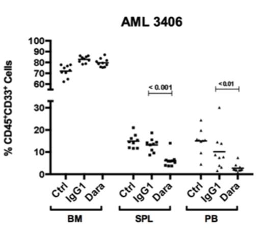
While we, and others, have shown that pre-incubation of primary AML cells with anti-CD38 antibodies inhibits engraftment in NSG mice, we aimed at evaluating the anti-leukemic activity of daratumumab in a therapeutic xenograft model using 3 different AML patients. NSG mice (10/group/patient) were transplanted with T cell-depleted AML cells and BM aspirates were collected 4-6 weeks later to assess leukemia burden in each mouse prior to treatment. Animals were untreated (Ctrl) or received daratumumab (10 mg/kg), or IgG1 isotype once a week for five weeks. We assessed AML burden (% huCD45+ CD33+) in BM, spleen (SPL) and peripheral blood (PB) within 5 days after the last treatment. First, we evaluated an AML (#3406, FLT3-ITD, see figure) with high expression of CD38 (13,445 molecules/cell) and low CD64 (489/cell) was evaluated. Daratumumab significantly reduced leukemia burden in SPL and PB, but had no effect in BM. The same daratumumab-induced reduction in peripheral blasts and lack of effect in BM was observed in 2 other AML patient xenografts (#7577, M1 IDH mutant/FLT3-ITD with 6,529 CD38 molecules/cell; #8096, M2 with 335 CD38 molecules/cell). Interestingly, we observed that daratumumab treatment led to a drastic reduction in CD38 surface expression in AML blasts including in BM, indicating that daratumumab efficiently targeted CD38 in bone marrow blasts. Our results suggest that the bone marrow microenvironment can impair the anti-leukemic activity of daratumumab observed in other tissues. Ongoing xenograft studies are testing whether induction with chemotherapy (Ara-C+doxorubicin), or with other agents disrupting the bone marrow microenvironment, can enhance the anti-leukemic activity of daratumumab.
Dos Santos:Janssen R&D: Research Funding. Xiaochuan:Janssen R&D: Research Funding. Doshi:Janssen R&D: Employment. Sasser:Janssen R&D: Employment. Danet-Desnoyers:Janssen R&D: Research Funding.
Author notes
Asterisk with author names denotes non-ASH members.


This feature is available to Subscribers Only
Sign In or Create an Account Close Modal