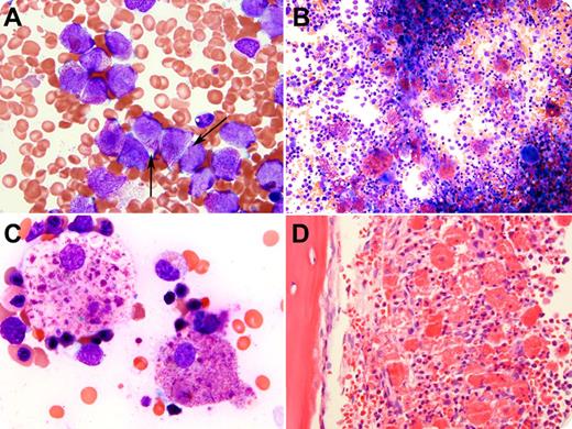
A 16-year-old Hispanic girl presented to our hospital after a 3-week history of bruising and gingival bleeding. She was found to be pancytopenic and diagnosed with acute promyelocytic leukemia (APL), with cytogenetics, fluorescence in situ hybridization, and reverse-transcriptase polymerase chain reaction confirming t(15;17)(q24;q21) and PML-RARA fusion. The initial bone marrow aspirate is depicted in the figure (panel A), with arrows indicating the cells with Auer rods. Chemotherapy with all-trans retinoic acid and arsenic trioxide was commenced and, before the end of induction, no abnormal promyelocytes or Auer rods within more mature cells in the neutrophil series could be identified in the peripheral blood. Bone marrow examination upon count recovery revealed hematologic remission. However, abundant phagocytic histiocytes were present, containing crystals (possibly representing Auer rods), granules, and globules, which appeared fuchsia on Wright-Giemsa stain (panels B-C), were eosinophilic with hematoxylin and eosin stain (panel D), and were strongly positive for CD68 and myeloperoxidase, the latter by enzyme cytochemistry and immunhistochemistry (not shown). There was no evidence of differentiation syndrome or hemophagocytic lymphohistiocytosis.
Although we did not directly observe phagocytosis of intact immature or differentiated APL cells (it is possible they underwent apoptosis before the second bone marrow procedure), these findings provide insight into a potential mechanism of clearance of neoplastic cells in APL by bone marrow histiocytes.
For additional images, visit the ASH IMAGE BANK, a reference and teaching tool that is continually updated with new atlas and case study images. For more information visit http://imagebank.hematology.org.


This feature is available to Subscribers Only
Sign In or Create an Account Close Modal