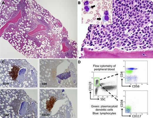
A 63-year-old man with a history of end-stage renal disease and hypertension underwent a bone marrow biopsy for leukocytosis (white blood cells 19.5 × 10⁹/L, monocytes 2.9 × 10⁹/L) and anemia (hemoglobin 96 g/L). The bone marrow was hypercellular with features of a myelodysplastic/myeloproliferative neoplasm consistent with chronic myelomonocytic leukemia (CMML). In addition, there were several focal paratrabecular infiltrates of mononuclear cells (panel A) that were small to medium-sized with round-to-oval nuclei and delicate chromatin patterns (panel B). The peripheral blood (panel B, inset arrows) and bone marrow aspirate contained a small percentage of similar cells with lightly basophilic cytoplasm, delicate chromatin, and occasional indistinct nucleoli. Immunohistochemistry showed that the paratrabecular infiltrates were positive for CD45, CD4 (dim), CD68, and CD123 (panel C), and were negative for CD34, CD117, myeloperoxidase, tryptase, CD3, CD5, CD7, CD20, PAX5, CD56, and CD79a. In addition, flow cytometric studies of the peripheral blood and bone marrow aspirate identified a small population of plasmacytoid dendritic cells that were positive for CD123 (panel D, gated), CD45 (dim), CD33, and CD4, and were negative for CD34, CD117, TdT, CD56, CD13, and CD11c.
Although plasmacytoid dendritic cell infiltrates in myeloid neoplasms, especially CMML, are well known, the bone marrow paratrabecular infiltration pattern similar to lymphoma and the documented peripheral blood involvement in this case are unusual.
For additional images, visit the ASH IMAGE BANK, a reference and teaching tool that is continually updated with new atlas and case study images. For more information visit http://imagebank.hematology.org.


This feature is available to Subscribers Only
Sign In or Create an Account Close Modal