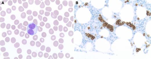
An asymptomatic 49-year-old female presented with lymphocytosis (11.1 × 10⁹/L lymphocytes) and thrombocytopenia (128 × 109/L platelets), with no additional cytopenias or physical findings. The blood film revealed lymphocytes of varying morphology, including unusual cells displaying bilobed nuclei (panel A). Bone marrow biopsy revealed mild interstitial B-cell lymphocytosis that included foci of intrasinusoidal localization highlighted by CD20 (panel B). Flow cytometry revealed a predominance of polyclonal B cells lacking additional antigenic aberrancies (peripheral blood and bone marrow).
A diagnosis was made of persistent polyclonal B-cell lymphocytosis (PPBL), a rare type of lymphocytosis. Associations include female gender, cigarette smoking, and polyclonal increase in immunoglobulin M (IgM) (both found in this case), as well as an HLA DRB1*07 haplotype (not assessed). There is potential for confusion with lymphoid neoplasia, given the presence of lymphocytosis, unusual lymphocyte morphology (bilobed lymphocytes characteristic), and splenomegaly (may be progressive). Bone marrow findings may also be misleading if they show an intrasinusoidal pattern of B-cell localization more commonly seen with a lymphoma such as splenic marginal zone lymphoma. Chromosomal abnormalities may be seen, including IgH-BCL2 rearrangement (not found in this case). However, B cells are polyclonal by flow cytometry, and clonal Ig gene rearrangements are not found in B cells. Growing experience with PPBL has not suggested an increased risk of evolution to lymphoma.
For additional images, visit the ASH IMAGE BANK, a reference and teaching tool that is continually updated with new atlas and case study images. For more information visit http://imagebank.hematology.org.


This feature is available to Subscribers Only
Sign In or Create an Account Close Modal