On page 1777 in the 1 March 2000 issue, there is an error in the Figure 2 legend. The legend omits the final sentence, which should read, “The mitochondrial swelling measurements were done concomitantly with 2 other drugs, namely C1 and C2, and therefore the upper 2 control panels are the same as in Figure 6A of Ref. 19.” On page 1777, Figure 3B includes incorrect flow histograms. On page 1778, Figure 5B includes an incorrect blot. The interpretation of the data and the message in the figures remain unchanged. The corrected Figures 3B and 5B are shown.
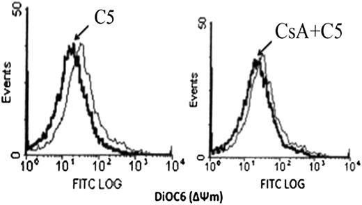
C5-triggered Cyt C release and drop in mitochondrial ΔΨmis dependent on opening of the MPT pore. (A) 0.5 mg purified rat liver mitochondria was exposed to 150 μg/mL C5 in the presence or absence of CsA (10 μmol/L) for 30 minutes at 30°C. Mitochondria were then pelleted, and supernatants were subjected to SDS-PAGE and western blot analysis for Cyt C, as described in Materials and methods. (B) Mitochondria were treated with C5 in the presence or absence of CsA as above and stained with membrane potential–sensitive dye DiOC6 (40 nmol/L) at 37°C for 30 minutes, washed, and analyzed by flow cytometry for ΔΨm.
C5-triggered Cyt C release and drop in mitochondrial ΔΨmis dependent on opening of the MPT pore. (A) 0.5 mg purified rat liver mitochondria was exposed to 150 μg/mL C5 in the presence or absence of CsA (10 μmol/L) for 30 minutes at 30°C. Mitochondria were then pelleted, and supernatants were subjected to SDS-PAGE and western blot analysis for Cyt C, as described in Materials and methods. (B) Mitochondria were treated with C5 in the presence or absence of CsA as above and stained with membrane potential–sensitive dye DiOC6 (40 nmol/L) at 37°C for 30 minutes, washed, and analyzed by flow cytometry for ΔΨm.
C5 does not activate caspase 8, 2, or 9. (A) HL60 cells were treated with 150 μg/mL C5 or 100 μg/mL C2 for 12 hours, and lysates were analyzed for caspase 8, 2, or 9 activation by fluorometric assays designed to detect cleavage of the AFC-conjugated specific substrate (IETD-AFC for caspase 8, VDVAD-AFC for caspase 2, and LEHD-AFC for caspase 9), as described in Materials and methods. Caspase activity is expressed as fold increase (x increase) over the activity obtained in untreated cells, shown here as 1 X. (B) Lysates obtained from C5- and C2-treated HL60 cells were also analyzed by SDS-PAGE and western blotting for cleavage of PARP (85-kd fragment) using a monoclonal anti-PARP antibody.
C5 does not activate caspase 8, 2, or 9. (A) HL60 cells were treated with 150 μg/mL C5 or 100 μg/mL C2 for 12 hours, and lysates were analyzed for caspase 8, 2, or 9 activation by fluorometric assays designed to detect cleavage of the AFC-conjugated specific substrate (IETD-AFC for caspase 8, VDVAD-AFC for caspase 2, and LEHD-AFC for caspase 9), as described in Materials and methods. Caspase activity is expressed as fold increase (x increase) over the activity obtained in untreated cells, shown here as 1 X. (B) Lysates obtained from C5- and C2-treated HL60 cells were also analyzed by SDS-PAGE and western blotting for cleavage of PARP (85-kd fragment) using a monoclonal anti-PARP antibody.


This feature is available to Subscribers Only
Sign In or Create an Account Close Modal