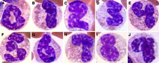
A 32-year-old male presented with a 1-month history of fever, weight loss, and worsening fatigue. Physical examination was remarkable for a temperature of 39.5°C and a massive splenomegaly. A complete blood count showed mild normochromic normocytic anemia and marked leukocytosis (∼90 000/μL), with basophilia (3500/μL), eosinophilia (5000/μL), and neutrophila/bandemia (80 000/μL). No increase in blast count was noted on peripheral blood or bone marrow examination. Cytogenetic analysis showed translocation t(9;22)(q34;q11), so-called Philadelphia chromosome, resulting in BCR-ABL1 fusion. The patient was diagnosed with chronic myeloid leukemia (CML) and began imatinib therapy.
The peripheral smear demonstrated unusually rich polynuclear segmentation generating a complete Arabic numeral list. Panels A through J represent the numbers 0 through 9. This segmentation is secondary to the accelerated myeloid cell production characteristic of CML and other myeloproliferative diseases.
For additional images, visit the ASH IMAGE BANK, a reference and teaching tool that is continually updated with new atlas and case study images. For more information visit http://imagebank.hematology.org.


This feature is available to Subscribers Only
Sign In or Create an Account Close Modal