In this issue of Blood, Sperandio and colleagues report the use of a unique intravital microscopic system to characterize an ontogenic process of blood cell and yolk sac endothelial maturation that is required to display full adult-type inflammation-induced leukocyte recruitment.1 They report that murine fetal blood neutrophil rolling, adhesion, and extravasation from inflamed yolk sac vessels is apparent late in development, but that before embryonic day (E) 15, fetal blood neutrophils display little ability to roll or adhere to inflamed vascular endothelial cells. Similar behavior was displayed when fetal blood cells were tested in vitro on immobilized recombinant adhesion molecules.
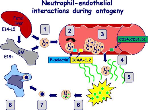
Simplified overview of proposed neutrophil–endothelial interactions during ontogeny. Before E14 to E15, hemogenic endothelial activity predominates, whereas after this time, HSC and progenitor activity concentrates extravascularly within the fetal liver, followed by seeding of bone marrow. (1) Neutrophil egress to the vasculature from the fetal liver (E14-E15) or the bone marrow (after E18). (2) Neutrophil rolling on activated endothelium occurs under conditions of low shear stress and is mediated by adhesive mechanisms such as neutrophil P-selectin glycoprotein ligand 1 interacting with activated endothelial P-selectin. (3) Firm arrest/adhesion involves interactions between activated neutrophils that express surface adhesion molecules, including Mac-1 and lymphocyte function-associated antigen 1, and cognate endothelial ligands such as the ICAMs. (4) Transendothelial migration/extravasation. Activated neutrophils migrate through endothelial gap junctions using adhesive interactions that include CD18 and CD31. (5) Chemotaxis. Activated neutrophils migrate toward inflammatory tissue in a directed process mediated by chemokines such as interleukin 8. (6) Phagocytosis and bacterial killing. Activated tissue neutrophils ingest pathogens and promote their intracellular demise. (7) Apoptosis. Tissue neutrophils undergo programmed cell death in preparation for their removal by the reticuloendothelial system by efferocytosis (8).
Simplified overview of proposed neutrophil–endothelial interactions during ontogeny. Before E14 to E15, hemogenic endothelial activity predominates, whereas after this time, HSC and progenitor activity concentrates extravascularly within the fetal liver, followed by seeding of bone marrow. (1) Neutrophil egress to the vasculature from the fetal liver (E14-E15) or the bone marrow (after E18). (2) Neutrophil rolling on activated endothelium occurs under conditions of low shear stress and is mediated by adhesive mechanisms such as neutrophil P-selectin glycoprotein ligand 1 interacting with activated endothelial P-selectin. (3) Firm arrest/adhesion involves interactions between activated neutrophils that express surface adhesion molecules, including Mac-1 and lymphocyte function-associated antigen 1, and cognate endothelial ligands such as the ICAMs. (4) Transendothelial migration/extravasation. Activated neutrophils migrate through endothelial gap junctions using adhesive interactions that include CD18 and CD31. (5) Chemotaxis. Activated neutrophils migrate toward inflammatory tissue in a directed process mediated by chemokines such as interleukin 8. (6) Phagocytosis and bacterial killing. Activated tissue neutrophils ingest pathogens and promote their intracellular demise. (7) Apoptosis. Tissue neutrophils undergo programmed cell death in preparation for their removal by the reticuloendothelial system by efferocytosis (8).
Intravital microscopy and in vitro flow chambers were used to characterize yolk sac–derived endothelium and enhanced green fluorescent protein (EGFP)+ neutrophils of LysEGFP+ mice from gestational ages E14 to E18. Neutrophil proportions were marginal (<2%) in nucleated fetal blood populations at E14, increasing daily to nearly 20% at E18, whereas the number of circulating progenitors decreased concomitantly. Functional in vivo studies showed absent rolling flux of neutrophils on yolk sac endothelium at E14, with chronologic increases through E18. In contrast, no rolling of EGFP− cells containing progenitor cell populations including hematopoietic stem cells (HSCs) was observed. Firm adhesion did not increase after superperfusion with inflammatory formyl peptide even at E18, although perivascular extravasation increased at later stages. In vitro studies confirmed a progressive increase in neutrophil adherence to immobilized ligands, although levels were still well below those of newborns or adults.
Studies to dissect developmentally impaired adherence mechanisms showed chronologic increases in the expression of P-selectin glycoprotein ligand 1 and CXCR2 (the interleukin 8 receptor that triggers firm arrest of neutrophils to endothelium after activation) in EGFP+ neutrophils when comparing E13 with E18 embryos. In contrast, expression of the CD18 molecule involved in firm endothelial adhesion (Mac-1), a cognate ligand for intercellular adhesion molecule (ICAM), was unchanged between E14 and E18. Injection of anti-P-selectin antibody abrogated neutrophil rolling in vivo, despite the detected absence of P- and E-selectin on yolk sac vessels. Although endothelial CD31, CD34, and β1 integrin were constitutively displayed in yolk sac vessels at all stages examined, ICAM-1 and ICAM-2 increased with development. Thus, murine embryonic and fetal neutrophils and yolk sac endothelium display a developmental sequence that permits significant proinflammatory leukocyte rolling, adhesion, and extravasation only late in gestation (see figure).
The murine extraembryonic yolk sac is a unique tissue that plays critical roles in early hematovascular development.2 In the adult mouse, the stem cell theory of hematopoiesis predicts that all lineages of mature blood cells are generated from oligo- or bipotent progenitor precursors that, in kind, are derivatives of the long-term repopulating HSCs. During murine embryogenesis, however, multiple waves of hematopoietic progenitors emerge in the yolk sac before adult repopulating HSC production from the aorto-gonad-mesonephros (AGM) region of the embryo proper. Not only do primitive erythroblasts, macrophages, and megakaryocytes uniquely arise as a first wave of blood cells, but adult-type erythro-myeloid progenitors also emerge from hemogenic endothelial cells as a second wave in the yolk sac.
Recently, autonomous concomitant emergence of B1- and T-cell progenitor cells has also been identified in the yolk sac and early AGM region from hemogenic endothelial cells.3 Although HSCs emerge at E10.5 in the AGM, HSCs are detectable in the yolk sac, liver, placenta, vitelline, and umbilical arterial vessels. Some estimates of completion of arterial–venous specification and the intact circuit of the systemic circulation occur as late as E10.5.
Despite apparent free circulation of erythrocytes after this stage, HSC and progenitor cells are differentially enriched in the yolk sac, AGM, placenta, and some embryonic blood vessels for several days, as these precursors associate as clusters of cells attached to the hemogenic endothelial cells from which they emerge. It is not completely clear when these sites of blood cell production decline; however, most estimates would predict that hemogenic endothelial activity declines before E15. At this time, most HSC and progenitor activity has become concentrated extravascularly within the fetal liver, and cells remobilize to enter the circulation to seed the nascent bone marrow medullary cavity. By E18.5, the bone marrow compartment possesses the majority of HSCs and clonogenic hematopoietic progenitor cells, and all other sites begin to decline.
In sum, the early yolk sac and embryonic vascular endothelial cells serve as an intravascular site of blood cell production, and numerous vessels are chimeric with nonhemogenic and hemogenic endothelial cells that are responsible for forming all of the elements of the hematopoietic system, from erythro-myeloid progenitors and lymphoid cells to HSCs. Given these specific developmental functions, one might anticipate that the properties of yolk sac endothelial cells would be dramatically different from the mature tissue–specific properties of arterial and venous endothelial cells of adult mice. This raises the question of when embryonic or fetal vascular endothelial cells begin to attract and retain activated leukocytes at sites of inflammation.
In contrast to murine ontogenic processes, hematopoiesis from hemogenic endothelium in humans is essentially complete by the end of the first trimester.4 However, the findings of Sperandio et al in their mouse system likely have clinical relevance, as the ontogenic changes in murine neutrophil rolling and adhesion were paralleled by this group’s observations of intrinsic differences of the same functions in human preterm neonates.5 The present report also confirms in vitro observations of impaired adhesive function in human newborns.6,7 Thus, intrinsic developmental alterations in neutrophil–endothelial interactions are likely to be critical factors underlying the greater incidence of sepsis in extremely preterm newborns.8
Chronologic maturation of fetal neutrophil–endothelial interactions may be of teleological importance in minimizing the activation of potentially injurious inflammatory mechanisms during normal parturition. Furthermore, evidence suggests that infected neonates may in fact be susceptible to hyperinflammatory responses that set the stage for chronic inflammatory disorders, particularly in extremely preterm human infants.9,10 Thus, continued characterization of fetal and neonatal immune mechanisms, particularly in the context of infection, will be critical to the development of modulatory therapeutic approaches for this fragile population.
Conflict-of-interest disclosure: The authors declare no competing financial interests.


This feature is available to Subscribers Only
Sign In or Create an Account Close Modal