Abstract
Abstract  3607
3607
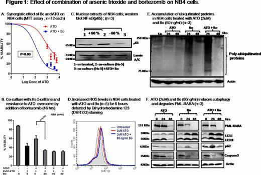
Bortezomib (Bo) has been shown to have direct cytotoxicity against malignant promyelocytes. There remain concerns about combining it with ATO. A major concern is that proteasomal inhibition by Bo could decrease ATO induced PML-RARA degradation and reduce its efficacy. In an in-vitro experiment we noted a significant synergistic cytotoxic effect when Bo (80ng/ml; a pharmacologically relevant concentration) was combined with ATO (1– 6uM) on NB4 cells (n = 12), P=0.03 (Fig 1a). We also noted that co-culture of NB4 or primary APL cells with stromal cells (Hs-5 cell line) protects them from ATO induced apoptosis and Bo can overcome this protective effect (Fig 1b). We undertook a series of experiments to address the mechanism of these observations.
We observed an increased activation of NF-κB pathway in malignant promyelocytes (NB4 cells) co-cultured with stromal cells. There was a synergistic inhibition of NF-κB when a combination of ATO and Bo was used (Fig 1c). Considering potent inhibition of NF-κB pathway and the known effect of this on reactive oxygen species (ROS), we checked the level of ROS in the malignant promyelocytes and observed that there was an increase in ROS levels when compared to ATO or Bo alone treated cells (n=5; treatment for 6 hours) (Fig 1d). When the ROS was abrogated by pre-treating the NB4 cells with N-Acetyl Cysteine (NAC; 5mM for 2 hrs) there was a protective effect against the ATO combined with Bo induced apoptosis at 24 hours (mean increase of 73% from 65.5%; p=0.03; n=4).
Degradation of PML-RARA was seen in ATO or Bo alone treated cells at 24 hours and a similar degradation was also seen when ATO and Bo were combined (Fig 1f; n=3). Bo at the concentration used (80ng/ml) was able to block 26s proteasome complex, as a result accumulation of ubiquitinated products was seen at 24 hours followed by degradation at the end of 48 hours (Fig 1e). We then looked at autophagy as a possible alternate mechanism to explain PML-RARA degradation when a combination of ATO and Bo was used in the setting of effective proteasomal inhibition. At 24 hours, there was a evidence of induction in autophagy as shown by LC3II formation using western blot technique and this was maximum at the end of 48 hours, this time point coincides with time at which maximum PML-RARA degradation happens in this experimental setting (Fig 1f). We have also observed that there is an accumulation of p62 (ubiquitinated protein binding protein) at 24 hours and degraded by 48 hours suggests that accumulated ubiquitinated products were cleared by autophagy via p62 (Fig 1f). The induction of autophagy was further validated by real time-PCR where autophagy genes atg5 and beclin1 levels were significantly increased by 3.1 fold (p=0.01) and 2.1 fold (p=0.03) respectively. Blocking autophagy by 3-methyl adenine or chloroquine did not enhance survival but paradoxically appeared to further enhance cell death (n= 3; p=0.02) though there was partial inhibition in the degradation of PML-RARA (data not shown).
We have also observed that there was a synergistic and more rapid increase in apoptosis when ATO and Bo were combined as evidenced by an enhanced degradation of caspase3 (Fig 1f).
Preliminary phase I data in 4 patients is summarized in table 1. Patients received Bo in induction and in consolidation once a week for 4 weeks along with ATO. Two cases, due to financial constraints, could not have a stem cell transplant (SCT) in molecular remission. In summary the combination of these drugs was well tolerated and durable CR3 and CR2 was obtained even in cases that did not have a SCT in remission. None of these cases have relapsed at a median follow up of 14.5 months.
In conclusion blocking 26s proteasomal complex by Bo does not alter the efficacy of ATO, instead Bo synergizes with ATO. The mechanism of this synergy is multi-factorial and appears to be predominantly due to increase in ROS activity and increased apoptosis. Additionally stromal cell mediated protection against ATO induced apoptosis is overcome by addition of Bo. Observed degradation of PML-RARA by ATO+Bo could be explained by induction of autophagy in these cells.
Summary of cases and duration of CR prior and post remission induction with arsenic trioxide and bortezomib
| Case . | Age . | Sex . | Relapse number . | Duration of last CR (months) . | Prior autologous SCT . | Post remission SCT . | Duration of current CR (months) . |
|---|---|---|---|---|---|---|---|
| RS | 25 | M | R2 | 19 | Yes | No | 19 |
| BJ | 31 | M | R1 | 15 | No | Yes (auto) | 15 |
| TK | 35 | M | R2 | 24 | Yes | Yes (MUD) | 14 |
| SS | 34 | F | R1 | 19 | No | No | 13 |
No relevant conflicts of interest to declare.
Author notes
Asterisk with author names denotes non-ASH members.

This icon denotes a clinically relevant abstract


This feature is available to Subscribers Only
Sign In or Create an Account Close Modal