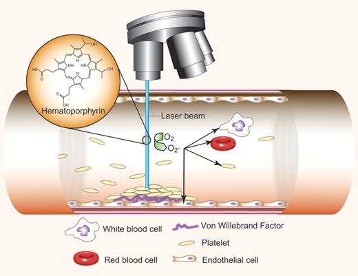
In this issue of Blood, Nishimura and colleagues present a novel in vivo murine model of thrombosis triggered on the undisrupted, activated endothelium in response to reactive oxygen species (ROS)–producing free radicals derived from oxygen (namely O2̄), which can be present in the vasculature as a result of inflammation1 (see figure). This new in vivo model involving a novel injury, minimally invasive vessel preparation, and advanced data collection may be particularly useful in dissecting the role of inflammation in thrombosis.
Study of details of thrombus formation at the single-cell level. Irradiation of hematoporphyrin with a 488-nm laser beam results in production of reactive oxygen species (ROS), mainly O2−. Free radicals activate all cells present in the irradiated field—white blood cells, red blood cells, and platelets, but maily endothelial cells. Activated endothelial cells release von Willebrand factor (VWF), regulated through inflammatory cytokine signaling. Discoid platelets adhere to the activated endothelium through GP1bα-VWF interaction and form a platelet-rich, occlusive thrombus. Single-cell dynamics and thrombus formation details are visualized during laser excitation and ROS production. Professional illustration by Paulette Dennis.
Study of details of thrombus formation at the single-cell level. Irradiation of hematoporphyrin with a 488-nm laser beam results in production of reactive oxygen species (ROS), mainly O2−. Free radicals activate all cells present in the irradiated field—white blood cells, red blood cells, and platelets, but maily endothelial cells. Activated endothelial cells release von Willebrand factor (VWF), regulated through inflammatory cytokine signaling. Discoid platelets adhere to the activated endothelium through GP1bα-VWF interaction and form a platelet-rich, occlusive thrombus. Single-cell dynamics and thrombus formation details are visualized during laser excitation and ROS production. Professional illustration by Paulette Dennis.
Experimental models of thrombosis that mimic human vascular disease are essential to unravel the complex factors involved in pathologic thrombus formation, but all have advantages and limitations.2 Most of these models are based on initiating thrombosis on artificially disrupted endothelium, exposing the prothrombotic, subendothelial components, including tissue factor and collagen, in different vascular beds. Certainly, thrombosis frequently occurs on such injured vessels after trauma and on ruptured atherosclerotic plaque, involving collagen and tissue factor exposure. Nevertheless, platelets adhere to activated endothelium, including the endothelium overlying pro-atherogenic lesions, and actively contribute to the development of vascular disease.3 Oxidative stress can cause this endothelial dysfunction, supporting proinflammatory, prothrombotic, proliferative, and vasoconstrictor processes. The molecular mechanism(s) underlying the consequences of oxidative stress and accompanying endothelial injury are still being elucidated, and the system presented here offers a new in vivo model to study this process.
There are two classic in vivo injury models involving oxidative stress with the generation of free radicals. FeCl3-induced outside-in injury is the most commonly used model to study factors involved in thrombus growth, stabilization, and lysis, but the exact underlying injury mechanism and physiologic consequences of externally applied FeCl3 are unclear. Recently it was shown that FeCl3-mediated vascular injury may be dependent on erythrocyte hemolysis and hemoglobin oxidation4 or may involve formation of spherical bodies on the endothelial cells that are filled with FeCl3 and expose large amounts of tissue factor.5 In the rose bengal photochemical injury model, thrombosis is triggered on endothelium irradiated by green light after intravenous injection of the dye.2 While it is believed that the damage is caused by ROS generated at the site of rose bengal excitation, the precise mechanism is not known. Rose bengal presumably accumulates in the membranes of endothelial and other cells. Electron microscopy studies showed that after this photochemical reaction, endothelial cells first contract and then detach from the vessel wall, with their cell membrane being destroyed,6 but the contribution of activation of circulating blood cells has not been studied. The extent of the endothelial cell injury is dependent on the intensity and length of irradiation, the dose of dye, and the type of the vessel studied.
In the model presented by Nishimura et al, free radicals, mainly O2̄, are derived from hematoporphyrin excited by low-power blue 488nm laser.1 This experimental system mimics more closely the involvement of free radicals in the inflammation that is at the root of cardiovascular disease. Furthermore, the surgery employed by these authors to expose the vessels studied causes minimal activation of the clotting cascade, in contrast to most other commonly used murine models. In this case, thrombosis occurs on undisrupted, activated endothelium. Like the rose bengal injury, not only endothelial cells, but all cells present in the imaging field, including platelets and leucocytes, are activated, so whether this hematoporphyrin-activation system provides an alternative outcome to the rose bengal system will need to be tested in the future.
What can be learned with such a system? Nishimura et al show that endothelial cells are stimulated to release von Willebrand factor (VWF) and to express P-selectin without affecting E-selectin expression, while platelets also express P-selectin and up-regulate αIIβ3 integrin. The key components in this model appear to be the VWF mobilization from endothelial cells modulated by the inflammatory cytokines TNF-α and IL-1. Initial platelet adhesion is mediated through GPIbα interaction, talin-dependent αIIβ3 activation, and partially through P-selectin interaction. Subsequently, the thrombus is stabilized by integrin activation and linkage to the actin cytoskeleton regulated by Rac1. Most of these steps have been described before as potentially contributing to a growing thrombus, although these studies describe their importance and sequence during in situ thrombosis as well as the role played by the inflammatory cytokines.
Likewise, it is not a radical finding in this radical system that thrombi were composed mainly of discoid platelets, because the translocation of discoid platelets as a first step of thrombus formation in vivo has been thoroughly described.7 Nevertheless, while work of Jackson's group focuses on the rheology of blood at the site of atherosclerotic plaque, this model emphasizes the single-platelet kinetics of adhesion caused by oxidative stress in the early stages of vascular disease using a state-of-the-art imaging system.
The intact, unactivated endothelium normally prevents platelet adhesion and pathologic thrombus generation. This paper by Nishimura et al presents a model system in which endothelial denudation is not an absolute prerequisite to allow platelet attachment to the arterial wall and to initiate early events of vascular disease before atherosclerotic plaque formation. This model has the potential for further studies of the important link between endothelial cell activation, inflammation, and thrombosis in vivo. Employment of such an advanced imaging technique in conjunction with transgenic mice will enable quantitative analysis of the kinetics of single platelets and other vascular and circulating cells in various-sized vessels and simultaneous monitoring of different components of thrombus formation in vivo under conditions of potential physiologic relevance in inflammation.
Conflict-of-interest disclosure: The author declares no competing financial interests. ■
REFERENCES
National Institutes of Health


This feature is available to Subscribers Only
Sign In or Create an Account Close Modal