Abstract
Alterations of the BM microenvironment have been shown to occur after chemoradiotherapy, during aging, and after genetic manipulations of telomere length. Nevertheless, whether BM stromal cells adopt senescent features in response to these events is unknown. In the present study, we provide evidence that exposure to ionizing radiation (IR) leads murine stromal BM cells to express senescence markers, namely senescence-associated β-galactosidase and increased p16INK4a/p19ARF expression. Long (8 weeks) after exposure of mice to IR, we observed a reduction in the number of stromal cells derived from BM aspirates, an effect that we found to be absent in irradiated Ink4a/arf-knockout mice and to be mostly independent of the CFU potential of the stroma. Such a reduction in the number of BM stromal cells was specific, because stromal cells isolated from collagenase-treated bones were not reduced after IR. Surprisingly, we found that exposure to IR leads to a cellular nonautonomous and Ink4a/arf-dependent effect on lymphopoiesis. Overall, our results reveal the distinct sensitivity of BM stromal cell populations to IR and suggest that long-term residual damage to the BM microenvironment can influence hematopoiesis in an Ink4a/arf-dependent manner.
Introduction
Exposure to ionizing radiation (IR) or high-dose chemotherapy leads to long-term residual damage of hematopoietic stem cells (HSCs).1-3 Not surprisingly, IR also induces damage to the BM microenvironment, reducing the CFU capacity of stromal cells by as much as 90%.4-6 A reduction in the number of mitotically competent stromal progenitor cells is likely permanent, because CFU capacity does not fully recover for at least several months/years after transplantation.4,5,7 It is not known whether irradiated BM stromal cells are eliminated and then only partly regenerated or whether they adopt a senescent phenotype within the BM niche. Indeed, exposure to DNA-damaging agents has been shown to induce senescence of several cell types both in vitro and in vivo.3,8-10 Hematopoietic progenitors isolated from irradiated mice have also been shown to express senescence markers such as 16INK4a.3 Therefore, we hypothesized that BM stromal cells could adopt senescent features and lose their regenerative potential in a p16INK4a-dependent manner after exposure to IR.
Various stimuli have the potential to induce senescence by inducing DNA damage.8,11-13 Common features of the senescent phenotype include permanent loss of proliferation potential, and in most cases, the expression of senescence-associated β-galactosidase (SA–β-gal).14,15 p16INK4a is an important regulator of senescence and arguably represents the most robust marker for in vivo detection of senescent cells.16-19
p16INK4a and p19ARF are 2 distinct tumor suppressor genes that originate from the Ink4a/arf locus (also called CDKN2a/b). p16INK4a is a cyclin-dependent kinase inhibitor the expression of which increases with age. By inhibiting cyclin-dependent kinase 4, it ultimately prevents the phosphorylation of Rb.20 Through its ability to inhibit the MDM2 protein, p19ARF acts as an activator of p53.21 Therefore, both proteins act as direct effectors of cellular senescence by preventing cell-cycle progression.
There are numerous studies supporting the importance of the stromal microenvironment in the success of BM transplantation. For example, old mice were shown to be deficient in supporting engraftment of HSCs compared with younger animals, suggesting an important role for the BM microenvironment in the engraftment process.22 Similarly, genetically induced telomere dysfunction leading to microenvironmental alterations of the BM niche was also shown to limit the function and engraftment of HSCs.23 Likewise, the BM stromal niche from Fancg−/− mice that are impaired in their ability to support adhesion and engraftment of hematopoietic progenitors can be rescued by the injection of stromal cells isolated from wild-type mice.24 These examples strongly point to a very important role for the BM stroma in hematopoiesis.
In the present study, we aimed to determine whether exposure to IR induces senescent features within the BM stromal compartment and whether this could affect its functions as a niche. Our results provide the first evidence for a cellular nonautonomous role of Ink4a/arf in regulating lymphopoiesis and reveal a distinct sensitivity of stromal cell populations, either BM- or bone-derived, to IR-induced long-term residual damage.
Methods
Mice
Eight- to 12-week-old female C57BL/6J or B6.SJL-PtrcaPep3b/BoyJ mice were purchased from The Jackson Laboratory. Ink4a/arf-null mice on a C57BL/6J background were bred on site under a material transfer agreement from the National Cancer Institute Mouse Models of Human Cancers Consortium (strain code 01XB1). All mice were allowed to acclimate at least 1 week before their use for experimentation. All in vivo manipulations were approved by the Comité Institutionnel des Bonnes Pratiques Animales en Recherche of Centre Hospitalier Universitaire Ste-Justine (protocol number S10-32).
Isolation, purification, and characterization of stromal cells
BM was collected by flushing tibias, femurs, and iliacs from C57BL/6J mice in PBS. Cleaned bones were next cut in small fragments of 1-2 mm and digested for 45 minutes with 0.25% collagenase type 1 (Sigma-Aldrich). For in vitro experiments, BM-derived stromal cells (BM-SCs) and bone-derived osteoblast-like stromal cells (OB-SCs), derived from BM or bone chips respectively, were isolated based on their property to attach to polystyrene after culture for 7 days in α-MEM containing 10% FBS and 1% penicillin/streptomycin. Stromal populations were then expanded for 2-4 passages (corresponding to 4-12 population doublings) in 3% oxygen and their phenotype confirmed by flow cytometry using the following Abs: Ter119, CD31, CD45, CD44, CD90, and CD105 (all from BioLegend). Otherwise, stromal cell populations were isolated using the EasySep negative selection kit for mesenchymal progenitors (StemCell Technologies) or sorted by flow cytometry using the Abs listed in the previous sentence at a purity of 80%-90% for BM-SCs and 99% for OB-SCs. Adipocytic and osteogenic differentiation potentials of stromal cell populations were determined as described previously.25
Cell proliferation, SA-β-gal detection, and BrdU incorporation
To assess cell proliferation, stromal cells were plated at a density of 7000 cells/cm2 and population doubling was evaluated after each cell passage (every 3-4 days). Where indicated, trypsinized cells were induced to senesce by exposure to 10 Gy of irradiation (using a Gammacell 220 and cobalt-60 as a source) without any interference in their ability to re-adhere to the culture dish. SA–β-gal detection was performed in vitro as described previously by Dimri et al.15 Briefly, 10 days after exposure or not to 10 Gy of irradiation, cells were fixed and stained at pH 5.0 for 16 hours at 37°C. Cell cycle progression into the S-phase, as determined by bromodeoxyuridine (BrdU) incorporation, was assessed by pulsing cells over a 3-day period ending on day 10 after IR with 10 μM BrdU (Roche) and by subsequent immunodetection using an Ab specific for BrdU (mouse mAb conjugated to FITC, clone BMC 9318; Roche). The proportion of BrdU+ cells to the total number of cells was determined by counting cells manually in random fields seen under the microscope (Zeiss Axiovert 35M and Olympus Q color 3 camera).
RNA isolation and real-time quantitative PCR
RNA was extracted from stromal cell populations using the RNeasy Mini or Micro Kit (QIAGEN). RNA was reverse-transcribed using the QuantiTect Reverse Transcription Kit (QIAGEN). Quantitative differences in gene expression were determined by real-time quantitative PCR using SensiMix SYBR Low-ROX (Quantace) and MxPro QPCR Version 4.01 software (Stratagene). Primers used for the amplification of p16INK4a, p19ARF, runx2, sp7, opn, bglap, ibsp and 18S RNA are available in Table 1. Values are presented as the ratio of target mRNA to 18S rRNA obtained using the relative standard curve method of calculation.
Quantitative PCR primers
| Gene . | Primers . | Accession no. . |
|---|---|---|
| p16INK4a | Forward: AACTCTTTCGGTCGTACCCC | AF332190 |
| Reverse: GCGTGCTTGAGCTGAAGCTA | ||
| p19ARF | Forward: GCCGCACCGGAATCCT | L76092 |
| Reverse: TTGAGCAGAAGAGCTGCTACGT | ||
| runx2 | Forward: GCCGGGAATGATGAGAACTA | NM_009820.2 |
| Reverse: GGACCGTCCACTGTCACTTT | ||
| sp7 | Forward: TCTCCATCTGCCTGACTCCT | NM_130458.2 |
| Reverse: AGCGTATGGCTTCTTTGTGC | ||
| opn | Forward: TCAGCTGGATGAACCAAGTCTGGA | BC002113 |
| Reverse: ACTAGCTTGTCCTTGTGGCTGTGA | ||
| bglap | Forward: AAGCAGGAGGGCAATAAGGT | NM_007541.2 |
| Reverse: TGCCAGAGTTTGGCTTTAGG | ||
| ibsp | Forward: AAAGTGAAGGAAAGCGACGA | NM_008318.3 |
| Reverse: GTTCCTTCTGCACCTGCTTC | ||
| 18S | Forward: TCAACTTTCGATGGTAGTCGCCGT | X00686 |
| Reverse: TCCTTGGATGTGGTAGCCGTTTCT |
CFUs
CFUs were determined by plating 2-5 × 10⁶ cells collected from BM aspirate or 2-5 × 10⁵ cells from collagenase-treated bones in 6-well dishes and allowing them to grow in α-MEM containing 10% FBS and 1% penicillin/streptomycin in a humidified atmosphere with 6% CO2 and 3% O2 at 37°C. The medium was changed 7 days after culture was initiated, and cells were fixed with 0.5% methylene blue in 50% methanol on day 14. Cell clusters visible to the naked eye (corresponding approximately to at least 50 stromal cells) were scored as a CFU colony. Femoral BM aspirates were processed independently and analyzed as replicates (n = 2) for each mouse, whereas cleaned bones from a single mouse were pooled together before collagenase treatment.
Homing
Wild-type and Ink4a/arf-deficient mice were exposed or not to a unique sublethal dose of 6 Gy of total body irradiation. Eight weeks after exposure to IR, 5 × 10⁶ nucleated BM cells stained according to the manufacturer's instructions with 0.5μM CFSE (Invitrogen) were injected into the tail veins without further irradiation. Sixteen hours after injection, recipient mice were killed and femoral BM was harvested for flow cytometric analysis of the absolute number of CFSE+ cells per femur using CountBright absolute counting beads (Invitrogen).
Hematopoietic reconstitution assays
For long-term hematopoietic reconstitution, 2 month-old Ly 5.2/CD45.2 wild-type mice were first exposed or not to 6 Gy of total body irradiation and allowed to recover for up to 11 months without BM transplantation. The proportions of B cells (B220-FITC, clone RA3-6B2; BioLegend) and myeloid cells (CD11b-APC, clone M1/70; BioLegend) in blood were evaluated 3 and 11 months after irradiation. Mice were then killed and 2 × 10⁶ unfractionated BM cells collected from wild-type mice exposed or not to IR 11 months previously were transplanted into Ly 5.1/CD45.1 lethally irradiated (10 Gy) recipient mice. The proportions of donor-derived B cells and myeloid cells in blood were determined in blood in recipient mice 3 months after transplantation. For short-term engraftment assays, 2 × 10⁶ unfractionated BM cells obtained from Ly 5.1/CD45.1 wild-type donor mice were injected into the tail veins of Ly 5.2/CD45.2 wild-type or ink4a/arf-deficient recipient mice exposed or not to 6 Gy of total body irradiation 8 weeks before transplantation. Mice were killed 2 weeks after transplantation, and the blood, BM, and spleens were analyzed by flow cytometry using the following Abs: B220-biotinylated, B220–Pe-Cy5, or B220-FITC (clone RA3-6B2); CD3-biotinylated or CD3-FITC (clone 145-2C11); CD11b-biotinylated, CD11b-APC, or CD11b-FITC (clone M1/70); Sca-1–PE-Cy7 (clone D7); CD19-PerCP-Cy5.5 (clone eBio1D3); and IL-7R–Pe-Cy5 (clone B12-1; all from BD Biosciences); Thy1.2-FITC (clone 30-H12), FcϵRIα-biotinylated or FcϵRIα-FITC (clone MAR-1), Gr-1-biotinylated, Gr-1–Pe-Cy7, or Gr-1–FITC (clone RB6-8C5); Ter119-biotinylated or Ter119-FITC (clone TER-119); c-Kit–PE-Cy5.5 or c-Kit–APC (clone 2B8); CD48-APC (clone HM48-1); streptavidin-eFluor710 (49-4317-82; all from eBioscience); and CD150–PE-Cy5 (clone TC15-12F12.2) and CD45.1-PE (clone a20; both from BioLegend).
Statistical analysis
Statistical analyses were performed with the Prism Version 5.0 software (GraphPad) using the unpaired Student t test. Results were considered statistically significant with P values < .05.
Results
Exposure to IR induces p16INK4a/p19ARF expression in distinct BM stromal cell populations
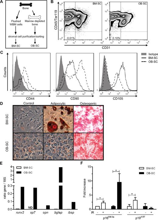
Previous studies have suggested that murine BM stromal cells are not eliminated after exposure to IR,6,7 making it conceivable that these cells adopt a senescent phenotype in response to DNA damage. To verify this hypothesis, we subjected C57BL/6J mice to total body irradiation at the sublethal dose of 6 Gy and analyzed their BM stromal cells for the induction of the senescence markers p16INK4a and p19ARF. We chose this dose because it was shown previously to induce senescence markers in other mouse tissues and because it does not require BM transplantation for 100% of the mice to survive the irradiation.26 Using a 2-step extraction procedure (Figure 1A), we purified stromal cell populations from mice femurs and measured p16INK4a and p19ARF expression levels. The BM was first flushed from bones and stromal cells within this fraction were identified as BM-SCs. Flushed bones were then treated with collagenase and stromal cells in this fraction were referred to as OB-SCs. In both cases, stromal cell populations were negative for CD45, Ter119, CD31, and 7-amino-actinomycin D and purified either by magnetic columns or flow cytometric cell sorting. Phenotypical, molecular, and functional characterization of these stromal cells confirmed the purification of distinct populations. Indeed, phenotypic characterization revealed that BM-SCs were CD90dim and CD105bright, whereas OB-SCs contained a mixture of cells that were either CD90dim or bright and CD105negative or dim (Figure 1C). Moreover, these populations were functionally different, because only BM-SCs were able to differentiate into adipocytes after adipocytic induction (Figure 1D). Finally, only OB-SCs expressed relatively high mRNA levels of runx2 and sp7 (encoding transcription factors) and of opn, bglap, and ibsp (encoding bone extracellular matrix proteins), which are all associated with the osteoblastic phenotype. In comparison, BM-SCs did not or only weakly express these genes (Figure 1E).
IR-induced p16INK4a/p19ARF expression is dependent on BM stromal cell populations. (A) Scheme representing the strategy used for the isolation of BM-SCs and OB-SCs. (B) Representative flow cytometry profiles of BM-SCs and OB-SCs identified as Ter119− (erythroid lineage), CD45− (hematopoietic lineage), and CD31− (endothelial lineage) isolated from a nonirradiated mouse are shown. (C) Representative flow cytometry profiles of CD44, CD90, and CD105 revealing distinct expression levels in BM-SC and OB-SC populations. (D) Osteogenic and adipocytic differentiation potential of BM-SC and OB-SC populations. (E) Differential mRNA expression levels in BM-SCs (□) and OB-SCs (■) of 5 osteoblast-associated genes normalized to 18S (runx2, sp7, opn, bglap, and ibsp). Each sample is a pool of 2 mice. (F) Quantitative PCR performed on mRNA extracted from purified/sorted BM-SCs and OB-SCs of 8-week-irradiated (+) or not (−) mice expressed as the fold increase in p16INK4a and p19ARF normalized to 18S. Up to 6 independent stromal cell purifications were performed using a minimum of 2-3 mice per group. *P < .05 by Student t test.
IR-induced p16INK4a/p19ARF expression is dependent on BM stromal cell populations. (A) Scheme representing the strategy used for the isolation of BM-SCs and OB-SCs. (B) Representative flow cytometry profiles of BM-SCs and OB-SCs identified as Ter119− (erythroid lineage), CD45− (hematopoietic lineage), and CD31− (endothelial lineage) isolated from a nonirradiated mouse are shown. (C) Representative flow cytometry profiles of CD44, CD90, and CD105 revealing distinct expression levels in BM-SC and OB-SC populations. (D) Osteogenic and adipocytic differentiation potential of BM-SC and OB-SC populations. (E) Differential mRNA expression levels in BM-SCs (□) and OB-SCs (■) of 5 osteoblast-associated genes normalized to 18S (runx2, sp7, opn, bglap, and ibsp). Each sample is a pool of 2 mice. (F) Quantitative PCR performed on mRNA extracted from purified/sorted BM-SCs and OB-SCs of 8-week-irradiated (+) or not (−) mice expressed as the fold increase in p16INK4a and p19ARF normalized to 18S. Up to 6 independent stromal cell purifications were performed using a minimum of 2-3 mice per group. *P < .05 by Student t test.
In contrast, p16INK4a expression levels were found to be increased in both populations after exposure to IR, although at a much higher level in OB-SCs compared with BM-SCs (9.5- vs 2.5-fold, respectively). However, only BM-SCs had a significant 4-fold increase in p19ARF levels (Figure 1F). Intriguingly, the expression of p16INK4a and p19ARF was not increased immediately after IR (data not shown), only 6-8 weeks later, a phenotype reminiscent of what we observed previously in other irradiated mouse tissues.13,26 These results demonstrate that stromal cell populations derived from the BM microenvironment activate senescence markers after IR-induced DNA damage in vivo.
IR-induced senescence of BM stromal cells is Ink4a/arf- dependent
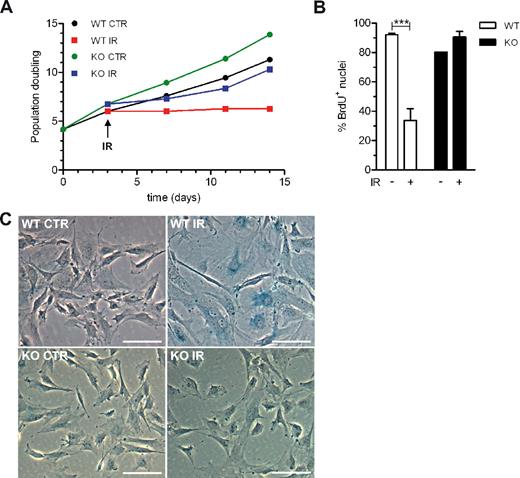
p16INK4a and p19ARF are well-established senescence markers.27 However, because their expression is delayed after damage, it is unclear whether their expression is causative of the senescence phenotype, and therefore if inhibition of their expression would be sufficient to prevent IR-induced senescence. To address these questions, we purified stromal cell populations from wild-type and Ink4a/arf-knockout mice and evaluated their susceptibility to undergo cellular senescence after exposure to IR in vitro. We observed a striking difference in the growth rate of wild-type versus Ink4a/arf-null OB-SCs under steady-state conditions and after exposure to IR. In fact, whereas cells derived from Ink4a/arf-null mice resumed proliferation after a short (approximately 5-7 days) interruption after exposure to IR, cells isolated from wild-type mice did not (Figure 2A). Such transient inhibition in the growth rate of Ink4a/arf-deficient stromal cells immediately after IR is not surprising, because these cells have intact and presumably functional DNA damage checkpoints.
Ink4a/arf is necessary to maintain IR-induced senescence of BM stromal cells. (A) OB-SC populations derived from wild-type (WT) or Ink4a/arf-deficient (KO) mice were established in vitro and their proliferation after exposure (IR) or not (CTR) to 10 Gy of IR is shown. A representative experiment (n = 3 independent stromal cell isolations) of cumulative population doubling determined over time is shown. (B) Proportion of WT or ink4a/arf-null OB-SCs in culture that have incorporated BrdU (3-day pulse) 7 days after exposure or not to IR. Up to 16 independent stromal cell isolations were performed. ***P < .001 by Student t test. (C) Representative photographs showing SA-β-gal (blue) of OB-SCs 10 days after exposure or not to 10 Gy of IR. Scale bar represents 100 μm.
Ink4a/arf is necessary to maintain IR-induced senescence of BM stromal cells. (A) OB-SC populations derived from wild-type (WT) or Ink4a/arf-deficient (KO) mice were established in vitro and their proliferation after exposure (IR) or not (CTR) to 10 Gy of IR is shown. A representative experiment (n = 3 independent stromal cell isolations) of cumulative population doubling determined over time is shown. (B) Proportion of WT or ink4a/arf-null OB-SCs in culture that have incorporated BrdU (3-day pulse) 7 days after exposure or not to IR. Up to 16 independent stromal cell isolations were performed. ***P < .001 by Student t test. (C) Representative photographs showing SA-β-gal (blue) of OB-SCs 10 days after exposure or not to 10 Gy of IR. Scale bar represents 100 μm.
SA-β-gal staining and BrdU incorporation studies confirmed that senescence induction was specific to stromal cells derived from wild-type mice (Figure 2B-C). Indeed, 10 days after exposure to IR, only irradiated wild-type stromal cells had a flattened morphology and exhibited accumulation of SA–β-gal, 2 characteristics that were absent in irradiated Ink4a/arf-deficient stromal cell cultures. A 3-day BrdU pulse confirmed this enhanced growth arrest in irradiated wild-type stromal cells, in contrast to stromal cells isolated from knockout animals (34% vs 91% BrdU+ nuclei, respectively). These data demonstrate that IR-induced senescence of OB-SCs is dependent on Ink4a/arf, at least in vitro.
Cellularity of BM stroma after exposure to IR is dependent on stromal cell type and Ink4a/arf status
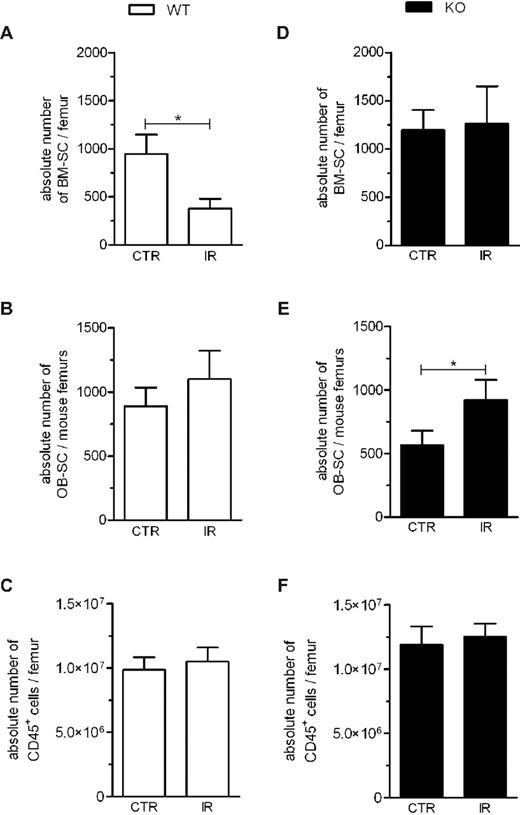
We next determined the impact of IR on BM stromal cells in vivo by evaluating the absolute number of BM-SCs and OB-SCs present per femur after exposure to IR. Using fluorescently labeled beads and flow cytometry, we found that the number of BM-SCs per femur was reduced 8 weeks after exposure to IR compared with the number found in control, nonirradiated femurs. This is in contrast to OB-SC and hematopoietic (CD45+) cell populations, in which numbers were not reduced during that same period (compare Figure 3A with panels B and C). This depletion in the number of BM-SCs long after exposure to IR was not observed in Ink4a/arf-deficient mice (Figure 3D). We also observed that the number of BM-SCs was not reduced 1 week after exposure to IR (supplemental Figure 1), which is in agreement with the observation that the expression of p16INK4a and p19ARF was not increased until several weeks after IR. These results reveal a difference in sensitivity to IR between the OB-SC and BM-SC populations and suggest that long-term maintenance of the BM-SC population is compromised in irradiated wild-type mice, an effect that can be reversed by the loss of Ink4a/arf.
IR-induced changes in the cellularity of the stroma are dependent on their stromal cell type and ink4a/arf status. Absolute cell counts of BM-SC (A,D), OB-SC (B,E), and hematopoietic cell (C,F) populations collected from femurs of wild-type (WT) and Ink4a/arf-deficient (KO) mice were determined by flow cytometry (see “Methods”) after exposure (IR) or not (CTR) to 6 Gy of IR. BM-SC and hematopoietic cell counts were evaluated in each femur OB-SC cell counts were evaluated from both femurs collected from the same mouse. n = 11-21 mice per group. *P < .05 by Student t test.
IR-induced changes in the cellularity of the stroma are dependent on their stromal cell type and ink4a/arf status. Absolute cell counts of BM-SC (A,D), OB-SC (B,E), and hematopoietic cell (C,F) populations collected from femurs of wild-type (WT) and Ink4a/arf-deficient (KO) mice were determined by flow cytometry (see “Methods”) after exposure (IR) or not (CTR) to 6 Gy of IR. BM-SC and hematopoietic cell counts were evaluated in each femur OB-SC cell counts were evaluated from both femurs collected from the same mouse. n = 11-21 mice per group. *P < .05 by Student t test.
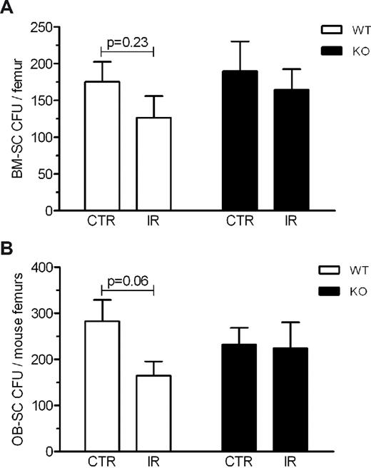
We also determined whether the expression of p16INK4a and p19ARF had an impact on the CFU potential of BM-SC and OB-SC populations isolated from the femurs of wild-type and ink4a/arf-deficient mice exposed or not to IR. We found the number of CFUs to be only modestly reduced 8 weeks after exposure to IR in both the BM-SC and OB-SC populations (P = .23 and P = .06, respectively), an effect that appeared to be Ink4a/arf-dependent (Figure 4). These results suggest that the ability to form CFUs ex vivo after exposure to IR may not be linked to the capacity to maintain stromal cells in vivo.
Loss of CFU potential after exposure to IR is dependent on Ink4a/arf status. BM-SCs (A) and OB-SCs (B) were isolated from wild-type (WT) or Ink4a/arf-deficient (KO) mice exposed (IR) or not (CTR) to 6 Gy of IR 8 weeks before being killed and cells were fixed after 2 weeks of culture to determine CFUs (average ± SEM). n = 9-19 mice per group. P values from Student t tests are shown.
Loss of CFU potential after exposure to IR is dependent on Ink4a/arf status. BM-SCs (A) and OB-SCs (B) were isolated from wild-type (WT) or Ink4a/arf-deficient (KO) mice exposed (IR) or not (CTR) to 6 Gy of IR 8 weeks before being killed and cells were fixed after 2 weeks of culture to determine CFUs (average ± SEM). n = 9-19 mice per group. P values from Student t tests are shown.
Ink4a/arf expression does not interfere with homing and engraftment processes
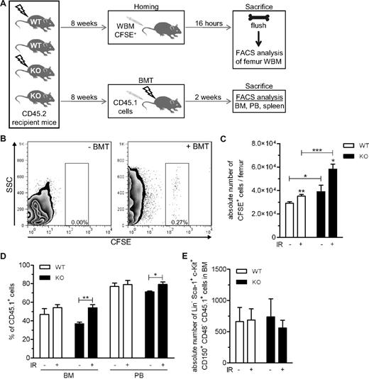
Shortly after irradiation, homing to the BM of hematopoietic cells was severely compromised (supplemental Figure 2), which is consistent with results published by Collis et al.28 However, to address the question of whether damage induced by IR can hamper long-term homing, recipient mice were irradiated 8 weeks before the transplantation of fluorescently labeled BM cells (Figure 5A). In addition, we assessed the role of p16INK4a and p19ARF in this process and found that their expression within the BM stroma and the consequent reduction in the size of the BM-SC population did not interfere with homing long after exposure to IR (Figure 5C). Similarly, we observed that IR-induced residual damage had no or little impact on engraftment in either the BM or blood of wild-type recipient mice (Figure 5D). However, engraftment was found to be significantly decreased in Ink4a/arf-null mice that had not been exposed to IR 8 weeks before transplantation; this effect likely stems from the higher replicative potential of Ink4a/arf-null hematopoietic progenitors, an advantage that they seem to have lost long after exposure to IR. In support of our observation that engraftment, at least in the short term, is not reduced by long-term residual damage induced by IR, we found that the number of donor-derived putative stem cells (Lin−, Sca-1+, c-Kit+, CD150+, CD48−) in the BM was equivalent in all groups of mice (Figure 5E).
Homing and engraftment processes are independent of long-term residual damage induced by IR. (A) Schematic of the experiments. Wild-type (WT) and Ink4a/arf-deficient (KO) mice were sublethally irradiated (6 Gy) or not and injected 8 weeks later, before homing or engraftment were determined. (B) Representative flow cytometric profiles of hematopoietic cells found in the BM of flushed femurs 16 hours after BM transplantation or not of 5 × 10⁶ cells previously stained with CFSE. (C) Absolute number (average ± SEM) of CFSE+ cells present in femur whole BM (WBM) of WT or KO mice exposed (+) or not (−) to IR 8 weeks before being killed. n = 6-14 mice per group. (D) BM and peripheral blood (PB) engraftment 2 weeks after transplantation of 2 × 10⁶ CD45.1 unfractionated BM cells in WT or KO CD45.2 mice exposed (+) or not (−) to IR 8 weeks before transplantation. n = 4-5 mice per group. (E) Absolute number of donor-derived SLAM (Lin−, Sca-1+, c-Kit+, CD150+, CD48−) CD45.1+ in BM 2 weeks after transplantation in WT or KO CD45.2 mice exposed (+) or not (−) to IR 8 weeks before transplantation. n = 4-5 mice per group. *P < .05, **P < .01, and ***P < .001 by Student t test.
Homing and engraftment processes are independent of long-term residual damage induced by IR. (A) Schematic of the experiments. Wild-type (WT) and Ink4a/arf-deficient (KO) mice were sublethally irradiated (6 Gy) or not and injected 8 weeks later, before homing or engraftment were determined. (B) Representative flow cytometric profiles of hematopoietic cells found in the BM of flushed femurs 16 hours after BM transplantation or not of 5 × 10⁶ cells previously stained with CFSE. (C) Absolute number (average ± SEM) of CFSE+ cells present in femur whole BM (WBM) of WT or KO mice exposed (+) or not (−) to IR 8 weeks before being killed. n = 6-14 mice per group. (D) BM and peripheral blood (PB) engraftment 2 weeks after transplantation of 2 × 10⁶ CD45.1 unfractionated BM cells in WT or KO CD45.2 mice exposed (+) or not (−) to IR 8 weeks before transplantation. n = 4-5 mice per group. (E) Absolute number of donor-derived SLAM (Lin−, Sca-1+, c-Kit+, CD150+, CD48−) CD45.1+ in BM 2 weeks after transplantation in WT or KO CD45.2 mice exposed (+) or not (−) to IR 8 weeks before transplantation. n = 4-5 mice per group. *P < .05, **P < .01, and ***P < .001 by Student t test.
IR-induced damage alters lymphopoiesis in a cellular nonautonomous and Ink4a/arf-dependent manner
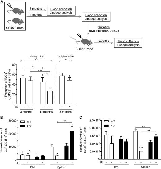
We next determined whether exposure to IR and expression of Ink4a/arf could interfere with long-term hematopoietic reconstitution. We therefore monitored the hematopoietic profiles of mice up to 11 months after exposure to sublethal irradiation (Figure 6A). As expected, Ink4a/arf-null mice exposed to IR developed lymphoma starting approximately 3 months after exposure to IR, whereas wild-type mice remained cancer free for the duration of the study (data not shown). However, wild-type mice showed a drastic reduction in the number of circulating B cells (identified as B220+) 11 months after exposure to IR (Figure 6A) that was accompanied by an increased myeloid (CD11b+) fraction (supplemental Figure 3). This endogenous recovery did not allow us to distinguish between a cell-autonomous or microenvironment-dependent effect on the B lineage. To address this question, we transplanted BM from mice previously exposed or not to IR (the 11-months group) into recipient mice (Figure 6A) and found that the number of circulating B cells derived from irradiated donors and non irradiated donors were comparable in transplanted recipients (Figure 6A). These results suggest that reduction in B-cell lymphopoiesis was mostly dependent on the effect of IR on the microenvironment.
IR-induced long-term residual damage alters lymphopoiesis in a nonautonomous, Ink4a/arf-dependent manner. (A) Schematic of the experiment. Mice were exposed (+) or not (−) to IR and allowed to recover without transplantation. Eleven months later, mice were killed and BM cells were used for the transplantation of CD45.1 recipient mice. The proportion of donor-derived B cells (B220+ CD45.2+) was analyzed at different times in the blood before and after transplantation. n = 3-9 mice per group. (B-C) As described in Figure 5A, wild-type (WT) and Ink4a/arf-deficient (KO) CD45.2 mice exposed (+) or not (−) to 6 Gy of IR were transplanted 8 weeks later with unfractionated CD45.1 BM cells and allowed to reconstitute for 2 weeks before BM and spleen analysis of donor-derived CLP (B) and B220+ (C) cell counts. n = 3-5 mice per group. *P < .05, **P < .01, ***P < .001, and °P < .05 by Student t test (compared with primary mice).
IR-induced long-term residual damage alters lymphopoiesis in a nonautonomous, Ink4a/arf-dependent manner. (A) Schematic of the experiment. Mice were exposed (+) or not (−) to IR and allowed to recover without transplantation. Eleven months later, mice were killed and BM cells were used for the transplantation of CD45.1 recipient mice. The proportion of donor-derived B cells (B220+ CD45.2+) was analyzed at different times in the blood before and after transplantation. n = 3-9 mice per group. (B-C) As described in Figure 5A, wild-type (WT) and Ink4a/arf-deficient (KO) CD45.2 mice exposed (+) or not (−) to 6 Gy of IR were transplanted 8 weeks later with unfractionated CD45.1 BM cells and allowed to reconstitute for 2 weeks before BM and spleen analysis of donor-derived CLP (B) and B220+ (C) cell counts. n = 3-5 mice per group. *P < .05, **P < .01, ***P < .001, and °P < .05 by Student t test (compared with primary mice).
IR-induced long-term residual damage did not affect homing and overall engraftment even though circulating B cells were impaired. We therefore monitored whether common lymphoid progenitors (CLPs, defined as Lin−, Sca-1dim, c-Kitdim, IL7R+)29 may be affected by IR. The absolute number of donor-derived CLPs was not found to be diminished in the BM of mice exposed to IR 8 weeks before the transplantation (Figure 6B), whereas the number of CLPs in the spleen was significantly reduced, a phenotype that was also associated with a reduction in the number of B220+ cells (Figure 6B-C). This effect was specific to the B lineage, because the absolute counts of CD3+ cells were not significantly changed (supplemental Figure 4). Remarkably, a reduction in the number of CLPs was not observed in previously irradiated Ink4a/arf-null mice (Figure 6B), and instead were significantly increased, suggesting that Ink4a/arf expression impairs B-cell homeostasis in a cell-nonautonomous manner. Further supporting these findings, we also performed secondary transplantations using irradiated wild-type and Ink4a/arf-null primary donor mice. We found that only a transient (2 weeks) exposure of progenitors to an irradiated wild-type, but not Ink4a/arf-null, microenvironment was sufficient to decrease B-cell formation in secondary recipient mice (supplemental Figure 5).
Discussion
Long-term residual damage induced by total body irradiation is a growing medical concern, with many cancer survivors showing treatment-related late effects.30,31 In the context of BM transplantation, radiation-based conditioning is suspected to impair hematopoiesis and to have an impact on the success of the graft itself by inducing damage to the BM stroma. Indeed, the importance of the stromal compartment as a niche for HSCs has been demonstrated in several ways, for example, after genetic manipulations regulating the osteoblastic pool size.32 Likewise, co transplantation of BM-derived stromal cells along with HSCs can be performed safely in humans and has been shown to improve engraftment in both humanized mouse and nonhuman primate models.33-35 The extent and nature of the damage to BM stroma after exposure to IR remain elusive. In the present study, we provide evidence that BM stromal cell populations express markers of senescence in response to IR. We have also identified for the first time a cellular nonautonomous role for Ink4a/arf in mediating IR-induced long-term residual damages to the hematopoietic stroma.
We found that the expression of p16INK4a increased after exposure to IR in both the BM-SC and OB-SC populations, albeit at a much higher level in the latter. Conversely, p19ARF expression, which is believed to be primarily induced after the oncogenic disruption of the cell cycle, was only increased within the BM-SC population after irradiation (Figure 1E).36 We hypothesize that p19ARF expression could be induced as a tumor-suppressive mechanism specifically in BM-SCs, given that these cells, by acting as progenitors for OB-SCs,37 are likely to undergo more cell divisions and therefore accumulate oncogenic mutations at a higher frequency.
One consequence of increased p16INK4a expression with age is a decrease in tissue-regenerative potential at the progenitor/stem cell level.38-40 Therefore, one could expect that IR-induced p16INK4a expression could also result in a regenerative defect in BM stromal cell progenitors. Indeed, 8 weeks after exposure to IR, we observed a significant reduction in the number of BM-SCs present per femur (Figure 3A). Surprisingly, this defect was specific to BM-SCs; it was not observed in the OB-SC population, which still expressed high levels of p16INK4a (Figure 1E). We believe that the inability to maintain BM-SCs may arise from the fact that these cells, by acting as progenitors for OB-SCs,37 are facing a higher replicative pressure and consequently a potential exhaustion. Along with this hypothesis, we could detect an increase in the expression of several genes specific to the osteoblastic lineage in BM-SCs isolated 8 weeks after exposure of mice to IR, suggesting the commitment of BM-SC progenitors toward differentiation in response to damage (supplemental Figure 6). Alternatively, maintenance of the OB-SC population may be explained by the work of Dominici et al, who showed that osteoblasts can be regenerated by megakaryocytes migrating to the endosteal surface shortly after radioablation.41
Intriguingly, the ability of the BM-SC and OB-SC populations to form CFUs was only slightly reduced long-term after exposure to IR despite high expression levels of p16INK4a/p19ARF in these populations. These results were unexpected given that others had shown IR-induced reduction in the CFUs of BM stromal cells up to several months/years in mice and humans, respectively.4,5 We believe that such discrepancies may be attributed to species and strain differences, a hypothesis supported by the work of Rombouts and Ploemacher.7 Our results also entail that stromal cells expressing p16INK4a/p19ARF are not likely to be found within the subpopulation of cells with CFU potential. However, further refinement in the identification of positive markers specific for these cells will be necessary to verify this hypothesis. For example, Méndez-Ferrer et al showed a direct relationship between homing and the proportion of Nestin+ BM stromal cells, a population that they have identified as having strong CFU capabilities.42
We also noticed a reduction in the number of circulating B cells in irradiated mice, a phenotype reminiscent of what is observed in aged mice.43,44 In fact, during the course of our 11-month study, we observed a reduction in the size of the B-cell population in non- irradiated mice (compare control groups at 3 and 11 months in Figure 6A), a reduction that was much less severe than in mice previously exposed to IR. Such slow progression of the phenotype in blood made it impossible to address the role of Ink4a/arf in knockout mice developing lymphoma within only few months after exposure to IR. Nevertheless, using groups of mice previously exposed or not to IR and subsequently transplanted, we could identify an important role of the microenvironment in B-cell lymphopoiesis. Analysis of donor-derived CLPs revealed a profound reduction in the spleen, but not in the BM, 2 weeks after transplantation, the latest time point at which mice were analyzed before Ink4a/arf-null mice developed lymphoma (Figure 6C). Because we did not analyze the number of lymphoid progenitors in the BM of wild-type mice more than 10 weeks after their exposure to IR, it remains to be determined whether a reduction in the number of CLPs in the spleen is sufficient to induce a reduction in the number of circulating B cells or if a reduction in the number of CLPs in the BM is eventually required for the phenotype to develop in blood. Nonetheless, we found that a 2-week exposure to an irradiated microenvironment was sufficient to decrease B-cell formation in secondary transplanted mice, an effect that was also INK4a/ARF-dependent. We hypothesized that such a profound and long-lasting impact on B-cell formation may arise from the action of the recently characterized senescence-associated secretory phenotype, which may be sustained by the expression of INK4a/ARF.9,45 Obviously, more work remains to be done to understand exactly how the microenvironment influences lymphoid progenitors.
It is reasonable to predict that BM-conditioning protocols that induce less damage to the hematopoietic niche may have a lesser impact in the long term. To begin addressing this idea, we tested whether an increase in p16INK4a expression, which is used as a marker of damage to the niche, is proportionally linked to the intensity of the irradiation dose or the type of drug used. We found that both low-dose IR (2.5 and 5 Gy) and administration of busulfan (35 mg/kg, the highest tolerated dose) resulted in an increase in p16INK4a expression at levels similar to the ones observed after exposure to 8 Gy of IR (data not shown). This suggests either that the increase in p16INK4a expression is not correlated with the intensity of the damage induced or that low levels of damage are sufficient to harm the BM stroma. In conclusion, future efforts should be made at identifying drugs and conditioning regimen therapies that will aim to better maintain the stromal niche.
The online version of this article contains a data supplement.
The publication costs of this article were defrayed in part by page charge payment. Therefore, and solely to indicate this fact, this article is hereby marked “advertisement” in accordance with 18 USC section 1734.
Acknowledgments
The authors thank Vimal Krishnan for his critical reading of the manuscript and Denise Carrier for her help with the mice.
This work was supported by the Canadian Institute of Health Research (grant MOP-102709 to C.M.B.) and the Canadian Cancer Society Research Institute (grant 019222 to T.H.). S.R.S. and G.D. have been supported by a student fellowship from the Fonds de recherche en Santé du Québec. C.L.C., A.F., and C.M.B. have been supported, respectively, by student fellowships from the Canadian Institutes of Health Research, the Fondation des étoiles, and a scientist award by the Fonds de recherche en Santé du Québec.
Authorship
Contribution: C.L.C., G.D., and C.M.B. conceived and designed the experiments; C.L.C., G.D., S.R.-S., A.F., and O.L. performed the experiments; C.L.C., G.D., S.R.-S., T.H., and C.M.B. analyzed the data; and C.L.C., T.H., and C.M.B. wrote the manuscript.
Conflict-of-interest disclosure: The authors declare no competing financial interests.
Correspondence: Christian M. Beauséjour, Centre Hospitalier Universitaire Ste-Justine, 3175 Côte Sainte-Catherine, Montréal, QC, Canada, H3T 1C5; e-mail: christian.beausejour@recherche-ste-justine.qc.ca.

This feature is available to Subscribers Only
Sign In or Create an Account Close Modal