Abstract
Deletion of Runx1 in adult mice produces a myeloproliferative phenotype. We now find that Runx1 gene deletion increases marrow monocyte while reducing granulocyte progenitors and that exogenous RUNX1 rescues granulopoiesis. Deletion of Runx1 reduces Cebpa mRNA in lineage-negative marrow cells and in granulocyte-monocyte progenitors or common myeloid progenitors. Pu.1 mRNA is also decreased, but to a lesser extent. We also transduced marrow with dominant-inhibitory RUNX1a. As with Runx1 gene deletion, RUNX1a expands lineage−Sca-1+c-kit+ and myeloid cells, increased monocyte CFUs relative to granulocyte CFUs, and reduced Cebpa mRNA. Runx1 binds a conserved site in the Cebpa promoter and binds 4 sites in a conserved 450-bp region located at +37 kb; mutation of the enhancer sites reduces activity 6-fold in 32Dcl3 myeloid cells. Endogenous Runx1 binds the promoter and putative +37 kb enhancer as assessed by ChIP, and RUNX1-ER rapidly induces Cebpa mRNA in these cells, even in cycloheximide, consistent with direct gene regulation. The +37 kb region contains strong H3K4me1 histone modification and p300-binding, as often seen with enhancers. Finally, exogenous C/EBPα increases granulocyte relative to monocyte progenitors in Runx1-deleted marrow cells. Diminished CEBPA transcription and consequent impairment of myeloid differentiation may contribute to leukemic transformation in acute myeloid leukemia cases associated with decreased RUNX1 activity.
Introduction
The Runx1 transcription factor contributes to formation of pluripotent adult HSCs from hemogenic endothelium during embryogenesis.1-3 Deletion of floxed Runx1 alleles in adult mice preserves pluripotent HSCs and erythroid cells but leads to thrombocytopenia and a marked reduction in B- and T-lymphoid cells and their precursors.4-6 In addition, these mice develop an expansion of myeloid progenitors, with increased numbers of lineage−Sca-1+ c-kit+ (LSK) cells, common myeloid progenitors (CMPs), granulocyte-monocyte progenitors (GMPs), myeloid CFUs, and mature myeloid cells.4,5 This myeloproliferative phenotype is transplantable and so intrinsic to the hematopoietic system.5
The myeloid expansion associated with absence of Runx1 may be relevant to myeloid leukemogenesis. Reduced RUNX1 activity occurs commonly in acute myeloid leukemia (AML), often because of expression of the RUNX1-ETO or core binding factor β (CBFβ)–SMMHC oncoproteins or as a result of RUNX1 genomic mutation.7-9 It has been suggested that these and other “type II” alterations, including CEBPA mutation, contribute to myeloid transformation by inhibiting myeloid differentiation, with coincident “type I” alterations, for example, activation of N-Ras or receptor tyrosine kinases, inhibiting apoptosis while stimulating proliferation.10
In this study, we further assess the effect of Runx1 gene deletion on myeloid differentiation in adult mice. Comparison of Runx1-deleted myeloid CFUs with those of control mice uncovered a marked increase in monocyte CFU (CFU-M) progenitors and a reduction in granulocyte CFUs (CFU-Gs), with an increase in mature monocytes evident also in total marrow cells. CEBPα and PU.1 are key transcriptional regulators of myeloid lineage determination,11 and Runx1 directly activates Pu.1 gene transcription.12 We find that both Pu.1 and Cebpa mRNA levels are reduced in marrow myeloid progenitors from Runx1-deleted mice, with a greater relative reduction of Cebpa mRNA. Altered myelopoiesis and reduced Cebpa mRNA was also seen when the Runx1 genes were deleted in vitro by Cre transduction, consistent with a marrow intrinsic effect. Decreased PU.1 levels in mice impairs monopoiesis while sparing granulopoiesis,13-15 suggesting that diminished Cebpa rather than Pu.1 expression predominantly accounts for the impairment in granulopoiesis and elevated monopoiesis we observed on Runx1 gene deletion. Consistent with this idea, exogenous Runx1 rescued granulopoiesis while reducing monopoiesis in Runx1-deleted marrow cells, associated with induction of C/EBPα but not PU.1, and exogenous C/EBPα also stimulated granulopoiesis relative to monopoiesis in marrow cells lacking Runx1. As an additional approach, we extended our study to in vivo analysis of marrow cells transduced with the dominant-inhibitory RUNX1a isoform, again finding impaired granulopoiesis and reduction in Cebpa and to a lesser extent Pu.1 mRNA. Runx1 binds an evolutionarily conserved site in the Cebpa promoter and 4 sites in a highly conserved, 450-bp region located at 37 kb. Mutation of the promoter site reduces activity 2-fold in 32Dcl3 myeloid cells, and mutation of the distal sites reduces activity 6-fold. The 37 kb element binds p300 strongly and contains abundant H3K4me1 histone modifications relative to the promoter, consistent with its potential role as an enhancer. Finally, Runx1-ER induces endogenous Cebpa mRNA even in cycloheximide, consistent with direct gene activation. Our findings suggest that in human AML cases reduction in RUNX1 activity impairs granulocytic differentiation to contribute to myeloid transformation via reduction in CEBPA gene transcription.
Methods
Cell culture and murine marrow transduction and transplantation
293T cells were cultured in DMEM with 10% heat-inactivated FBS (HI-FBS). 32Dcl3 cells16 were cultured in (IMDM with 10% HI-FBS and 1 ng/mL murine IL-3. C57BL/6 Runx1(f/f) mice harboring loxP sites surrounding Runx1 exon 4 were bred to Mx1-Cre transgenic C57BL/6 mice (The Jackson Laboratory) to obtain Runx1(f/f);Mx1-Cre mice.5 At 12-16 weeks of age these or Runx1(f/f) mice were injected intraperitoneally with 500 μg of pIpC every other day for 6 doses. Marrow was isolated 4 weeks later for analysis. For in vitro studies, marrow isolated from mice injected intraperitoneally with 150 mg/kg 5-fluorouracil (5-FU) was subjected to red cell lysis and cultured for 1 day in 10 ng/mL murine IL-3, 10 ng/mL murine IL-6, and 10 ng/mL murine SCF (PeproTech) followed by the addition of 4 μg/mL Polybrene and retroviral supernatants obtained from 293T cells transduced with 12 μg of pBabePuro, pBabePuro-RUNX1-ER, pBabePuro-Cre, or pBabePuro-C/EBPα-ER17,18 ; 3 μg of pkat2ecopac19 ; and 35 μL of Lipofectamine 2000 (Invitrogen) per 100-mm dish. Three days later, 2 μg/mL puromycin was added, and after 2 additional days viable cells isolated with Lympholyte M (Cedarlane Labs) were plated in methylcellulose or in liquid culture with IMDM, 10% HI-FBS, IL-3, IL-6, and SCF. Hematopoietic colonies were enumerated 8 days later on the basis of colony structure. Viable cell counts were obtained with the use of a hemocytometer and Trypan Blue dye. In some experiments 5E6 mononuclear marrow from wild-type (WT) C57BL/6 mice exposed to 5-FU were placed in IMDM with 10% HI-FBS, 10 ng/mL IL-6, and 100 ng/mL SCF for 1 day, followed by transduction via spinoculation at 830g for 2 hours at 4°C with 6 μg/mL Polybrene and MIG or MIG-RUNX1a retroviral vectors that had been concentrated at 2000g for 15 minutes with the use of Amicon-Ultra filters (Millipore). Transduced cells were cultured for an additional day under the same conditions and analyzed for green fluorescent protein (GFP) expression. 1E6 cells were introduced by tail vein injection into syngeneic WT recipients irradiated to 950 cGy, and marrow was isolated 4 weeks later for analysis.
FACS and cell structure
Cells were subjected to FACS analysis for GFP or after staining with PE– or peridinin chlorophyll protein complex/cyanine 5.5–anti-Mac-1, FITC- or allophycocyanin (APC)–anti-Gr-1, PE–anti-Sca-1, APC–anti-c-kit, PE–anti-FcγR, PE–anti-B220, PE–anti-CD3, PE–anti-Ter119, or biotin-conjugated mouse Lineage Cocktail (BD PharMingen), or peridinin chlorophyll protein complex/cyanine 5.5–streptavidin, FITC–anti-CD34, PE–anti-F4/80, biotin–anti-CD115/MCSFR, APC-streptavidin, or PE-Cy7–anti-Sca-1 (eBiosciences). Cell sorting for GFP+ cells or for HSCs, CMPs, GMPs, and megakaryocyte-erythroid progenitors (MEPs) was conducted with the use of a FACSAria II cell sorter (BD Biosciences). For cell cycle analysis, cells fixed with 70% methanol and treated with DNase-free RNase were stained with 25 μg/mL propidium iodide and analyzed by FACS. Cell structure was assessed by Wright-Giemsa staining of cytospun cells. Photomicrographs were taken with a Zeiss Axiophot microscope (Carl Zeiss), a Kontron Electronik Progress 3012 camera (Kontron), and a 63×/1.40 NA oil objective.
RNA analysis and Western blot analysis
Total cellular RNA was prepared with the NucleoSpin RNA II kit, including use of RNase-free DNase (Machery-Nagel). First-strand cDNA was prepared with AMV reverse transcriptase (Promega) and oligodT primer at 42°C for 1 hour. Quantitative PCR was performed with 5 ng of each cDNA with the use of iQ SYBR Green supermix (Bio-Rad). Oligonucleotides used were as follows: Cebpa-F, 5′-TGGATAAGAACAGCAACGAG, and Cebpa-R, 5′-TCACTGGTCAACTCCAGCAC; Pu.1-F, 5′-CAGAAGGGCAACCGCAAGAA, and Pu.1-R, 5′-GCCGCTGAACTGGTAGGTGA; Runx1-F, 5′-CACCGTCATGGCAGGCAAC, and Runx1-R, 5′-GGTGATGGTCAGAGTGAAGC; mS16-F, 5′-CTTGGAGGCTTCATCCACAT, and mS16-R, 5′-ATATTCGGGTCCGTGTGAAG; and actin-F, 5′-GACCTCTATGCCAACACAGT, and actin-R, 5′-AGTACTTGCGCTCAGGAGGA.
Cells were washed with PBS, and cellular proteins were prepared by adding Laemmli sample buffer. Extracts corresponding to 5E5 cells were subjected to PAGE with the use of 8%-10% gels and transferred to Hybond-P membrane (Amersham), followed by probing with primary Abs. Abs used were C/EBPα (14AA), PU.1 (D-19), or α-tubulin (Sigma-Aldrich), Runx1 (Active Motif), or β-actin (AC-15; from Sigma-Aldrich). After adding HRP-conjugated secondary Ab and subsequent washes, a signal was generated with the use of HyGlo chemiluminescence reagents (Denville Scientific).
Gel shift, ChIP, and reporter assays
Nuclear extract preparation from transfected 293T cells and gel shift assays were performed as described,20 using Runx1, H3K4me1 (ab8895; Abcam), or p300 (C-20; Santa Cruz Biotechnology) antisera or rabbit Ig control. Oligonucleotide probes containing 5′TCGA overhangs were radio-labeled to similar specific activity with the use of Klenow enzyme and α-P32-dCTP. Sense strands of the WT probes used, with binding sites underlined, were as follows: mCEBPA-promWT1 + 2, 5′-GGCGCCTAACCACGGACCACGTGTGTGC; hCEBPA-promWT1 + 2, 5′-CTACCGACCACGTGGGCTGCCGCGTGGTTCGCCG; hCEBPA-promWT1, 5′-CCGCTACCGACCACGTGGGCGCGG; mCEBPA-enhR1, 5′-GACTACCGGCGACCACAGGAAGTGCTG; mCEBPA-enhR2, 5′-TTCTTGCCACAACCACACATCAGTTAT; mCEBPA-enhR3, 5′-CTGCTCCAACGACCACACTCCTGTTCCC; and mCEBPA-enhR4, 5′-GGGCCTGCTCACCACATCACATAGAGG.
Mutant variants of these probes are described in the figures. ChIP was conducted as described21 with the use of the following genomic DNA PCR primers: CEBPA-323F, 5′-CCTAGTGTTGGCTGGAAGTG; CEBPA-210R, 5′-GGCGAACCCGGCTTGCAG; CEBPA-2500F, 5′-TTCCTTATCCTTCCAGAGACTTCC; CEBPA-2500R, 5′-GATTTCCAGCCTCCCGTGTGATG; CEBPA-enhR1-F, 5′-TTCCCGTTTCTGAAATCTGC; CEBPA-enhR2-R, 5′-GGTTGTGGCAAGAAGGTCAC; CEBP-enhR3-F, 5′-AACAGGAAAGATGGCACCAG; CEBP-enhR4-R, 5′-CCACACCCCTCTATGTGATG; Actin-1500F, 5′-GGGAAAGTTCTCTCAGGGTTGG; and Actin-1500R, 5′-TGCTGTGAACTGGAAACACACC.
CEBPA-LUC was constructed by inserting a murine DNA PCR fragment extending from −720 bp to 125 bp, just upstream of the initiating Cebpa ATG, into pREP4-LUC.22 Point mutations were introduced by PCR mutagenesis with the use of the QuickChange kit (Stratagene). A 497-bp DNA segment containing the putative 37 kb enhancer flanked by KpnI and NotI sites was isolated by genomic PCR and positioned upstream of the Cebpa promoter in pREP4-LUC. A version of this DNA segment with the 4 Runx1 sites mutated was synthesized (Origene) and subcloned similarly. All constructs were confirmed by DNA sequencing. 2E6 32Dcl3 cells were transduced with 5 μg of either of these DNAs and 0.25 μg of CMV-βGal with the use of diethyl aminoethyl–dextran as described,23 and luciferase and β-galactosidase assays were conducted 2 days later.
Statistical comparisons were conducted with the Student t test.
Results
Deletion of Runx1 increases monopoiesis and impairs granulopoiesis
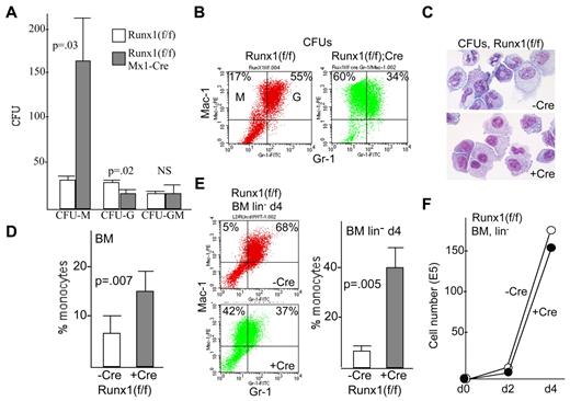
Twelve- to 16-week-old Runx1(f/f);Mx1-Cre or Runx1(f/f) mice were subjected to 6 pIpC injections and allowed to recover for 4 weeks to ensure maximal deletion, which requires ≥ 2 weeks,5 and to ensure recovery from IFN induction and reestablishment of steady-state hematopoiesis. Marrow mononuclear cells were then analyzed for myeloid CFUs in methylcellulose culture with IL-3, IL-6, and SCF (Figure 1A). Consistent with prior findings, total myeloid CFU numbers were increased on Runx1 gene deletion. Of note, CFU-Ms were increased on average 5-fold in 3 independent assessments, whereas CFU-Gs were reduced 2-fold. FACS analysis of pooled CFUs confirmed that Runx1 deletion increased Mac-1+Gr-1− monocytes at the expense of Mac-1+Gr-1+ granulocytes (Figure 1B), and structural analysis further confirmed increased monocyte/macrophages in CFUs on Runx1 deletion, without accumulation of granulocytic precursors (Figure 1C). Most of these Mac-1+Gr-1− cells also express the monocyte/macrophage markers F4/80 and MCSFR (supplemental Figure 1, available on the Blood Web site; see the Supplemental Materials link at the top of the online article). Immediately after harvest, total marrow from 6 control or Runx1-deleted mice had equivalent proportions of mature, Mac-1+ myeloid cells (72% ± 13% vs 71% ± 6%); among these cells there was a significant increase in the percentage of Mac-1+Gr-1− monocytes on Runx1 deletion, from 7% to 15% (Figure 1D). Most of the granulocytic cells had mature structure, as previously seen.5 In addition, when lineage-depleted marrow from these mice was placed in liquid culture with IL-3, IL-6, and SCF and analyzed by FACS for myeloid differentiation 4 days later, a marked increase in the proportion of Mac-1+Gr-1− monocytes among total myeloid cells was evident compared with similarly cultured cells from control mice (Figure 1E), again with a corresponding decrease in granulocytes. No difference was observed in the rate of in vitro myeloid cell expansion with or without Runx1 gene deletion during this 4-day period (Figure 1F). In conclusion, absence of Runx1 in vivo increases monopoiesis at the expense of granulopoiesis in murine marrow progenitors.
Absence of Runx1 favors monopoiesis over granulopoiesis. (A) Marrow mononuclear cells from Runx1(f/f) or Runx1(f/f);Mx1-Cre mice exposed to pIpC were assessed for myeloid progenitor numbers in IL-3, IL-6, and SCF per 1E4 mononuclear cells plated (mean and SE from 3 determinations). (B) Pooled CFUs were subjected to FACS analysis for Mac-1 and Gr-1. The proportions of Mac-1+Gr-1− monocytes (M) and Mac-1+Gr-1+ granulocytes (G) are indicated. (C) CFUs were cytospun and subjected to Wright-Giemsa staining. (D) Marrow mononuclear cells from Runx1(f/f) or Runx1(f/f);Mx1-Cre mice exposed to pIpC were subjected to similar FACS analysis immediately after isolation. The proportion of Mac-1+Gr-1− monocytes relative to the sum of monocytes and granulocytes is shown (mean and SE from 6 determinations). (E) Control or Runx1-deleted marrow cells were lineage-depleted and placed in liquid culture with IL-3, IL-6, and SCF and subjected to FACS analysis 4 days later. Representative data (left panels) and a summary of 3 determinations (right graph) are shown. (F) Viable cell numbers were enumerated immediately after lineage depletion of control or Runx1-deleted cells and 2 or 4 days later. A representative growth curve is shown.
Absence of Runx1 favors monopoiesis over granulopoiesis. (A) Marrow mononuclear cells from Runx1(f/f) or Runx1(f/f);Mx1-Cre mice exposed to pIpC were assessed for myeloid progenitor numbers in IL-3, IL-6, and SCF per 1E4 mononuclear cells plated (mean and SE from 3 determinations). (B) Pooled CFUs were subjected to FACS analysis for Mac-1 and Gr-1. The proportions of Mac-1+Gr-1− monocytes (M) and Mac-1+Gr-1+ granulocytes (G) are indicated. (C) CFUs were cytospun and subjected to Wright-Giemsa staining. (D) Marrow mononuclear cells from Runx1(f/f) or Runx1(f/f);Mx1-Cre mice exposed to pIpC were subjected to similar FACS analysis immediately after isolation. The proportion of Mac-1+Gr-1− monocytes relative to the sum of monocytes and granulocytes is shown (mean and SE from 6 determinations). (E) Control or Runx1-deleted marrow cells were lineage-depleted and placed in liquid culture with IL-3, IL-6, and SCF and subjected to FACS analysis 4 days later. Representative data (left panels) and a summary of 3 determinations (right graph) are shown. (F) Viable cell numbers were enumerated immediately after lineage depletion of control or Runx1-deleted cells and 2 or 4 days later. A representative growth curve is shown.
Exogenous Runx1 stimulates granulocyte lineage specification in Runx1−/− marrow cells
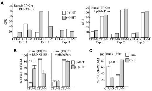
We next sought to determine whether exogenous Runx1 would correct defective granulopoiesis evident on Runx1 deletion. Runx1(f/f):Mx1-Cre mice injected with pIpC and allowed to recover for 4 weeks were injected with 5-FU. Five days later marrow mononuclear cells were isolated and transduced 1 day later with pBabePuro-RUNX1-ER or with empty pBabePuro vector, followed by puromycin selection and subsequent isolation of viable cells, which were then plated in methylcellulose with IL-3, IL-6, and SCF, with or without 4-HT. In 3 independent experiments, activation of RUNX1-ER increased both the absolute number and proportion of CFU-Gs relative to CFU-Ms, whereas 4-HT had no effect on the number or distribution of myeloid CFUs generated from vector-transduced marrow cells (Figures 2A-B). On average, total CFU-G increased 5-fold, whereas total CFU-Ms decreased 2-fold in response to RUNX1-ER activation. This finding supports the idea that Runx1 helps direct granulopoiesis from bipotent, immature myeloid cells. In addition, this result obtained on in vitro transduction of isolated marrow cells indicates that the impairment in granulopoiesis evident on Runx1 gene deletion is intrinsic to the isolated myeloid progenitors. Consistent with this conclusion, transduction of marrow isolated from Runx1(f/f) mice with pBabePuro-Cre resulted in reduced granulopoiesis relative to monopoiesis in methylcellulose culture compared with cells transduced with empty vector (Figure 2C).
Exogenous Runx1 restores granulopoiesis and diminishes monopoiesis in Runx1-deleted marrow cells. (A) Marrow mononuclear cells isolated from Runx1(f/f);Mx1-Cre mice exposed to pIpC and subsequently to 5-FU were transduced with RUNX1-ER or with the pBabePuro vector, selected with puromycin, and then plated in methylcellulose with IL-3, IL-6, and SCF with or without 4HT. The absolute number of CFU-Gs or CFU-Ms per 1E4 cells plated in 3 independent transduction experiments is shown. (B) The proportion of CFU-Gs or CFU-Ms among these myeloid CFUs is shown (mean and SE from 3 determinations). (C) Marrow isolated from Runx1(f/f) mice exposed to 5-FU was transduced with pBabePuro or pBabePuro-Cre for 3 days, followed by puromcyin selection for 2 days. Cells were then plated in methylcellulose with IL-3, IL-6, and SCF, and CFUs were enumerated 8 days later. Shown is the percentage of CFU-Gs and the percentage of CFU-Ms among CFU-Gs plus CFU-Ms (mean and SE from 3 determinations).
Exogenous Runx1 restores granulopoiesis and diminishes monopoiesis in Runx1-deleted marrow cells. (A) Marrow mononuclear cells isolated from Runx1(f/f);Mx1-Cre mice exposed to pIpC and subsequently to 5-FU were transduced with RUNX1-ER or with the pBabePuro vector, selected with puromycin, and then plated in methylcellulose with IL-3, IL-6, and SCF with or without 4HT. The absolute number of CFU-Gs or CFU-Ms per 1E4 cells plated in 3 independent transduction experiments is shown. (B) The proportion of CFU-Gs or CFU-Ms among these myeloid CFUs is shown (mean and SE from 3 determinations). (C) Marrow isolated from Runx1(f/f) mice exposed to 5-FU was transduced with pBabePuro or pBabePuro-Cre for 3 days, followed by puromcyin selection for 2 days. Cells were then plated in methylcellulose with IL-3, IL-6, and SCF, and CFUs were enumerated 8 days later. Shown is the percentage of CFU-Gs and the percentage of CFU-Ms among CFU-Gs plus CFU-Ms (mean and SE from 3 determinations).
Dominant inhibition of Runx1 increases monopoiesis relative to granulopoiesis in vivo
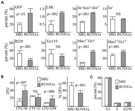
RUNX1a is a naturally occurring splice variant of RUNX1 that lacks the major C-terminal transactivation domain but retains the DNA-binding domain, allowing RUNX1a to dominantly inhibit transactivation by the full-length RUNX1b or RUNX1c isoforms.24 Transduction of murine marrow cells with MIG-RUNX1a, expressing RUNX1a and GFP, followed by transplantation into irradiated, syngeneic recipients led to a 2- to 3-fold in vivo expansion of GFP+ cells 4-8 weeks later compared with cells transduced with the empty MIG retroviral vector (Figure 3A top left), similar to results reported previously.25 Analysis of the vector- or RUNX1a-transduced, GFP+ populations found that expression of RUNX1a not only increased the total number of GFP+ cells but also increased the proportion of LSK HSCs 2.4-fold and lin−Sca-1− c-kit+ progenitors 1.9-fold (Figure 3A). The proportion of B220+ B-lymphoid cells was reduced 2.2-fold, whereas the Ter119+ erythroid population was only minimally affected. Expansion of LSK and myeloid progenitors, reduction in B cells, and an unaltered erythroid population parallel results obtained previously on FACS analysis of marrow cells from Runx1-deleted versus control mice.4,5 In addition, exogenous RUNX1a induced a 4-fold increase in the proportion of Mac-1+Gr-1− monocytes among total marrow cells but only a 1.3-fold increase in Mac-1+Gr-1+ granulocytes (Figure 3A) and thus a 3-fold increase in the proportion of monocytes among marrow myeloid cells.
Dominant inhibition of Runx1 by RUNX1a favors monopoiesis over granulopoiesis. (A) WT marrow cells transduced with RUNX1a or with the empty MIG retroviral vector were transplanted into irradiated, syngeneic recipients, and the proportion of marrow cells expressing GFP was determined 4 weeks later. The proportions of LSK, lin−c-kit−Sca-1+, lin−, B220+, Ter119+, Mac-1+Gr-1−, or Mac-1+Gr-1+ cells among the GFP+ cells were also assessed by FACS analysis (mean and SE from 3 determinations). (B) GFP+ cells were sorted and assessed for myeloid CFUs per 1E4 cells plated after culture in methylcellulose with IL-3, IL-6, and SCF (left; mean and SE from 3 determinations). The proportion of CFU-Gs relative to CFU-Gs plus CFU-Ms among these CFUs is also shown (right). (C) Cells isolated 4 weeks after transplantation were stained with propidium iodide, and the proportion of cells in the G1, S, or G2/M cell cycle phases was enumerated among GFP+ cells (mean and SE from 3 determinations).
Dominant inhibition of Runx1 by RUNX1a favors monopoiesis over granulopoiesis. (A) WT marrow cells transduced with RUNX1a or with the empty MIG retroviral vector were transplanted into irradiated, syngeneic recipients, and the proportion of marrow cells expressing GFP was determined 4 weeks later. The proportions of LSK, lin−c-kit−Sca-1+, lin−, B220+, Ter119+, Mac-1+Gr-1−, or Mac-1+Gr-1+ cells among the GFP+ cells were also assessed by FACS analysis (mean and SE from 3 determinations). (B) GFP+ cells were sorted and assessed for myeloid CFUs per 1E4 cells plated after culture in methylcellulose with IL-3, IL-6, and SCF (left; mean and SE from 3 determinations). The proportion of CFU-Gs relative to CFU-Gs plus CFU-Ms among these CFUs is also shown (right). (C) Cells isolated 4 weeks after transplantation were stained with propidium iodide, and the proportion of cells in the G1, S, or G2/M cell cycle phases was enumerated among GFP+ cells (mean and SE from 3 determinations).
As an additional comparison, GFP+ cells sorted from mice engrafted with vector- or RUNX1a-transduced marrow cells were subjected to colony assay in methylcellulose culture with IL-3, IL-6, and SCF (Figure 3B left). Expression of RUNX1a led to a 3-fold increase in the number of CFU-Ms and granulocyte macrophage CFUs without altering the number of CFU-Gs per 1E4 cells plated. In contrast to Runx1 gene deletion, the robust hematopoietic expansion induced by RUNX1a potentially obviated an absolute reduction in total CFU-Gs; however, the percentage of CFU-Gs among CFU-Gs and CFU-Ms was reduced 1.5-fold (Figure 3B right). These data obtained via transduction with RUNX1a followed by transplantation into WT recipients provide further evidence for the cell autonomous nature of the myeloid expansion and shift of myeloid development from granulopoiesis toward monopoiesis that occurs consequent to reduced Runx1 activity. Comparison of vector- versus RUNX1a-transduced cells isolated 4 weeks after transplantation found an equivalent distribution of cells in the G1, S, or G2/M cell cycle phases (Figure 3C).
Deletion or dominant-inhibition of Runx1 reduces Cebpa expression
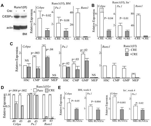
Total marrow mononuclear cells isolated from Runx1(f/f) or Runx1(f/f);Mx1-Cre mice exposed 4 weeks earlier to pIpC were subjected to Western blot analysis for C/EBPα and β-actin (Figure 4A). C/EBPα protein levels were reduced ∼ 2-fold. In this same cell population, Cebpa mRNA levels were reduced 3-fold and Pu.1 mRNA 2-fold (Figure 4A). In lineage− marrow cells, predominantly consisting of myeloid progenitors, Cebpa mRNA was reduced 5-fold and Pu.1 mRNA 2-fold, again averaging results from 3 separate RNA preparations (Figure 4B). Marrow cells from these mice also underwent FACS into HSC, CMP, GMP, and MEP subsets as described.26 Representative sorting data are shown (supplemental Figure 2). Of note, our findings confirm that Runx1 deletion increases HSCs, CMPs, and GMPs and reduces MEPs, consistent with prior findings.4,5 Runx1 mRNA expression was markedly reduced in each of these stem/progenitor populations on pIpC induction of Cre expression, just as it was in total and lineage− marrow cells (Figure 4A-C). In the absence Cre, both Cebpa and Pu.1 RNA levels increased as cells progressed from HSC to CMP to GMP and were barely detected in MEPs, as expected (Figure 4C). Runx1 gene deletion did not reduce the low level of Cebpa mRNA evident in HSCs and only minimally reduced Pu.1 mRNA. In CMPs, Cebpa mRNA was reduced 2-fold and Pu.1 mRNA 1.5-fold, and in GMPs, Cebpa mRNA was reduced 1.6-fold and Pu.1 mRNA 1.4-fold.
Absence or dominant inhibition of Runx1 reduces Cebpa expression. (A) Marrow mononuclear cells from Runx1(f/f) or Runx1(f/f);Mx1-Cre mice exposed to pIpC were subjected to Western blot analysis for C/EBPα or β-actin (left panel; representative of 3 experiments), and total cellular RNA from these cells were assessed for expression of Cebpa, Pu.1, or Runx1 mRNAs, relative to the RNA encoding ribosomal protein mS16, via quantitative RT-PCR (right graphs; mean and SE from 3 determinations). (B) Similar RNA analysis was conducted with lineage− marrow cells from these mice. (C) Marrow cells from control or Runx1-deleted mice were sorted into HSCs, CMPs, GMPs, and MEPs. Total cellular RNAs from these populations were then analyzed for Cebpa, Pu.1, or Runx1 mRNA expression (mean and SE from 3 determinations). (D) RNA isolated from Runx1(f/f) marrow cells transduced with pBabePuro or pBabePuro-Cre, either immediately after puromycin selection (d0) or 3 days later (d3), were analyzed form Cebpa, Pu.1, and Runx1 expression (mean and SE from 3 determinations). (E) WT marrow cells transduced with RUNX1a or with the empty MIG retroviral vector were transplanted into irradiated, syngeneic recipients. Four weeks later, RNAs isolated from total BM or lineage-negative (lin−) marrow cells were analyzed for expression of Cebpa or Pu.1 relative to β-actin mRNA (mean and SE from 3 determinations).
Absence or dominant inhibition of Runx1 reduces Cebpa expression. (A) Marrow mononuclear cells from Runx1(f/f) or Runx1(f/f);Mx1-Cre mice exposed to pIpC were subjected to Western blot analysis for C/EBPα or β-actin (left panel; representative of 3 experiments), and total cellular RNA from these cells were assessed for expression of Cebpa, Pu.1, or Runx1 mRNAs, relative to the RNA encoding ribosomal protein mS16, via quantitative RT-PCR (right graphs; mean and SE from 3 determinations). (B) Similar RNA analysis was conducted with lineage− marrow cells from these mice. (C) Marrow cells from control or Runx1-deleted mice were sorted into HSCs, CMPs, GMPs, and MEPs. Total cellular RNAs from these populations were then analyzed for Cebpa, Pu.1, or Runx1 mRNA expression (mean and SE from 3 determinations). (D) RNA isolated from Runx1(f/f) marrow cells transduced with pBabePuro or pBabePuro-Cre, either immediately after puromycin selection (d0) or 3 days later (d3), were analyzed form Cebpa, Pu.1, and Runx1 expression (mean and SE from 3 determinations). (E) WT marrow cells transduced with RUNX1a or with the empty MIG retroviral vector were transplanted into irradiated, syngeneic recipients. Four weeks later, RNAs isolated from total BM or lineage-negative (lin−) marrow cells were analyzed for expression of Cebpa or Pu.1 relative to β-actin mRNA (mean and SE from 3 determinations).
Transduction of marrow from Runx1(f/f) mice with pBabePuro-Cre led to an 87% reduction of Runx1 mRNA after 3 days of transduction and 2 days of puromycin selection (d0), and 99% reduction 3 days later (d3), and at these times Cebpa mRNA was reduced ∼ 2-fold, whereas Pu.1 mRNA was not reduced (Figure 4D). In addition, we assessed the effect of dominant-inhibitory RUNX1a on expression of Cebpa or Pu.1 in total or lineage− marrow cells isolated 4 weeks after transplantation of transduced WT marrow cells into syngeneic recipients. In comparison with empty MIG vector, RUNX1a reduced Cebpa mRNA 1.6-fold and Pu.1 mRNA 1.3-fold in total marrow mononuclear cells and reduced Cebpa mRNA 2.2-fold and Pu.1 mRNA 1.3-fold in lineage− marrow cells (Figure 4E); each result was the average of data obtained from 3 independent transductions.
Runx1 binds and activates the Cebpa promoter
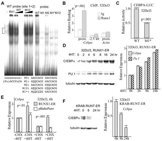
The WT murine Cebpa promoter contains tandem sites, 5′-AACCACG GACCACG, centered at −285 bp that match the consensus for Runx1 binding, 5′-(Pu)ACCPuCA, except that each of the 2 sites has a G instead of an A in the last position, both purines. Gel shift analysis was conducted with a radiolabeled probe containing these sites, using nuclear extracts from 293T cells transfected with empty CMV vector or with the same vector expressing Runx1 and its DNA-binding partner, CBFβ (Figure 5A left panel). Binding of Runx1 to this probe was evident, as a doublet, and this interaction was prevented by 25-fold excess of unlabeled probe but not by similar excess of the same probe harboring point mutations in the 2 Runx1 consensus sites. Endogenous Runx1 in myeloid cells is also detected as a doublet on gel shift assay.27 When WT or mutant probes were radiolabeled and assessed for binding to Runx1, mutation of the more upstream site 1 (M1) obviated binding and mutation of site 2 (M2) greatly reduced binding, suggesting that Runx1 binds these sites cooperatively (Figure 5A right panel lanes 1-3). An alternative set of clustered mutations in site 1 (M1*), or mutation of both sites (M12), also completely prevented binding (Figure 5A right panel lanes 4 and 5). The human CEBPA promoter retains 5′-GACCACG at −273 bp. A double-stranded oligonucleotide containing the −273-bp human site binds RUNX1 or RUNX1-ETO in a gel shift assay, and mutation of the Runx1 consensus within human site 1 prevents binding (supplemental Figure 3A). In addition to interacting with oligonucleotides derived from the murine or human Cebpa promoters in gel shift assays, interaction of endogenous Runx1 with the cellular Cebpa promoter was detected in 32Dcl3 myeloid cells, in 4 separate experiments with independent cell extracts, via the ChIP assay with the use of a genomic probe centered at −260 bp (Figure 5B), whereas no interaction was evident at −2.5 kb or with the β-actin promoter.
Runx1 binds and activates the Cebpa promoter. (A) Gel shift assay was conducted with radiolabeled WT probe from the murine Cebpa promoter containing 2 Runx1 consensus binding sites and nuclear extracts from 293T cells transfected with 6 μg of empty CMV vector (−) or with 3 μg of CMV-CBFβ and 3 μg of CMV-RUNX1c (RX1), in the absence of competitor, with 5- or 25-fold excess unlabeled WT competitor, or with 5- or 25-fold excess M12 competitor mutant in both sites (left panel). Gel shift assay was also conducted after incubating the indicated radiolabeled probes with a 293T cell extract expressing exogenous Runx1 (right panel). Bracket on the left denotes specific Runx1 gel shift complexes. The sequence of the 2 adjacent Runx1 sites and of mutant variants is shown below. (B) 32Dcl3 cells were subjected to ChIP with the use of rabbit anti-Runx1 antiserum or normal rabbit IgG, followed by genomic DNA PCR with the use of oligonucleotides centered at −260 bp (prom.) or −2.5 kb of the Cebpa gene or within the β-actin promoter. Data representative of 4 experiments are shown. Binding was quantified relative to input, and this value was set to 1.0 for ChIP with IgG on the Cebpa promoter. (C) 32Dcl3 cells were transduced with 5 μg of CEBPA-LUC or CEBPA(M1*)–LUC harboring 4-bp changes in the more upstream Runx1 consensus site at −285 bp, together with 0.25 μg of CMV-βGal. Luciferase and β-galactosidase activities were assessed 2 days later. Normalized luciferase activity of each reporter relative CMV-βGal activity is shown, with activity of CEBPA-LUC set to 1.0 in each experiment (mean of SE of 3 determinations, each done in triplicate). (D) 32Dcl3-RUNX1-ER cells proliferating in IL-3 were exposed to 4HT for the indicated times. Total cellular proteins were assessed for C/EBPα, PU.1, or α-tubulin expression by Western blot analysis (left panel), and total cellular RNAs were assessed for Cebpa and Pu.1 mRNA expression, relative to mS16 mRNA (right panel; mean and SE from 3 determinations). (E) 32Dcl3-RUNX1-ER or 32Dcl3-pBabePuro cells were cultured without or with 50 μg/mL cycloheximide for 30 minutes, followed by continued culture with or without 4HT for 6 hours. Total cellular RNAs were then analyzed for Cebpa expression (mean and SE from 3 determinations). (F) 32Dcl3-KRAB-RUNT-ER cells exposed to 4HT for 0, 8, or 24 hours were assessed for C/EBPα and α-tubulin expression by Western blot analysis (left panel) and for Cebpa mRNA expression (right graph; mean and SE from 3 determinations).
Runx1 binds and activates the Cebpa promoter. (A) Gel shift assay was conducted with radiolabeled WT probe from the murine Cebpa promoter containing 2 Runx1 consensus binding sites and nuclear extracts from 293T cells transfected with 6 μg of empty CMV vector (−) or with 3 μg of CMV-CBFβ and 3 μg of CMV-RUNX1c (RX1), in the absence of competitor, with 5- or 25-fold excess unlabeled WT competitor, or with 5- or 25-fold excess M12 competitor mutant in both sites (left panel). Gel shift assay was also conducted after incubating the indicated radiolabeled probes with a 293T cell extract expressing exogenous Runx1 (right panel). Bracket on the left denotes specific Runx1 gel shift complexes. The sequence of the 2 adjacent Runx1 sites and of mutant variants is shown below. (B) 32Dcl3 cells were subjected to ChIP with the use of rabbit anti-Runx1 antiserum or normal rabbit IgG, followed by genomic DNA PCR with the use of oligonucleotides centered at −260 bp (prom.) or −2.5 kb of the Cebpa gene or within the β-actin promoter. Data representative of 4 experiments are shown. Binding was quantified relative to input, and this value was set to 1.0 for ChIP with IgG on the Cebpa promoter. (C) 32Dcl3 cells were transduced with 5 μg of CEBPA-LUC or CEBPA(M1*)–LUC harboring 4-bp changes in the more upstream Runx1 consensus site at −285 bp, together with 0.25 μg of CMV-βGal. Luciferase and β-galactosidase activities were assessed 2 days later. Normalized luciferase activity of each reporter relative CMV-βGal activity is shown, with activity of CEBPA-LUC set to 1.0 in each experiment (mean of SE of 3 determinations, each done in triplicate). (D) 32Dcl3-RUNX1-ER cells proliferating in IL-3 were exposed to 4HT for the indicated times. Total cellular proteins were assessed for C/EBPα, PU.1, or α-tubulin expression by Western blot analysis (left panel), and total cellular RNAs were assessed for Cebpa and Pu.1 mRNA expression, relative to mS16 mRNA (right panel; mean and SE from 3 determinations). (E) 32Dcl3-RUNX1-ER or 32Dcl3-pBabePuro cells were cultured without or with 50 μg/mL cycloheximide for 30 minutes, followed by continued culture with or without 4HT for 6 hours. Total cellular RNAs were then analyzed for Cebpa expression (mean and SE from 3 determinations). (F) 32Dcl3-KRAB-RUNT-ER cells exposed to 4HT for 0, 8, or 24 hours were assessed for C/EBPα and α-tubulin expression by Western blot analysis (left panel) and for Cebpa mRNA expression (right graph; mean and SE from 3 determinations).
Mutation of site 1 within an 845-bp murine Cebpa promoter segment (−720 to 125, just preceding the ATG) linked to a luciferase reporter reduced promoter activity 2-fold in 32Dcl3 myeloid cells, in 3 independent experiments each conducted in triplicate (Figure 5C). To assess the ability of Runx1 to induce expression of the endogenous Cebpa gene, we used a previously described 32Dcl3 cell line expressing RUNX1-ER.18 Activation of RUNX1-ER by 4HT induced expression of C/EBPα protein or mRNA within several hours (Figure 5D). RUNX1-ER also induced expression of PU.1 protein and mRNA. Neither protein was induced by the addition of 4HT to 32Dcl3 cells stably transduced with the empty pBabePuro vector (not shown). Induction of Cebpa mRNA by 4HT at 6 hours occurred even in the presence of cycloheximide, a translational inhibitor, consistent with direct activation of the Cebpa gene by preexisting RUNX1-ER (Figure 5E). Pu.1 mRNA was also induced (1.5-fold) by RUNX1-ER in the absence or presence of cycloheximide (not shown), consistent with the prior finding that Runx1 binds and activates the Pu.1 −14 kb enhancer.12 We also took advantage of a previously described 32Dcl3 cell line expressing KRAB-RUNT-ER, containing the KRAB repression domain, the Runx1 DNA-binding domain, and the ER ligand-binding domain.18 Activation of KRAB-RUNT-ER rapidly depleted 32Dcl3 cells of C/EBPα protein and mRNA (Figure 5F); Pu.1 mRNA was not reduced (not shown). RUNX1-ER and KRAB-RUNT-ER each bound the Cebpa promoter in the presence but not the absence of 4HT, as assessed by the ChIP assay that used ERα antiserum (supplemental Figure 3B). Overall, these several DNA-binding and functional assays indicate that Runx1 directly binds and activates the Cebpa promoter to stimulate C/EBPα expression.
Runx1 binds and activates a conserved 450-bp Cebpa +37 kb distal element
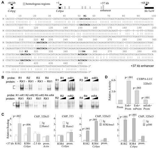
ChIP-Seq for Runx1 in the murine HPC-7 hematopoietic progenitor cell line identified 2 peaks in the vicinity of the Cebpa gene, at 35 and 37 kb.28 We used Blast (blast.ncbi.nlm.nih.gov) to search for homologous regions between −64 kb and 68 kb relative to the Cebpa transcription start site, encompassing the regions between the adjacent Cebpg and Slc7a10 genes (Figure 6A top). Aside from the immediate promoter regions and the Cebpa RNA coding segment, 8 blocks with ≥ 79% homology were identified, at 2.5, 5, 8, 19, 20, 26, 35, and 37 kb. Among these regions, the murine 5, 8, and 20 kb segments have a single consensus Runx1 site, but these are not conserved in the human sequence, and the 2.5, 19, 26, and 35 kb segments lack Runx1 sites. By contrast, the 37 kb region contains 4 conserved Runx1 sites, designated R1-R4 (Figure 6A bottom). Each of these sites binds Runx1 in gel shift assay, but not if the Runx1 consensus is mutated (Figure 6B left panels), and mutation of each Runx1 consensus site prevents or greatly diminishes competition with radiolabeled probe for Runx1 binding (Figure 6B right panels). Endogenous Runx1 in 32Dcl3 myeloid cells binds the 37 kb element in the ChIP assay; as a control, NIH 3T3 cells, which lack C/EBPα and Runx1, did not show binding to the enhancer or promoter (Figure 6C). H3K4me1 and p300 were strongly associated with the 37 kb region relative to the promoter (Figure 6C), typical of mammalian enhancers.29 When the 497-bp DNA segment containing the potential 37 kb enhancer was positioned upstream of the Cebpa promoter and the luciferase cDNA, luciferase activity was stimulated 6-fold; mutation of the Cebpa promoter Runx1 site 1 reduced activity 2-fold; and mutation of the 4 Runx1-binding sites in the 37 kb segment reduced activity 6-fold, eliminating activation by the enhancer (Figure 6D).
Runx1 binds and activates transcription via a 450-bp conserved 37 kb Cebpa enhancer. (A) Comparison of the murine and human Cebpa genomic loci identifies 8 regions of homology upstream of the single Cebpa exon (top). Alignment of a 453-bp region from 37 kb of the murine Cebpa locus (M) with a related region from 41 kb in the human CEBPA locus (H) is also shown. The 4 conserved Runx1 sites (R1-R4) are indicated in bold (bottom). (B) Double-stranded DNA probes containing sites R1-R4, or mutant variants in which the core Runx1 consensus 5′-ACCACA was mutated to 5′-TGCACA, were radiolabeled and subjected to gel shift analysis with the use of nuclear extracts from 293T cells transduced with empty CMV vector (−) or with CMV-RUNX1c and CMV-CBFβ (RX1, left panels). Radiolabeled sites R1-R4 were also subjected to gel shift analysis alone or in the presence of 5- or 25-fold excess unlabeled WT or mutant oligonucleotides (right panels). (C) 32Dcl3 cells were subjected to ChIP with the use of 2 μg of rabbit anti-Runx1 antiserum or normal rabbit IgG, followed by genomic DNA PCR with the use of oligonucleotides centered between R1 and R2 or surrounding R3 and R4, at −2.5 kb of the Cebpa promoter, or within the β-actin promoter. Binding was quantified relative to input, and this value was set to 1.0 for ChIP with IgG on the R1-R2 region of the Cebpa enhancer (panel 1). NIH 3T3 were subjected to ChIP with the use of 2 μg of Runx1 antiserum, followed by enhancer or promoter PCR (panel 2). 32Dcl3 cells were also subjected to ChIP with 0.5 μg of H3K4me1 or 1 μg of p300 antisera (panels 3 and 4). Data are mean and SE of 3 determinations. (D) 32Dcl3 cells were transduced with 5 μg of luciferase reporters containing the murine Cebpa promoter alone (Prom), the promoter with the conserved 37 kb region positioned upstream (Enh+Prom), or the later construct harboring either mutation of the promoter Runx1 site 1 (Enh+mProm) or mutation of sites R1-R4 in the enhancer region (mEnh+Prom), together with 0.25 μg of CMV-βGal. Luciferase and β-galactosidase activities were assessed 2 days later. Normalized luciferase activity of each reporter relative to CMV-βGal activity is shown, with activity of CEBPA(Prom)–LUC set to 1.0 in each experiment (mean of SE of 3 determinations, each done in triplicate).
Runx1 binds and activates transcription via a 450-bp conserved 37 kb Cebpa enhancer. (A) Comparison of the murine and human Cebpa genomic loci identifies 8 regions of homology upstream of the single Cebpa exon (top). Alignment of a 453-bp region from 37 kb of the murine Cebpa locus (M) with a related region from 41 kb in the human CEBPA locus (H) is also shown. The 4 conserved Runx1 sites (R1-R4) are indicated in bold (bottom). (B) Double-stranded DNA probes containing sites R1-R4, or mutant variants in which the core Runx1 consensus 5′-ACCACA was mutated to 5′-TGCACA, were radiolabeled and subjected to gel shift analysis with the use of nuclear extracts from 293T cells transduced with empty CMV vector (−) or with CMV-RUNX1c and CMV-CBFβ (RX1, left panels). Radiolabeled sites R1-R4 were also subjected to gel shift analysis alone or in the presence of 5- or 25-fold excess unlabeled WT or mutant oligonucleotides (right panels). (C) 32Dcl3 cells were subjected to ChIP with the use of 2 μg of rabbit anti-Runx1 antiserum or normal rabbit IgG, followed by genomic DNA PCR with the use of oligonucleotides centered between R1 and R2 or surrounding R3 and R4, at −2.5 kb of the Cebpa promoter, or within the β-actin promoter. Binding was quantified relative to input, and this value was set to 1.0 for ChIP with IgG on the R1-R2 region of the Cebpa enhancer (panel 1). NIH 3T3 were subjected to ChIP with the use of 2 μg of Runx1 antiserum, followed by enhancer or promoter PCR (panel 2). 32Dcl3 cells were also subjected to ChIP with 0.5 μg of H3K4me1 or 1 μg of p300 antisera (panels 3 and 4). Data are mean and SE of 3 determinations. (D) 32Dcl3 cells were transduced with 5 μg of luciferase reporters containing the murine Cebpa promoter alone (Prom), the promoter with the conserved 37 kb region positioned upstream (Enh+Prom), or the later construct harboring either mutation of the promoter Runx1 site 1 (Enh+mProm) or mutation of sites R1-R4 in the enhancer region (mEnh+Prom), together with 0.25 μg of CMV-βGal. Luciferase and β-galactosidase activities were assessed 2 days later. Normalized luciferase activity of each reporter relative to CMV-βGal activity is shown, with activity of CEBPA(Prom)–LUC set to 1.0 in each experiment (mean of SE of 3 determinations, each done in triplicate).
Exogenous C/EBPα favors granulocytic specification in Runx1-deleted marrow cells
In addition to inducing C/EBPα in 32Dcl3 cells, activation of exogenous RUNX1-ER after its transduction into Runx1-deleted marrow cells induced C/EBPα several fold, as was seen also in a second experiment, without affecting PU.1 protein expression (Figure 7A). To evaluate whether exogenous C/EBPα is sufficient to correct the granulocytic defect induced by Runx1 gene deletion, marrow mononuclear cells isolated from Runx1(f/f);Mx1-Cre mice exposed to pIpC and 4 weeks later to 5-FU were transduced with pBabePuro-C/EBPα-ER or the pBabePuro vector. After puromycin selection, viable cells were plated in methylcellulose with IL-3, IL-6, and SCF, with or without 1μM estradiol (E2). As we found previously,30 activation of C/EBPα-ER with this saturating dose of E2 reduced the total number of CFUs ∼ 8-fold, probably reflecting the cell cycle–inhibitory effect of C/EBPα. Although absolute numbers of both CFU-Gs and CFU-Ms were reduced, the proportion of CFU-Gs among these progenitors was increased 3-fold on average, from 8% to 24%, with a corresponding decrease in CFU-Ms (Figure 7B left panel). As we did previously to reduce cell cycle inhibition mediated by C/EBPα-ER, this experiment was repeated with 0.03μM E2 (Figure 7B center panel). Total colony numbers were now reduced < 2-fold, and the proportion of CFU-Gs still increased 1.5-fold. E2 at 1μM did not alter the number or proportion of myeloid CFUs obtained from Runx1-deleted marrow cells transduced with pBabePuro.
Exogenous C/EBPα restores granulopoiesis relative to monopoiesis in Runx1-deleted marrow cells. (A) Marrow mononuclear cells isolated from Runx1(f/f);Mx1-Cre mice exposed to pIpC and subsequently to 5-FU were transduced with RUNX1-ER or with the pBabePuro vector, selected with puromycin, and then placed in liquid culture with IL-3, IL-6, and SCF with or without 4HT. Total cellular proteins collected 2 days later were subjected to Western blot analysis with the use of C/EBPα, PU.1, ERα, or β-actin Abs. (B) Marrow mononuclear cells isolated from Runx1(f/f);Mx1-Cre mice exposed to pIpC and subsequently to 5-FU were transduced with C/EBPα-ER, selected with puromycin, and then plated in methylcellulose with IL-3, IL-6, and SCF with or without 1.0μM or 0.03μM E2. The proportion of CFU-Gs or CFU-Ms among these myeloid CFUs is shown (left and center panels; mean and SE from 3 determinations). Similar analysis was conducted after transduction with pBabePuro and culture with or without 1.0 μM E2 (right panel; mean and SE from 3 determinations).
Exogenous C/EBPα restores granulopoiesis relative to monopoiesis in Runx1-deleted marrow cells. (A) Marrow mononuclear cells isolated from Runx1(f/f);Mx1-Cre mice exposed to pIpC and subsequently to 5-FU were transduced with RUNX1-ER or with the pBabePuro vector, selected with puromycin, and then placed in liquid culture with IL-3, IL-6, and SCF with or without 4HT. Total cellular proteins collected 2 days later were subjected to Western blot analysis with the use of C/EBPα, PU.1, ERα, or β-actin Abs. (B) Marrow mononuclear cells isolated from Runx1(f/f);Mx1-Cre mice exposed to pIpC and subsequently to 5-FU were transduced with C/EBPα-ER, selected with puromycin, and then plated in methylcellulose with IL-3, IL-6, and SCF with or without 1.0μM or 0.03μM E2. The proportion of CFU-Gs or CFU-Ms among these myeloid CFUs is shown (left and center panels; mean and SE from 3 determinations). Similar analysis was conducted after transduction with pBabePuro and culture with or without 1.0 μM E2 (right panel; mean and SE from 3 determinations).
Discussion
Absence of Runx1 in adult mice leads to increased numbers of LSK cells, CMPs, GMPs, and myeloid CFUs.1-6 In human AMLs associated with expression of CBF oncoproteins or RUNX1 mutation, a related expansion of myeloid progenitors may contribute to transformation. Herein, we demonstrate that Runx1 gene deletion or dominant inhibition impairs Cebpa gene transcription and granulopoiesis, with a compensatory shift toward monopoiesis. Runx1 binds the Cebpa promoter and a putative 450-bp enhancer located at +37 kb in gel shift or ChIP assays, mutation of a conserved Runx1 binding site in the promoter or of 4 conserved RUNX1 binding sites in the distal region reduces reporter activity in a myeloid cell line, and RUNX1-ER rapidly induces transcription of the endogenous Cebpa gene in the presence or absence of cycloheximide. Runx1 gene deletion in vivo or in vitro or expression of the dominant-inhibitory RUNX1a in vivo reduces CFU-Gs relative to CFU-Ms and reduces Cebpa mRNA levels in CMPs, GMPs, lineage−, or total mononuclear marrow cells. In addition, exogenous RUNX1-ER or C/EBPα-ER rescues granulopoiesis in Runx1−/− marrow cells. Of note, in a large cohort of human AML cases, mutation of RUNX1 was associated on average with ∼ 2-fold reduced CEBPA mRNA expression.31
Besides inhibition of differentiation, a direct effect on cell cycle kinetics might also contribute to the myeloproliferative effect of Runx1 gene deletion or dominant-inhibition. Several lines of evidence suggest, however, that perturbation of Runx1 would slow rather than accelerate the cell cycle in myeloid progenitors. Runx1 gene deletion in adult mice slows G1 to S cell cycle progression of HSCs or multipotent progenitors,32 transduction of myeloid cells with CBFβ-SMMHC or AML1-ETO slows their cell cycle progression,33,34 and exogenous Runx1 accelerates the cell cycle.35 Of note, C/EBPα inhibits cell cycle progression in myeloid progenitors and in other lineages via interaction with E2F1.36-38 Reduced expression of C/EBPα in response to Runx1 deletion would probably accelerate cell proliferation, but this effect could be balanced by reduced expression of Runx1 genetic targets such as cdk4 or cyclin D2.18,39 In fact, Runx1-deleted myeloid cells expanded at a rate similar to control cells in vitro in this study, despite having reduced C/EBPα levels.
Herein, we demonstrate that RUNX1 binds and activates transcription via conserved sites in both the Cebpa promoter and via 4 sites in a highly conserved 450-bp segment located at 37 kb. Although future in vivo studies are required to further assess the role of the 37 kb segment in Cebpa gene regulation, our findings lend credence to the idea that Runx1 directly regulates Cebpa gene expression and that RUNX1 gene alterations in AML reduce CEBPA expression as a direct consequence, to impair myeloid differentiation and favor myeloid transformation. Of note, ChIP-Seq data indicate that SCL, LYL1, LMO2, GATA2, ERG, and FLI-1 colocalize with RUNX1 on the putative +37 kb enhancer, as these investigators find occurs commonly at other genomic locations as well,28 and we find that this region strongly binds the p300 coactivator and contains the enhancer-specific H3K4me1 histone modification.29
Runx1 also binds and activates the Pu.1 distal enhancer, and Cre-mediated deletion of floxed Runx1 alleles reduces total marrow expression of Pu.1.12 Consistent with these findings, we also find reduced Pu.1 mRNA levels in marrow cells lacking Runx1, with Pu.1 levels also reduced in GMP and CMP myeloid progenitors. Reduced PU.1 expression in AML cases with diminished Runx1 activity might contribute to myeloid transformation, because homozygous deletion of the Pu.1 distal enhancer leads to a highly penetrant AML in mice.14 Of note, deletion of the Pu.1 enhancer reduces PU.1 to 20% of control values; in contrast, heterozygous Pu.1+/− mice with 50% of normal PU.1 levels do not develop AML. However, reduced PU.1 expression probably does not account for the impaired granulopoiesis we find in Runx1-deleted mice or after RUNX1a expression, because diminished expression of PU.1 reduces monopoiesis while sparing granulopoiesis,13-15 and absence of one Pu.1 allele increases granulopoiesis in adult Runx1(f/f);Mx1-Cre mice exposed to pIpC.12
We instead postulate that reduced C/EBPα expression resulting from Runx1 deletion impedes granulopoiesis and provides the main impetus for the resulting myeloproliferative phenotype. Of note, we found a greater reduction in Cebpa than Pu.1 mRNA in marrow progenitor subsets lacking Runx1 or expressing RUNX1a. In 32Dcl3 cells, which are committed to the granulocyte lineage, exogenous C/EBPα substitutes for G-CSF signals to direct their maturation.17 However, in marrow myeloid progenitors, exogenous C/EBPα stimulates monopoiesis.29 Direct activation of the Pu.1 distal enhancer might contribute to C/EBPα-mediated monopoiesis; consistent with this idea, C/EBPα-ER instead increases CFU-Gs relative to CFU-Ms in Pu.1(kd/kd) marrow lacking the distal enhancer.40 Similarly, C/EBPα-ER may stimulate granulopoiesis in Runx1-deleted marrow, as shown herein, because of a diminished ability to induce Pu.1 transcription in the absence of Runx1. RUNX1-ER induced PU.1 in 32Dcl3 cells but not in Runx1-deleted marrow cells, perhaps reflecting inaccessibility of the PU.1 distal enhancer.41
C/EBPα may also contribute to normal monopoiesis via leucine zipper-mediated heterodimerization with AP-1 proteins to induce FosB and other target genes, with C/EBPα homodimers favoring granulopoiesis.42,43 Perhaps lower expression of C/EBPα resulting from Runx1 gene deletion allows sufficient C/EBPα:AP-1 heterodimer formation while greatly reducing C/EBPα:C/EBPα homodimerization, based on mass action considerations, to impede granulopoiesis. Of note, as with Runx1 gene deletion, adult mice lacking NF-κB p50 have a ∼ 3-fold reduction in marrow Cebpa expression and reduced granulopoiesis.44 Also connecting C/EBPα expression to granulopoiesis, G-CSF induces SHP2 phosphorylation more potently than M-CSF, and shRNA-mediated inhibition of SHP2 in marrow cells impairs Cebpa transcription and reduces granulopoiesis relative to monopoiesis.45,46 SHP2 dephosphorylates Runx1 tyrosine residues,47 and exogenous Runx1 partially rescues C/EBPα expression in 32Dcl3 cells harboring SHP2 shRNA,46 consistent with the idea that G-CSF signals stimulate Runx1 activity via SHP2 to allow increased Runx1-mediated activation of Cebpa transcription and thereby granulopoiesis.
The relevance of reduced C/EBPα activity to myeloid transformation is evident from the finding that CEBPA gene mutations occur in about 10% of human AML cases; in contrast, PU.1 mutations are apparently less common.48,49 In addition, RUNX1-ETO directly represses the CEBPA promoter.50 RUNX1-ETO may tether to the CEBPA promoter via bound C/EBPα, as we proposed, but in addition might bind to the promoter or +37 kb enhancer via the Runx1-binding sites we have now identified. RUNX1-ETO also potentially represses PU.1 gene transcription via interaction with its distal enhancer, and CEBPA gene mutation might secondarily reduce PU.1 expression because of the presence of functional, evolutionarily conserved C/EBPα-binding sites in the same enhancer.40 Thus, as diagrammed (supplemental Figure 4), RUNX1 mutation or dominant-inhibition, present in ∼ 30% of AML cases, probably reduces expression or activity of RUNX1, CEBPA, and PU.1; CEBPA mutation probably affects both CEBPA and PU.1; whereas PU.1 mutation may only reduce PU.1 activity, perhaps accounting for the rarity of such genetic lesions.
The online version of this article contains a data supplement.
The publication costs of this article were defrayed in part by page charge payment. Therefore, and solely to indicate this fact, this article is hereby marked “advertisement” in accordance with 18 USC section 1734.
Acknowledgments
This work was supported by National Institutes of Health grants R01 HL089176 and U01 HL099775/HL100397, and a grant from the Maryland Stem Cell Research Foundation to A.D.F. and U01 HL100405 to N.A.S.
National Institutes of Health
Authorship
Contribution: H.G. and O.M. performed the experiments; H.G., O.M., N.A.S., and A.D.F. designed the study, analyzed the data, and wrote the paper; all authors approved the final version of the manuscript.
Conflict-of-interest disclosure: The authors declare no competing financial interests.
Correspondence: Alan D. Friedman, Division of Pediatric Oncology, Johns Hopkins University School of Medicine, CRB I, Rm 253, 1650 Orleans St, Baltimore, MD 21231; e-mail: afriedm2@jhmi.edu.


This feature is available to Subscribers Only
Sign In or Create an Account Close Modal