Abstract
Alzheimer disease is characterized by the presence of increased levels of the β-amyloid peptide (Aβ) in the brain parenchyma and cerebral blood vessels. This accumulated Aβ can bind to fibrin(ogen) and render fibrin clots more resistant to degradation. Here, we demonstrate that Aβ42 specifically binds to fibrin and induces a tighter fibrin network characterized by thinner fibers and increased resistance to lysis. However, Aβ42-induced structural changes cannot be the sole mechanism of delayed lysis because Aβ overlaid on normal preformed clots also binds to fibrin and delays lysis without altering clot structure. In this regard, we show that Aβ interferes with the binding of plasminogen to fibrin, which could impair plasmin generation and fibrin degradation. Indeed, plasmin generation by tissue plasminogen activator (tPA), but not streptokinase, is slowed in fibrin clots containing Aβ42, and clot lysis by plasmin, but not trypsin, is delayed. Notably, plasmin and tPA activities, as well as tPA-dependent generation of plasmin in solution, are not decreased in the presence of Aβ42. Our results indicate the existence of 2 mechanisms of Aβ42 involvement in delayed fibrinolysis: (1) through the induction of a tighter fibrin network composed of thinner fibers, and (2) through inhibition of plasmin(ogen)–fibrin binding.
Introduction
Cerebrovascular dysfunction has been implicated as an early event in Alzheimer disease (AD) progression,1-4 but the origins and mechanisms of vascular dysfunction in AD are not clear. The β-amyloid peptide (Aβ) accumulates in the brain parenchyma and blood vessel walls of AD patients and has been genetically and clinically linked to AD. We have previously shown that fibrinogen, the main protein component of blood clots, can bind Aβ42 (henceforth designated Aβ) specifically with a Kd of 26.3 ± 6.7nM.5 We also found that fibrin clots formed in the presence of Aβ are structurally altered and more resistant to fibrinolysis than normal clots.6 However, the mechanism by which Aβ-fibrin(ogen) binding delays fibrin clot lysis has not been defined.
Fibrin clot lysis is mediated by plasmin, a serine protease that cleaves the fibrin network at specific sites. Plasmin is derived from plasminogen by tissue plasminogen activator (tPA) in the presence of fibrin, which itself enhances the rate of the reaction. One fibrin site initially involved in plasminogen activation by tPA includes residues 148-160 on the Aα-chain (reviewed by Medved and Nieuwewnhuizen7 ). This site becomes exposed and available for plasminogen binding after the conversion of fibrinogen to fibrin,8 but could remain hidden if clots are formed in the presence of Aβ, leading to delayed clot lysis. This hypothesis is derived from our finding that Aβ binds the fibrinogen β-chain near the β-hole,5 which is in close spatial proximity to residues 148-160 of the Aα-chain.9 Another potential explanation for delayed clot lysis is based on the relationship between fibrin structure and its susceptibility to fibrinolysis. Tighter fibrin networks composed of thin fibers are degraded less efficiently by plasmin than those composed of thick fibers10-13 because: (1) there are more fibers to be cleaved,11,14 requiring plasmin to detach from and move between fibers more frequently15 ; and (2) decreased network porosity of tighter fibrin networks results in impeded diffusion of fibrinolytic enzymes throughout the clot (reviewed by Lord16 ). Potential effects of Aβ on fibrin fiber thickness could thus be another mechanism of Aβ-mediated impaired fibrinolysis.
Here, we investigate the effects of Aβ itself and of the Aβ-influenced fibrin network on the activity and function of the fibrinolytic factors tPA, plasminogen, and plasmin. We show that generation of plasmin is slowed, and that plasmin-mediated degradation of clots is attenuated in clots formed with Aβ. This occurs through Aβ-mediated hindrance of plasmin(ogen)'s access to fibrin and from Aβ-induced tightening of the fibrin network, and not through the direct effect of Aβ on fibrinolytic enzyme activity.
Methods
Materials
Human plasminogen-free fibrinogen was from Calbiochem. Alexa Fluor 488–conjugated fibrinogen was from Invitrogen. Bovine TPCK trypsin was from Thermo Scientific. Streptokinase (SK), human α-thrombin, and human plasmin were from Sigma-Aldrich. tPA was generously provided by Genentech. Human plasminogen was purified from human plasma (New York Blood Center) using lysine-sepharose as described,17 and copurifying plasmin was inactivated with 10mM diisopropyl fluorophosphate. The absence of plasmin activity in our plasminogen preparation was confirmed by chromogenic substrate assay (not shown). FITC-labeled human Glu-plasminogen was from Oxford Biomedical. Monoclonal anti-plasminogen antibody l0A1 was from Santa Cruz Biotechnology. Aβ peptides, HiLyte Fluor 555–labeled Aβ peptides, and amylin were from Anaspec. Chromogenic substrates Pefa-5329 and S-2288 were from Centerchem and Diapharma, respectively.
Preparation of amyloid peptides
Aβ42 was reconstituted to 1 mg/mL in 50mM Tris pH 7.4, 0.1% NH4OH and stored at −80°C. Before use, Aβ was incubated at 37°C with shaking for 12 hours to generate a range of oligomeric species. Insoluble Aβ42 was removed by centrifugation at 12 000g for 10 minutes.18 The concentration of soluble Aβ42 was verified via bicinchoninic acid assay (BCA), and a representative transmission electron microscope (TEM) image of the Aβ species used is shown in supplemental Figure 1 (available on the Blood Web site; see the Supplemental Materials link at the top of the online article). HiLyte Fluor 555-Aβ42 and HiLyte Fluor 555-Aβ1-9 were reconstituted to 0.5 mg/mL in the same buffer and not incubated before use.
Amylin was reconstituted to 2 mg/mL in DMSO. Before use, amylin was diluted to 0.2 mg/mL with 50mM Tris pH 7.4 (10% DMSO final concentration) and incubated for 24 hours at 37°C with shaking. Presence of amyloid fibrils was confirmed by TEM.
TEM
Samples were diluted to 0.1 mg/mL, applied to glow discharged CF200-Cu grids (Electron Microscopy Sciences), washed 3 times with ultrapure water (UV-treated with a Millipore system), and negatively stained with 2% uranyl acetate. Images were acquired using a JEOL JEM 100CX transmission microscope at The Rockefeller University Electron Microscopy Resource Center.
Preparation of fibrin monolayers
Clot turbidity analysis
Assays were performed at room temperature (RT) in Fisherbrand High Binding 96-well plates (Fisher Scientific) in triplicate using a Molecular Devices Spectramax Plus384 reader. For clot formation and lysis, fibrinogen (1.5μM) with or without Aβ42 (3μM) was mixed with plasminogen (125nM-300nM), thrombin (0.5 U/mL), tPA (0.15nM-15nM), and CaCl2 (5mM) in 20 mM HEPES (N-2-hydroxyethylpiperazine-N′-2-ethanesulfonic acid) buffer (pH 7.4) with 140mM NaCl, in a volume of 150 μL.
For lysis of preformed clots, fibrinogen (2.2μM) with or without Aβ42 (5μM) was mixed with plasminogen (125nM), thrombin (0.5 U/mL), and CaCl2 (5mM) in 20mM HEPES buffer (pH 7.4) with 140mM NaCl, in a volume of 100 μL. Clots were incubated at 37°C for 1 hour, then overlaid with a 100 μL solution containing 10-50nM tPA, 250nM plasmin, or 1μM trypsin. Clots overlaid with plasmin and trypsin did not contain plasminogen. Before lysis, some clots were overlaid with 5μM Aβ42 for 1 hour, the overlays removed, and the clot surface washed 3 times with HEPES buffer.
The fibrinogen used (Calbiochem) is plasminogen-depleted by the manufacturer. We confirmed the absence of plasminogen by Western blot (not shown). No exogenous factor XIII was added to our clotting reactions. However, factor XIII copurifies with fibrinogen, and its presence and crosslinking activity in our clots was confirmed by SDS-PAGE of fibrin clot degradation products, which contained D-dimers (not shown). Trace amounts of fibronectin were also detected in the fibrinogen preparation (not shown), but would not affect the results because our Aβ preparation does not bind fibronectin.5
Enzyme activity
Assays were performed at RT in Fisherbrand 96-well plates with a reaction volume of 150 μL. For plasmin activity, chromogenic substrate Pefa-5329 (530μM) was added to plasmin (440nM) with various amounts of Aβ42. For tPA activity, chromogenic substrate S-2288 (530μM) was added to tPA (100nM) with various amounts of Aβ42. For tPA-mediated plasminogen activation, plasminogen (440nM) and tPA (100nM) were mixed with various amounts of Aβ42, and Pefa-5329 was added to monitor plasmin generation.
To monitor plasmin activity and clot turbidity simultaneously and under the same reaction conditions, parallel clots were prepared containing fibrinogen (1.5μM), plasminogen (300nM), tPA (0.15nM) or SK (0.75nM), thrombin (0.5 U/mL), CaCl2 (5mM) and either Pefa-5329 (400μM) or buffer only (modified from Longstaff et al13 and Mutch et al21 ). Readings were taken at dual wavelengths of 405 nm (A405) and 350 nm (A350). Plasmin activity was detected in the samples containing Pefa-5329 at A405, from which the signal arising from the changing turbidity (A405) of the forming and lysing clot without Pefa-5329 was subtracted. Clot turbidity and plasmin activity were plotted together to best visualize the temporal relationship of the 2 processes.
Fibrin monolayers were overlaid with 60 μL of Aβ42 (2μM) or vehicle in PBS and incubated for 18 hours at 4°C. The Aβ was removed, and the well surface washed 3 times with PBS with 0.05% Tween 20. Fibrin monolayers or control wells were then overlaid with 150 μL of plaminogen (300nM) and tPA (1.5nM), and plasmin activity measured with 530μM of the chromogenic substrate Pefa-5329.
Clot structure and binding studies: laser scanning confocal microscopy
Samples were visualized at RT using a Zeiss (Jena, Germany) LSM 510 confocal laser scanning system and a Zeiss Axiovert 200 microscope with a 40×-Axiovert 1.2/water objective (3× optical zoom). Laser scanning was done in multitrack scanning mode with excitation at 488 nm and emission at 500-530 nm (for Alexa Fluor 488 fibrinogen) and excitation at 543 nm and emission at 565-615 nm (for 555 HiLyte Fluor Aβ). All images were acquired using LSM 510 Version 3.2 software (Zeiss) 50 μm above the glass surface. Z-stacks of 5 μm with slices taken every 0.5 μm (11 slices per image) were projected 2-dimensionally to produce the final image.
Aβ binding to fibrin(ogen)
Clots (30 μL final volume) were formed in 1.5 mm glass-bottom dishes (Mattek). Fibrinogen (2.7μM) and Alexa Fluor 488 fibrinogen (0.3μM) were mixed with thrombin (0.5 U/mL) and CaCl2 (5 mM) in 20mM HEPES (pH 7.4) with 140mM NaCl with or without Aβ. For clots with Aβ, nonlabeled Aβ42 (1.5μM) was mixed with HiLyte Fluor 555 Aβ42 (1.5μM) or HiLyte Fluor 555 Aβ1-9 (1.5μM). Clots were incubated in the dark at 37°C for 1 hour before imaging. For overlay experiments, clots formed as described were overlaid with HiLyte Fluor 555 Aβ42 (3μM), HiLyte Fluor 555 Aβ1-9 (3μM) or vehicle for 1 hour, the overlays removed, and the clot surfaces washed with HEPES buffer. The fluorescent label does not alter Aβ's ability to delay clot lysis (supplemental Figure 2).
Plasminogen binding to fibrin
Clots (30 μL final volume) were formed in 1.5 mm glass-bottom dishes with fibrinogen (3μM), FITC labeled plasminogen (125nM), thrombin (0.5 U/mL), CaCl2 (5mM), with or without Aβ42 (5μM) in 20mM HEPES (pH 7.4) buffer with 140mM NaCl. Samples were visualized as previously described, but using single track mode with excitation at 488 nm and emission at 500-530 nm. Five sections were captured from random locations in 3 separate control and Aβ-influenced clots. Representative 5 μm z-stacks composed of 11 slices and projected 2-dimensionally were also acquired for each control and Aβ-containing clot. FITC-plasminogen binding to fibrin was analyzed using ImageJ Version 1.41o software (National Institutes of Health), with total fluorescence in each section used as a measure of plasminogen binding.
Plasminogen binding to fibrin monolayer: ELISA
Fibrin monolayers were incubated with Aβ42 (2μM) or vehicle for 18 hours at 4°C. Nonbound Aβ was removed and the monolayers washed with PBS with 0.05% Tween 20 three times. Plasminogen (500nM) was then applied to the monolayers in PBS with 4% milk and 0.01% Tween 20 for 2 hours at 37°C. Plasminogen was removed, and monolayers washed with PBS with 0.05% Tween 20 three times. Monoclonal plasminogen antibody 10A1 (1:2000) in PBS with 4% milk and 0.01% Tween 20 was applied to the monolayer for 1 hour at RT. After washing, HRP-conjugated anti–mouse antibody (1:5000) in the same buffer was applied for 1 hour at RT. After washing, the ELISA was developed using a tetramethylbenzidine peroxidase substrate (Vector) and reactions stopped using 1N H2SO4. Absorbance was measured at 450 nm. All conditions were tested in triplicate.
Statistical analysis
Data are presented as the mean ± SD of at least 3 separate experiments. Turbidity curves are presented as the mean without standard deviation for clarity. Statistical significance was determined by using the unpaired, 2-tailed t test (GraphPad Prism Version 5.0c software). P values less than .05 are considered significant.
Results
Aβ delays clot lysis but does not directly inhibit fibrinolytic enzyme activity
The effect of Aβ42 on clot lysis was evaluated in different conditions using 2 methods. In 1 method, tPA was added to the clotting mixture at the start of clot formation to simulate internal fibrinolysis, which is thought to approximate physiologic conditions.22 Lysis rates were compared using half-lysis times, which were calculated from the time when maximum turbidity was achieved to when the clot reached half its maximum turbidity. We tested the effect of Aβ on clot lysis over a wide range of tPA concentrations (Figure 1A-B; panel A shows only 1 tPA concentration for clarity), because tPA levels can increase dramatically from their base levels in response to injury. A significant delay in lysis relative to control was observed at all 3 tPA concentrations tested (Figure 1B), suggesting that the effect may be relevant in chronic as well as acute injury states. Furthermore, the delay in lysis is Aβ concentration-dependent, with concentrations from 500nM to 3μM producing significant and increasing delays in lysis (Figure 1C-D). In a complementary method, preformed clots made with or without Aβ were overlaid with tPA to initiate fibrinolysis, a system that is more relevant to the pharmacologic treatment of thrombosis.22 In this system, half lysis time was defined as the time from tPA overlay until the clot reached half its maximum turbidity, and was significantly increased for Aβ-containing clots (Figure 1E-F).
Clot lysis is delayed through a range of tPA concentrations in the presence of Aβ42 in a dose-dependent manner. Clot formation and lysis were monitored by turbidity assay. (A) Clot formation and lysis were initiated as described in “Clot turbidity analysis” by combining thrombin, fibrinogen, CaCl2, plasminogen, and 0.15nM, 1.5nM, or 15nM tPA with or without 3μM Aβ42 (0.15nM and 15nM curves not shown because of scale differences). (B) Half-lysis of Aβ clots was significantly longer than for control clots for all concentrations of tPA. (C) Clots were formed as in panel A with 0.5μM, 1.5μM, 3μM Aβ42, or vehicle and 1.5nM tPA. (D) Half-lysis of Aβ clots was significantly delayed in a dose-dependent manner. (E) Preformed clots prepared as described in “Clot turbidity analysis” were overlaid with 50nM tPA. (F) Half-lysis of Aβ clots was significantly longer than control. (G) Clotting and lysis of clots formed as in panel A but with 3μM amylin confirmed by TEM to be fibrillar (inset) did not differ from control. (H) Clots formed as in panel A but with 3μM Aβ1-28 did not differ from control clots (turbidity plots represent mean of 3 experiments; bar graphs represent mean ± SD of 3 experiments; statistical significance noted as *P < .05, **P < .005, and ***P < .0005).
Clot lysis is delayed through a range of tPA concentrations in the presence of Aβ42 in a dose-dependent manner. Clot formation and lysis were monitored by turbidity assay. (A) Clot formation and lysis were initiated as described in “Clot turbidity analysis” by combining thrombin, fibrinogen, CaCl2, plasminogen, and 0.15nM, 1.5nM, or 15nM tPA with or without 3μM Aβ42 (0.15nM and 15nM curves not shown because of scale differences). (B) Half-lysis of Aβ clots was significantly longer than for control clots for all concentrations of tPA. (C) Clots were formed as in panel A with 0.5μM, 1.5μM, 3μM Aβ42, or vehicle and 1.5nM tPA. (D) Half-lysis of Aβ clots was significantly delayed in a dose-dependent manner. (E) Preformed clots prepared as described in “Clot turbidity analysis” were overlaid with 50nM tPA. (F) Half-lysis of Aβ clots was significantly longer than control. (G) Clotting and lysis of clots formed as in panel A but with 3μM amylin confirmed by TEM to be fibrillar (inset) did not differ from control. (H) Clots formed as in panel A but with 3μM Aβ1-28 did not differ from control clots (turbidity plots represent mean of 3 experiments; bar graphs represent mean ± SD of 3 experiments; statistical significance noted as *P < .05, **P < .005, and ***P < .0005).
Aβ can adopt β-sheet structure, and it is possible that β-sheet structure alone was responsible for delayed fibrinolysis. However, clots formed with the amyloid peptide amylin, which had been aged and confirmed by TEM to contain fibrils (Figure 1G inset), did not exhibit delayed lysis (Figure 1G). Furthermore, a truncated Aβ peptide (Aβ1-28) had no effect on clot lysis (Figure 1H). Thus, β-sheet structure alone is not enough to elicit delayed clot lysis, and a specific interaction between Aβ and fibrin(ogen) is crucial for the effect.
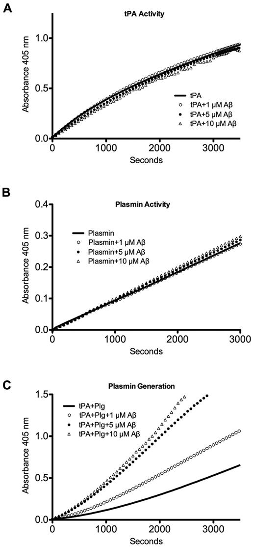
It is possible that Aβ interacts directly with fibrinolytic enzymes in solution and reduces their activity. We assessed the effect of a range of Aβ concentrations on the activities of tPA and plasmin. No effect of Aβ on tPA (Figure 2A) or plasmin (Figure 2B) activity was observed. It has been shown that the conversion of plasminogen to plasmin by tPA is enhanced in the presence of Aβ, particularly for aggregated forms of Aβ.23-25 Because Aβ preparations can vary dramatically, we wanted to exclude the possibility that our preparation would decrease the conversion of plasminogen to plasmin. In agreement with published results, we found that the generation of plasmin from plasminogen by tPA was increased in the presence of Aβ in solution (Figure 2C). These data indicate that the inhibitory effect of Aβ on clot lysis exists despite its ability to potentiate the generation of plasmin in solution.
Aβ does not directly inhibit tPA activity, plasmin activity, or plasmin generation from plasminogen. Aβ42 at 1μM, 5μM, and 10μM or vehicle was combined with (A) tPA and S-2288 to monitor tPA activity; (B) plasmin and Pefa-5329 to monitor plasmin activity; or (C) tPA, plasminogen (Plg), and Pefa-5329 to monitor plasmin generation from plasminogen. Representative results from ≥ 3 separate experiments.
Aβ does not directly inhibit tPA activity, plasmin activity, or plasmin generation from plasminogen. Aβ42 at 1μM, 5μM, and 10μM or vehicle was combined with (A) tPA and S-2288 to monitor tPA activity; (B) plasmin and Pefa-5329 to monitor plasmin activity; or (C) tPA, plasminogen (Plg), and Pefa-5329 to monitor plasmin generation from plasminogen. Representative results from ≥ 3 separate experiments.
Aβ-associated fibrin is a weaker enhancer of plasmin generation and a poorer substrate for plasmin cleavage
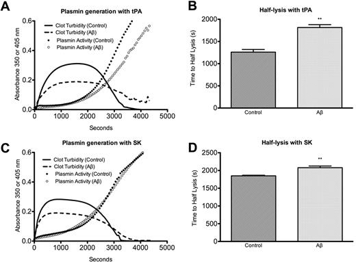
Fibrin enhances the activation of plasminogen by tPA, and any changes introduced into the fibrin network by Aβ could alter the efficiency of tPA/plasminogen interactions with fibrin. To evaluate this possibility, we included the plasmin activity-monitoring chromogenic substrate Pefa-5329 in clotting reactions, and the rate of plasmin generation was monitored during clotting and lysis. Clots formed with Aβ showed delayed lysis and had reduced plasmin activity compared with control clots (Figure 3A-B). Because the reduced activity in Figure 3A is not due to direct inhibition of plasmin activity by Aβ (in solution, Figure 2B; in clot overlay, supplemental Figure 3), it implies a decrease in the rate of plasmin generation and may reflect Aβ-mediated disruption of the interaction between fibrin and tPA/plasminogen. To determine whether the decrease in plasmin generation is fibrin-related, we used SK instead of tPA to activate plasminogen, since the rate of plasminogen activation by SK, unlike tPA, is not enhanced in the presence of fibrin. Accordingly, there was no difference in plasmin generation between Aβ and control when clots were lysed with SK and plasminogen (Figure 3C). Despite similar rates of plasmin generation, the lysis of Aβ-influenced clots was still delayed with SK-initiated lysis (Figure 3C-D). This result could be due to the reduced capability of SK-generated plasmin26 to bind or process fibrin fibers in Aβ-influenced clots versus control clots.
Plasmin generation by tPA, but not SK, is decreased in Aβ-influenced clots during clotting and lysis. (A) Fibrinogen, plasminogen, tPA, thrombin, CaCl2, and Pefa-5329 or vehicle were mixed with or without 3μM Aβ42 as described in “Enzyme activity.” Absorbance was measured at 350 nm to follow clot formation and lysis and at 405 nm to monitor plasmin activity. Curves of A405 without Pefa-5329 were subtracted from Pefa-5329 A405 curves to control for A405 arising from clot turbidity and not plasmin activity. (B) Half-lysis of tPA/plasminogen-lysed clots was delayed in the presence of Aβ compared with control (P = .011). (C) Same as panel A, except SK was substituted for tPA. (D) Half-lysis of SK/plasminogen-lysed clots was delayed in the presence of Aβ compared with control (P = .005).
Plasmin generation by tPA, but not SK, is decreased in Aβ-influenced clots during clotting and lysis. (A) Fibrinogen, plasminogen, tPA, thrombin, CaCl2, and Pefa-5329 or vehicle were mixed with or without 3μM Aβ42 as described in “Enzyme activity.” Absorbance was measured at 350 nm to follow clot formation and lysis and at 405 nm to monitor plasmin activity. Curves of A405 without Pefa-5329 were subtracted from Pefa-5329 A405 curves to control for A405 arising from clot turbidity and not plasmin activity. (B) Half-lysis of tPA/plasminogen-lysed clots was delayed in the presence of Aβ compared with control (P = .011). (C) Same as panel A, except SK was substituted for tPA. (D) Half-lysis of SK/plasminogen-lysed clots was delayed in the presence of Aβ compared with control (P = .005).
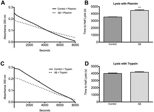
To test this possibility, we initiated clot lysis using preformed plasmin to bypass the plasminogen activation step. In agreement with the SK/plasminogen result, we observed an increase in half-lysis time of Aβ-influenced clots compared with control clots (Figure 4A-B). Plasmin interacts with fibrin through several binding domains and a catalytic domain.15 The serine protease trypsin is also able to dissolve fibrin clots,15 but in contrast to plasmin, it operates entirely through its catalytic domain. When preformed clots were overlaid with trypsin, no delay of lysis of Aβ-influenced clots was observed (Figure 4C-D), suggesting that Aβ interferes with plasmin's access to its binding site and not its cleavage site on fibrin.
Aβ-influenced clots are resistant to lysis by plasmin but not trypsin. (A) Preformed clots prepared as described in “Clot turbidity analysis” with or without 5μM Aβ42 were overlaid with 250nM plasmin. (B) Half-lysis was significantly slower in clots containing Aβ (P = .0001). (C) Preformed clots as in panel A were overlaid with 1μM trypsin. (D) There was no significant difference between half-lysis times of control and Aβ clots (P = .12).
Aβ-influenced clots are resistant to lysis by plasmin but not trypsin. (A) Preformed clots prepared as described in “Clot turbidity analysis” with or without 5μM Aβ42 were overlaid with 250nM plasmin. (B) Half-lysis was significantly slower in clots containing Aβ (P = .0001). (C) Preformed clots as in panel A were overlaid with 1μM trypsin. (D) There was no significant difference between half-lysis times of control and Aβ clots (P = .12).
Aβ is incorporated into fibrin fibers throughout the clot network
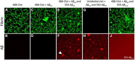
We have previously shown that Aβ-influenced clots are characterized by irregular clusters punctuating the fibrin network, and Congo red staining suggested that Aβ was confined to these clusters.6 Our hypothesis that Aβ interferes with plasmin(ogen)'s access to fibrin throughout the fibrin network requires its regular distribution along fibrin strands. This prompted us to more closely analyze Aβ localization within clots. Tracer amounts of Alexa Fluor 488–labeled fibrinogen (fibrinogen-488) were used to visualize the fibrin network, and HiLyte Fluor-555–labeled Aβ42 (Aβ42-555), was used to visualize Aβ. In agreement with previous results, clots formed in the absence of Aβ showed a regular fibrin network (Figure 5A), whereas clots formed in the presence of Aβ contained irregular clusters (Figure 5C). Aβ-555 labeling of irregular clusters (Figure 5F arrowhead) confirmed our previous conclusion that Aβ binds to fibrin aggregates. However, the uniform labeling of normal fibrin fibrils by Aβ (Figure 5F) showed that it is also distributed throughout the fibrin lattice. The striking colocalization between fibrin and Aβ is not due to the detection of fibrinogen-488 fluorescence in the 555 channel, since clots formed without Aβ-555 produced no signal in the 555 channel (Figure 5B-D). Furthermore, omitting labeled fibrinogen, but not labeled Aβ, reproduced the Aβ-covered fibrin lattice (Figure 5G-H). Replacing 555-Aβ42 with 555-Aβ1-9, which does not cause delayed fibrinolysis (supplemental Figure 2), eliminated the colocalization (Figure 5I-J), confirming that the labeling reflects specific Aβ-fibrin(ogen) binding and is not a result of nonspecific trapping of the 555-labeled peptides in the forming fibrin network. Clots made without unlabeled Aβ42 but still containing 555-Aβ1-9 did not have colocalization between fibrin and Aβ (supplemental Figure 4), precluding the possibility that unlabeled Aβ42 blocks access of 555-Aβ1-9 to fibrin. The specific Aβ-fibrin colocalization found in these experiments, together with the previously described spatial proximity of Aβ and plasminogen binding sites on fibrin(ogen),5,9 suggested that Aβ could block plasmin(ogen) from accessing its binding sites on fibrin. However, another potential mechanism became apparent as well: Aβ-influenced fibrin is composed of thinner fibers arranged in a tighter network than control fibrin (Figure 5A-C), which could make it more resistant to fibrinolysis.10-13
Confocal microscopy of fibrin clots show Aβ binding to fibrin fibrils. Fibrin clots were formed with or without Aβ42 as described in “Aβ binding to fibrin(ogen)” to determine the location of Aβ binding. (Top row) Fibrin visualized with Alexa Fluor 488–labeled fibrinogen (green); (bottom row) Aβ visualized with HiLyte Fluor 555–labeled Aβ (red); (A-B) control clot. (C-D) Clot with only unlabeled Aβ42 shows that fibrin fibers and irregular clusters do not produce signal in the red channel. (E-F) Clot formed with unlabeled Aβ42 and HiLyte Fluor 555–labeled Aβ42 shows colocalization between Aβ and fibrin fibers as well as Aβ and irregular clusters (arrowhead). (G-H) Clot formed without Alexa Fluor 488–labeled fibrinogen but with both unlabeled and labeled Aβ42 shows Aβ signal in the fibrin fiber pattern, confirming that Aβ signal is not Alexa Fluor 488 signal detected in the red channel. (I-J) Clot formed with unlabeled Aβ42 and HiLyte Fluor-555–labeled Aβ1-9 does not have Aβ signal along fibrin fibers or in aggregates, indicating that the Aβ42 signal represents specific Aβ-fibrin(ogen) binding and not fluorophore entrapment. Images are representative of ≥ 3 experiments.
Confocal microscopy of fibrin clots show Aβ binding to fibrin fibrils. Fibrin clots were formed with or without Aβ42 as described in “Aβ binding to fibrin(ogen)” to determine the location of Aβ binding. (Top row) Fibrin visualized with Alexa Fluor 488–labeled fibrinogen (green); (bottom row) Aβ visualized with HiLyte Fluor 555–labeled Aβ (red); (A-B) control clot. (C-D) Clot with only unlabeled Aβ42 shows that fibrin fibers and irregular clusters do not produce signal in the red channel. (E-F) Clot formed with unlabeled Aβ42 and HiLyte Fluor 555–labeled Aβ42 shows colocalization between Aβ and fibrin fibers as well as Aβ and irregular clusters (arrowhead). (G-H) Clot formed without Alexa Fluor 488–labeled fibrinogen but with both unlabeled and labeled Aβ42 shows Aβ signal in the fibrin fiber pattern, confirming that Aβ signal is not Alexa Fluor 488 signal detected in the red channel. (I-J) Clot formed with unlabeled Aβ42 and HiLyte Fluor-555–labeled Aβ1-9 does not have Aβ signal along fibrin fibers or in aggregates, indicating that the Aβ42 signal represents specific Aβ-fibrin(ogen) binding and not fluorophore entrapment. Images are representative of ≥ 3 experiments.
Binding of plasminogen to fibrin and plasmin generation are decreased in the presence of Aβ and are fibrin thickness-independent
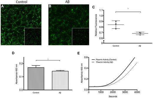
We next tested whether Aβ binding to fibrin affected the ability of plasminogen to bind to fibrin using 2 complementary approaches: confocal microscopy of clots formed with FITC labeled plasminogen and ELISA with an antibody against plasminogen. Confocal microscopy of clots formed with FITC-plasminogen showed plasminogen binding as fluorescence in the pattern of the fibrin network to which it was bound (Figure 6A). Fluorescence was decreased in clots containing Aβ (Figure 6B), suggesting that less plasminogen was bound to the fibrin network. The amount of plasminogen binding was quantified as total fluorescence intensity per slice, because fluorescence intensity is greater for FITC-labeled proteins bound to their target than for FITC-labeled proteins in solution.27 Total fluorescence was significantly lower for Aβ-containing clots (Figure 6C). These results demonstrate that Aβ-modified fibrin binds less plasminogen, but they do not prove that Aβ is blocking plasminogen's access to fibrin, since changes in fibrin fiber thickness may be an alternative explanation. To avoid the possible modification of fibrin fibers by Aβ during fibrin formation, we used an immobilized fibrin monolayer overlaid with Aβ or vehicle as a surface for plasminogen binding. Excluding Aβ from fibrin polymerization eliminates the influence of Aβ on fibrin thickness, and also changes the binding target for Aβ from fibrinogen to fibrin. The binding affinity of Aβ for preformed fibrin may be different (and possibly lower) than for fibrinogen because of conformational changes near the Aβ binding site on fibrinogen that accompany the fibrinogen-fibrin transition. Nonetheless, Aβ overlay of fibrin monolayers decreased the amount of plasminogen bound to fibrin (Figure 6D), suggesting that Aβ can inhibit plasminogen binding to fibrin by impeding its access to fibrin independently of fibrin fiber thickness.
Plasminogen binding to fibrin and plasmin generation is inhibited by Aβ. Fibrin clots were formed with FITC-plasminogen as described in “Plasminogen binding to fibrin,” and 5 μm z-stacks composed of 11 sections were acquired and projected 2-dimensionally for control (A) and Aβ42-containing (B) clots. Images of 15 random sections from 3 separate clots were also acquired and used for quantification (insets show representative single sections). (A) Control clot has formed with FITC-plasminogen shows plasminogen fluorescence in the pattern of the fibrin network. (B) Clot formed with Aβ has less FITC-plasminogen fluorescence. (C) Fluorescence intensity relative to maximum intensity recorded was significantly lower (P = .02) for Aβ-containing clots. (D) Plasminogen binding to fibrin monolayers exposed to 2μM Aβ42 or vehicle was measured by ELISA and normalized to samples not containing plasminogen. Plasminogen binding was decreased in the presence of Aβ (P = .04). (E) Plasmin generation was measured by overlaying tPA, plasminogen, and chromogenic substrate Pefa-5329 on fibrin monolayers exposed to 2μM Aβ42 or vehicle and recording absorbance at 405 nm. Plasmin generation on fibrin monolayers exposed to Aβ was attenuated.
Plasminogen binding to fibrin and plasmin generation is inhibited by Aβ. Fibrin clots were formed with FITC-plasminogen as described in “Plasminogen binding to fibrin,” and 5 μm z-stacks composed of 11 sections were acquired and projected 2-dimensionally for control (A) and Aβ42-containing (B) clots. Images of 15 random sections from 3 separate clots were also acquired and used for quantification (insets show representative single sections). (A) Control clot has formed with FITC-plasminogen shows plasminogen fluorescence in the pattern of the fibrin network. (B) Clot formed with Aβ has less FITC-plasminogen fluorescence. (C) Fluorescence intensity relative to maximum intensity recorded was significantly lower (P = .02) for Aβ-containing clots. (D) Plasminogen binding to fibrin monolayers exposed to 2μM Aβ42 or vehicle was measured by ELISA and normalized to samples not containing plasminogen. Plasminogen binding was decreased in the presence of Aβ (P = .04). (E) Plasmin generation was measured by overlaying tPA, plasminogen, and chromogenic substrate Pefa-5329 on fibrin monolayers exposed to 2μM Aβ42 or vehicle and recording absorbance at 405 nm. Plasmin generation on fibrin monolayers exposed to Aβ was attenuated.
To test whether the decrease in plasminogen binding translates to decreased plasmin generation, fibrin monolayers were exposed to Aβ or vehicle, and the rate of plasminogen activation by tPA was measured using chromogenic substrate Pefa-5329. Plasmin activity was decreased in fibrin monolayers that had been exposed to Aβ (Figure 6E). The activation of plasminogen by tPA was fibrin-dependent, since identical reactions in wells that did not contain fibrin monolayers produced negligible amounts of plasmin (supplemental Figure 5). This confirms that fibrin in the presence of Aβ is a weaker enhancer of plasminogen activation by tPA (Figure 3), but without the differences in clot structure as a confounding factor.
Aβ overlaid onto preformed clots delays fibrinolysis
We next tested whether Aβ can delay fibrinolysis independently of its effect on clot structure. Clots prepared without Aβ, and therefore having normal structure, were overlaid with a solution containing Aβ or vehicle for 1 hour, after which the solutions were removed and the clot surfaces washed. The clots were then overlaid with a tPA solution to initiate fibrinolysis. Aβ overlay significantly delayed half-lysis of clots compared with control (Figure 7A-B), indicating that Aβ-mediated alterations in clot structure are not necessary for Aβ-mediated clot lysis delay.
Preformed clots overlaid with Aβ are resistant to lysis and contain fibrin-bound Aβ. (A) Preformed clots (as described in “Clot turbidity analysis”) containing no Aβ were overlaid with 5μM Aβ42 (dashed line) or control buffer (solid line) for 1 hour, the overlays removed, and the clot surfaces washed. All clots were then overlaid with 10nM tPA to initiate lysis. (B) Half-lysis of clots that had been overlaid with Aβ was significantly delayed compared with control clots (P = .012). (C-H) Confocal microscopy of clots using Alexa Fluor 488–labeled fibrinogen and HiLyte Fluor-555–labeled Aβ prepared as described in “Aβ binding to fibrinogen.” (C-D) Normal clot overlaid with buffer. (E-F) Normal clot overlaid with 555-Aβ42 (3μM) for 1 hour contained fibrin-bound Aβ42 (G-H). Normal clot overlaid with 555-Aβ1-9 (3μM) for 1 hour did not show specific colocalization between fibrin and Aβ1-9. Images are representative of ≥ 3 experiments.
Preformed clots overlaid with Aβ are resistant to lysis and contain fibrin-bound Aβ. (A) Preformed clots (as described in “Clot turbidity analysis”) containing no Aβ were overlaid with 5μM Aβ42 (dashed line) or control buffer (solid line) for 1 hour, the overlays removed, and the clot surfaces washed. All clots were then overlaid with 10nM tPA to initiate lysis. (B) Half-lysis of clots that had been overlaid with Aβ was significantly delayed compared with control clots (P = .012). (C-H) Confocal microscopy of clots using Alexa Fluor 488–labeled fibrinogen and HiLyte Fluor-555–labeled Aβ prepared as described in “Aβ binding to fibrinogen.” (C-D) Normal clot overlaid with buffer. (E-F) Normal clot overlaid with 555-Aβ42 (3μM) for 1 hour contained fibrin-bound Aβ42 (G-H). Normal clot overlaid with 555-Aβ1-9 (3μM) for 1 hour did not show specific colocalization between fibrin and Aβ1-9. Images are representative of ≥ 3 experiments.
We examined whether the delay in lysis provoked by overlaid Aβ results from its ability to penetrate the clot and bind to fibrin after clot formation. Clots formed without Aβ were overlaid with Aβ42-555 and incubated for 1 hour. After removal of the overlay and washing, the interior of the clots was visualized. We found no structural alterations of the Aβ-overlaid fibrin network (Figure 7C-E). However, we observed Aβ42-555 labeling of the fibrin fibers (Figure 7F), showing that Aβ had penetrated the clot and accumulated on the fibrin lattice. Overlays with Aβ1-9-555 did not lead to specific 555-labeling of fibrin, but produced diffuse fluorescence corresponding to nonspecific penetration of Aβ1-9-555 into the clot (Figure 7G-H). The delay in lysis in these structurally normal clots could thus result from Aβ-mediated blockage of plasmin(ogen)'s access to fibrin (Figure 6A-C).
Discussion
This study defined 2 mechanisms responsible for the increased stability of Aβ-influenced clots in vitro: thinning/tightening of the fibrin network and Aβ-mediated hindrance of plasmin(ogen)'s access to fibrin. In our system, the amount of fibrinogen is 4- to 6-fold lower than in plasma (6-12μM)28 and than what we used in a previous investigation (10μM).6 The concentrations of other clotting and fibrinolytic factors have been adjusted to achieve lysis on a convenient time scale. Low fibrinogen concentration is known to increase the porosity of the fibrin network,29 and changes in clotting factor concentration can further modify fibrin structure.28,30,31 However, both at current conditions (Figure 1) and at those previously used,6 the lysis of Aβ-influenced clots by tPA/plasminogen was delayed, confirming the existence of the phenomenon at a broad range of parameters. We showed that a 1:3 Aβ:fibrinogen ratio produced delayed lysis (Figure 1B). Although plasma Aβ concentrations are low,32 local concentrations can be high due to release of Aβ by platelets at the site of thrombus formation32-34 and to the clearance of Aβ from CAA vessels into the circulation by LRP receptors (reviewed by Deane et al35 ). Furthermore, fibrinogen escaping the AD vasculature through a leaky blood-brain barrier36 can encounter high concentrations of Aβ in the brain parenchyma, resulting in persistent fibrin deposits.
We considered the possibility that Aβ could inhibit fibrinolysis by directly affecting the activity of fibrinolytic enzymes. Several synthetic peptides are known to delay clot lysis,37-39 including dodecapeptide γW12, which suppresses the generation of plasmin from plasminogen by tPA in solution.38 When Aβ was tested under similar conditions, the opposite results were observed, with Aβ promoting the generation of plasmin (Figure 3C). This result is in agreement with the established ability of Aβ peptides23-25 and other β-sheet forming peptides40 to bind tPA and substitute for fibrin as an enhancer of plasminogen activation. However, it does not explain the increased resistance of Aβ-influenced clots to lysis. Mechanisms capable of negating the positive effect of Aβ on plasmin generation must therefore exist to account for the reduced plasmin generation and delayed fibrinolysis in Aβ clots.
A tighter network of thinner fibers is reported to be more resistant to fibrinolysis than a network of thicker fibers.10-13 We observed an increase in fiber density and a decrease in fiber thickness in Aβ clots (Figure 5A,C), which is supported by their reduced maximum turbidity (Figure 1A,C,E). Although the mechanism behind the formation of thinner fibers in the presence of Aβ is not clear, we can provide a hypothesis based on the nature of fibrin fiber organization. Fibrin fibers are known to be twisted, and the stretching of twisted fibrin strands determines the limit of fiber thickness.41 Aβ-intercalated fibrin fibers (Figure 5E-F) may be less flexible and could therefore yield thinner fibers arranged in a tighter network.
However, tightening of the fibrin network cannot entirely account for the Aβ-influenced delay in fibrinolysis. Our combined data suggest that Aβ directly interferes with the binding of plasminogen and plasmin to fibrin fibers. The possibility of Aβ interference with fibrinolytic factor binding was supported by the uniform distribution of Aβ along fibrin strands (Figure 5F). We then demonstrated that structural changes are not required for Aβ-mediated delay of clot lysis by exposing normal clots to an Aβ overlay. The overlaid Aβ was able to penetrate into clots and colocalize with fibrin fibers without introducing aggregates or changes in fibrin fiber thickness (Figure 7C-D). Despite being structurally normal, these clots exhibited delayed lysis (Figure 7A-B), pointing to the importance of a direct role for Aβ in hindering plasmin(ogen)'s access to fibrin. This was also the first demonstration that Aβ can bind to preformed fibrin (and not just to fibrinogen), suggesting that fibrin deposits and thrombi could accumulate Aβ after their formation and thus become less prone to degradation over time.
The existence of the Aβ interference mechanism was further demonstrated in plasminogen-fibrin binding experiments. The presence of Aβ during clot formation reduced the binding of FITC-labeled plasminogen to fibrin (Figure 6A-B), which was reflected in a reduction of total fluorescence per slice (Figure 6C). To exclude the possible influence of thinner fibrin fibers on plasminogen binding, we used an immobilized fibrin monolayer overlaid with Aβ or vehicle. Decreased binding of plasminogen to fibrin monolayers that had been exposed to Aβ after their formation (Figure 6D) supported the mechanism of Aβ-mediated hindrance of plasminogen's access to fibrin. It is likely that Aβ only partially obstructs plaminogen's access to fibrin, because complete obstruction of the binding site by Aβ would result in more dramatic inhibition of plasminogen-fibrin binding and more dramatic delay of fibrinolysis than observed (Figures 6A-D and 1A-F). In agreement with decreased plasminogen binding to Aβ-fibrin, delayed tPA-mediated plasmin generation is observed in Aβ-influenced clots (Figure 3A-B) and on fibrin monolayers exposed to Aβ (Figure 6E). Delayed plasmin generation and impaired fibrinolysis were also demonstrated in clots modified by polyphosphate21 and B-knob related peptides.37,38 The basis for the impairment of plasmin generation in these studies, as in our experiments, appears to be the occlusion of binding sites for plasminogen and tPA on fibrin.21,37,38
Our results indicate that interference with plasmin generation is not the only level at which Aβ impedes fibrinolysis. Half-lysis of Aβ-influenced clots is delayed with SK/plasminogen, despite the fact that plasmin generation by SK, which occurs without fibrin enhancement, is not reduced (Figure 3C-D). Half-lysis is also delayed when clots are lysed with preformed plasmin (Figure 4A-B). The observed delay in lysis may be because of interference of fibrin-bound Aβ with plasmin binding or plasmin cleavage, because plasmin initially binds to fibrin at residues Aα148-160 via its kringle domains15 and cleaves fibrin at residues Aα101 or 124. Aβ blockage of plasmin binding sites appears more likely because trypsin, which (unlike plasmin) does not require binding to fibrin via specialized kringle domains,15 degrades Aβ-containing and control fibrin fibers at a similar rate (Figure 4C-D).
In summary, our data demonstrate that Aβ could be introduced into fibrin clots in 2 ways: (1) by intercalation into fibrin fibers during clot formation, and (2) by penetration into preformed clots. Aβ-fibrin binding can induce the formation of a tighter fibrin network and hinder plasmin(ogen)'s access to fibrin, contributing to delayed fibrinolysis. These mechanisms may act in the cerebrovasculature as well as in the brain parenchyma to support the accumulation of persistent fibrin, which could be obstructive, proinflammatory, and contribute to AD pathology.6,36
There is an Inside Blood commentary on this article in this issue.
The online version of this article contains a data supplement.
The publication costs of this article were defrayed in part by page charge payment. Therefore, and solely to indicate this fact, this article is hereby marked “advertisement” in accordance with 18 USC section 1734.
Acknowledgments
The authors thank The Rockefeller University Bio-Imaging Resource Center and Electron Microscopy Resource Center for assistance and equipment; Erin Norris, Marta Cortes-Canteli, Hyung Jin Ahn, Barry Coller, Seth Darst, and Deena Oren for discussion; Oleg Gorkun for advice regarding clot turbidity assay setup; and Charlotte Michaelcheck and Tu Pham for assistance.
This work was supported by National Institutes of Health grant NS50537, Alzheimer's Drug Discovery Foundation, Thome Memorial Medical Foundation, Litwin Foundation, May and Samuel Rudin Family Foundation, Blanchette Hooker Rockefeller Fund, and the Mellam Family Foundation.
National Institutes of Health
Authorship
Contribution: D.Z. performed research, analyzed data, and wrote the paper; and S.S. assisted in study design and critically reviewed the paper.
Conflict-of-interest disclosure: The authors declare no competing financial interests.
Correspondence: Sidney Strickland, PhD, The Rockefeller University, 1230 York Ave, New York, NY 10065; e-mail: strickland@rockefeller.edu.



This feature is available to Subscribers Only
Sign In or Create an Account Close Modal