Abstract
Abstract 1812
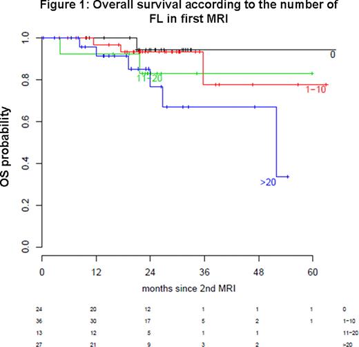
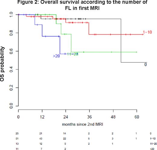
Magnetic resonance imaging (MRI) has been demonstrated to be the most sensitive imaging technique to detect both diffuse and focal infiltration of multiple myeloma cells in bone marrow. Whole body magnetic resonance imaging (WB-MRI) can be applied using affordable extensions to standard MRI scanners. Recent studies have shown that the presence and number of focal lesions (FL) in MRI are of prognostic significance in symptomatic as well as smoldering multiple myeloma. The aim of the present study was to investigate the significance of persisting FL after autologous stem cell transplantation. To answer this question WB-MRI of 100 patients eligible for high dose chemotherapy with symptomatic myeloma were retrospectively evaluated before the start of systemic treatment and after single or tandem autologous stem cell transplantation. Number of FL was assessed by two investigators in consensus for each time point separately. The analysis was approved by the institutional ethics committee. Patients were classified into different groups according to the number of FL in WB-MRI. At first MRI, 24 patients presented with 0, 36 with 1–10, 13 with 11–20 and 27 with more than 20 FL. At time of second MRI 25 patients had no, 51 patients 1–10, 13 patients 11–20 and 11 patients more than 20 FL. Overall survival was defined as time from second MRI until death of any cause. MRI-response according to the change of the number of FL was defined as focal complete remission (fCR) indicating total disappearance of FL, focal partial remission (fPR) being defined as reduction of the number of FL of 50% or more, focal stable disease (fSD) as unchanged number of FL and focal progressive disease (fPD) as any increase in number of focal lesions after therapy. At second MRI an fCR was found in 25; an fPR in 22; an fSD in 26 and an fPD in 27 patients. For evaluation of prognostic significance of the number of FL for overall survival a log rank test resulted in a p-value of 0.17 for the first and 0.02 for the second WB-MRI. Kaplan-Meier plots for overall survival according to the number of FL at first and second MRI are shown in Figure 1 and 2 respectively. Number of FL was not of prognostic significance for progression free survival (p = 0.6 and p = 0.8 for first and second MRI respectively). Serological and MRI-defined response concerning FL were significantly associated (p = 0.003; Kendall's Tau 0.26) with a trend to an underestimation of response in FL compared to serological response. The fact that the number of FL after high dose chemotherapy but not at first diagnosis of symptomatic myeloma had a significant impact on survival in our study confirms the importance of the measurement of residual disease after systemic treatment as it has been demonstrated for methods as flow cytometry and others. Thereby WB-MRI allows the evaluation of nearly the whole bone marrow compartment non-invasively and without the need of irradiation exposure. A limitation of MRI is the fact that it is not capable of differentiating between FL containing vital tumor cells and those representing osteolyses without active myeloma. New functional MRI sequences like dynamic contrast-enhanced MRI and diffusion weighted imaging may overcome this disadvantage. Furthermore, positron emission tomography has been demonstrated to be another possibility for detection of residual disease activity after therapy. Further studies will show which imaging technique is most useful in myeloma or if different clinical and scientific questions necessitate different methods.
Disclosures:
Goldschmidt:Janssen-Cilag: Consultancy; Celgene: Consultancy; Millennium Pharmaceuticals, Inc.: Consultancy.
Author notes
*
Asterisk with author names denotes non-ASH members.
© 2011 by The American Society of Hematology
2011


This feature is available to Subscribers Only
Sign In or Create an Account Close Modal