Abstract
Indoleamine 2,3-dioxygenase (IDO) is an immunoregulatory enzyme that is implicated in suppressing T-cell immunity in normal and pathologic settings. Here, we describe that spontaneous cytotoxic T-cell reactivity against IDO exists not only in patients with cancer but also in healthy persons. We show that the presence of such IDO-specific CD8+ T cells boosted T-cell immunity against viral or tumor-associated antigens by eliminating IDO+ suppressive cells. This had profound effects on the balance between interleukin-17 (IL-17)–producing CD4+ T cells and regulatory T cells. Furthermore, this caused an increase in the production of the proinflammatory cytokines IL-6 and tumor necrosis factor-α while decreasing the IL-10 production. Finally, the addition of IDO-inducing agents (ie, the TLR9 ligand cytosine-phosphate-guanosine, soluble cytotoxic T lymphocyte–associated antigen 4, or interferon γ) induced IDO-specific T cells among peripheral blood mononuclear cells from patients with cancer as well as healthy donors. In the clinical setting, IDO may serve as an important and widely applicable target for immunotherapeutic strategies in which IDO plays a significant regulatory role. We describe for the first time effector T cells with a general regulatory function that may play a vital role for the mounting or maintaining of an effective adaptive immune response. We suggest terming such effector T cells “supporter T cells.”
Introduction
Induction of tolerance, which is a central mechanism counteracting tumor-specific immunity and preventing effective anticancer immune therapy, requires a specific environment in which tolerogenic dendritic cells (DCs) play an essential role deviating the immune response away from effective immunity. It was recently shown that IDO provides a potential mechanism for the development of DC-mediated T-cell tolerance. IDO+ DCs inhibit T-cell proliferation because of tryptophan depletion and accumulation of toxic tryptophan metabolites.1,2 IDO+ DCs have been shown to induce T-cell anergy or generation of regulatory T cells (Tregs). In patients with cancer, IDO elevation occurs in a subset of plasmacytoid DCs in tumor-draining lymph nodes.3 In addition, most human tumors overexpress IDO.4 Activation of IDO in either tumor cells or nodal regulatory DCs each appears to be sufficient to facilitate tumoral immune escape.2 IDO may help in tilting the tumor microenvironment from hostile to supportive for tumor cells and also may elaborate a peripheral mechanism of immune escape that could facilitate tumor progression.5,6
Tregs have been defined as a specialized subpopulation of T cells that act to suppress activation of the immune system and thereby maintain immune system homeostasis and tolerance to self-antigens.7,8 Subsequently, they are additionally termed suppressor T cells. Tregs exist to down-regulate immune responses in various inflammatory circumstances and ultimately assure peripheral T-cell tolerance. The best-characterized subset of these immune suppressive cells are CD4+CD25highCD127−Foxp3+ T cells.9-11 Over the past years, additional regulatory T-cell subsets, including CD8+ suppressor T cells, have been described in humans and mice.12-14 Recently, we identified very potent antigen-specific CD8+ suppressor T cells in peripheral blood mononuclear cells (PBMCs) from patients with cancer.15 These natural-occurring human leukocyte antigen A2 (HLA-A2)–restricted CD8+ T cells were specific for the anti-inflammatory molecule Heme Oxygenase-1. The data linked the cellular stress response to the regulation of adaptive immunity and added a new dimension to the role of antigen-specific CD8+ T cells in the regulation of cellular immune responses.
We have recently described that IDO is spontaneously recognized by cytotoxic T cells (CTLs) in patients with cancer.16 Thus, IDO-specific T cells were present in peripheral blood as well as in the tumor microenvironment. These IDO-reactive T cells were able to recognize and kill tumor cells, including directly isolated acute myelocytic leukemia blasts, as well as IDO-expressing DCs, that is, one of the main immune-suppressive cell populations. We could not detect spontaneous responses against IDO in the control group of healthy persons. Thus, albeit IDO has an immune suppressive effect, the up-regulation of IDO expression seems to induce a specific cytotoxic T-cell response. However, we found it quite astonishing that the T cells in the patients did not exhibit tolerance toward IDO, because IDO is inducible under normal physiologic conditions. We speculated that this could suggest a more general role of IDO-specific T cells in the regulation of the immune system. IDO may play a critical role for the strength and duration of a given immune response because of its inflammation-induced counter-regulatory function. Hence, IDO-specific CD8+ T cells may play an important role in the early phase of an immune response by eliminating IDO+ cells, thereby delaying local immune suppression. With this hypothesis, we continued our analysis of possible IDO-specific T-cell responses in healthy donors as well as in patients with cancer and examined the role of IDO-specific T cells in the adaptive immune system.
Methods
Donors
PBMCs were collected from healthy persons and patients with cancer (renal cell carcinoma, melanoma, and breast cancer). Blood samples from patients with cancer were drawn a minimum of 4 weeks after termination of any kind of anticancer therapy. Most of the patients with renal cell carcinoma had previously been treated with interleukin-2 (IL-2) and interferon α (IFN-α), most patients with melanoma had received high-dose IL-2 and IFN-α, whereas all patients with breast cancer were pretreated with several kinds of chemotherapy, (eg, epirubicin, docetaxel, capecitabine), trastuzumab, or endocrine therapy. Informed consent was obtained from the patients before any of these measures in accordance with the Declaration of Helsinki. All protocols were approved by the Herlev University Hospital Ethics Committee.
Enzyme-linked immunospot assay
Major histocompatibility complex–tetramer staining
PBMCs were stained with phycoerythrin (PE)–coupled major histocompatibility complex (MHC) tetramers, followed by antibody staining with CD8-allophycocyanin (APC) and CD3-fluorescein isothiocyanate (FITC; BD Biosciences). 7-Amino-actinomycin D (BD Biosciences) was used for exclusion of nonviable cells in all samples. MHC tetramers were prepared as described.19,20 The MHC-tetramer complexes used were as follows: HLA-A2/IDO5 (IDO199-207; ALLEIASCL), HLA-A2/cytomegalovirus (CMV) pp65495-503 (NLVPMVATV), HLA-A2/CMV IE1316-324 (VLEETSVML), HLA-A2/influenza (Flu) matrix p58-66 (GILGFVFTL), HLA-A2/MART-126-35 (EAAGIGILTV), HLA-A2/HIV-1 pol476-484 (ILKEPVHGV), and HLA-A3/HIV-1 nef73-82 (QVPLRPMTYK). The samples were analyzed and occasionally sorted on FACSAria or FACSCanto II, using DIVA software (BD Biosciences).
Establishment of antigen-specific T-cell cultures and clones
PBMCs were stimulated with irradiated (25 Gy), IDO5 (IDO199-207; ALLEIASCL)–loaded autologous DC with β2-microglobulin, IL-12 (PeproTech), and IL-7 (in U/mL; PeproTech) in X-vivo with 5% human AB serum. The cultures were restimulated every 7-10 days with IL-2. After 4-5 weeks, growing cultures were tested for specificity for IDO5, and specific cultures were cloned by limiting dilution in the presence of IDO5-loaded PBMCs and IL-2. Growing clones were expanded with IL-2 and IDO5-loaded PBMCs or Dynabeads CD3/CD28 T-cell expander (Dynal).
Cytotoxicity assay
Conventional 4-hour 51Cr-release assays for CTL-mediated cytotoxicity was carried out as described elsewhere.21 Target cells were peptide-loaded T2 cells, the colon cancer cell lines HCT116 and SW480 (ATCC), the melanoma cell line FM55M,22 in vitro–generated autologous immature DCs and matured DCs, and allogeneic ex vivo–isolated CD14+ monocytes (isolated with the use of magnetically activated cell sorting [MACS] CD14+ microbeads). In some assays, CD14+ monocytes were treated with 100 U/mL IFN-γ for 2 days before analysis. Lysis was blocked with the use of the HLA class I–specific monoclonal antibody W6/32 (2 μg/100 μL).23
Down-regulation of IDO in cancer cells
Human SW480 cancer cells were transfected with indicated short hairpin RNA (ShRNA) plasmids obtained from SuperArray with the use of FuGene6 (Roche) according to the manufacturer's instructions. Blots were developed with the enhanced chemiluminescence system obtained from Amersham and a CCD camera (LAS-1000; Fujifilm). Antibodies used were anti-Cdk7 (MO-1; Santa Cruz Biotechnology) and anti-IDO (Millipore Corporation).
Coculturing with autologous IDO-specific T cells
PBMCs were stimulated in vitro with 50 μg/mL viral peptide (CMV pp65495-503 [NLVPMVATV], CMV IE1316-324 [VLEETSVML], or Flu matrix p58-66 [GILGFVFTL]). IL-2 (40 U/mL) was added on days 2 and 6. The PBMCs were either cultured alone or added to autologous IDO5-specific T cells (in a PBMC-to-IDO5-specific T-cell ratio of 2000:1) on day 6. On day 9, the cultures were stimulated with 120 U/mL IL-2. After 12 days in culture, the number of viral-specific T cells in the cultures, either cultured alone or added to IDO5-specific T cells, was compared by MHC-tetramer staining. The number of Tregs, IL-17A–producing T cells, and the CD4/CD8 cell ratio in the cultures were also compared. As a control, PBMCs were cocultured with autologous CD8+ T cells of irrelevant specificity.
Costimulation with IDO peptide
PBMCs were stimulated in vitro with 25 μg/mL viral or tumor-associated antigens (CMV pp65495-503 [NLVPMVATV], CMV IE1316-324 [VLEETSVML], or MART-126-35 [EAAGIGILTV]), either in coculture with 25 μg/mL IDO5 peptide or an irrelevant peptide (HIV-1 pol476-484 [ILKEPVHGV]). IL-2 (40 U/mL) was added every third day. Every 7 days, the cultures were stimulated with a mixture of CMV or MART-1 peptide plus IDO5 peptide, or a mixture of CMV or MART-1 peptide plus HIV-1 pol476-484 peptide, respectively. Cells were stimulated with 10-, 100-, and 1000-fold diluted peptides for the second, third, and fourth peptide stimulation, respectively. After 3-4 stimulations, the number of CMV- or MART-1–specific T cells in the cultures, either cocultured with IDO5 peptide or HIV-1 pol476-484 peptide, was compared by MHC-tetramer staining. The number of Tregs, IL-17A–producing T cells, and the CD4/CD8 cell ratio in the cultures were also compared.
Intracellular staining for CD4+CD25highCD127−Foxp3+ Tregs
Cells were stained with the following antibodies: CD3-APC-indocyanine 7, CD4–peridinin chlorophyll protein complex, CD25-APC, and CD127-FITC (BD Bioscience and eBioscience). After fixation and permeabilization, cells were stained with PE anti–human Foxp3 (eBioscience). Isotype controls were used to enable correct compensation and to confirm antibody specificity. Cells were analyzed with FACSCanto II flow cytometer (BD Bioscience).
Intracellular staining for IL-17–producing T cells
PBMCs were stimulated with Leukocyte Activation Cocktail containing phorbol myristate acetate, ionomycin, and Brefeldin A (BD Bioscience) for 5 hours. Cells were stained with FITC anti–human IL-17A (eBioscience) after fixation and permeabilization, according to the manufacturer's instructions, after surface staining with CD3-APC-indocyanine 7 and CD4–peridinin chlorophyll protein complex (BD Bioscience). Isotype controls were used to enable correct compensation and to confirm antibody specificity. Stained cells were analyzed with FACSCanto II flow cytometer (BD Bioscience).
Cytokine ELISA
Cell culture supernatants were collected and stored at −80°C. Amounts of IL-6, IL-10, IL-17A, IFN-γ, and tumor necrosis factor-α (TNF-α) were measured by standard sandwich enzyme-linked immunoabsorbent assay (ELISA) with the use of commercially available antibodies and standards, according to the manufacturer's protocols (eBioscience).
ELISA for quantitative determination of tryptophan
Cell culture supernatants were collected and stored at −80°C. After precipitation and derivatization, tryptophan concentrations were quantitatively determined by competitive ELISA, according to the manufacturer's instructions (Labor Diagnostika Nord). Quantification of unknown samples was achieved by comparing their absorbance with a reference curve prepared with known standards.
Induction of IDO-specific T cells by IFN-γ, CTLA4-IgG, or cytosine-phosphate-guanosine oligodeoxynucleotide
PBMCs were stimulated with 100 U/mL IFN-γ, 1 μg/mL cytosine-phosphate-guanosine(CpG) ODN (Type B CpG oligodeoxynucleotide specific for human TLR9; InvivoGen), or 1 μg/mL cytotoxic T lymphocyte–associated antigen 4–immunoglobulin G2a (CTLA4-IgG2a) fusion protein (Research Diagnostics) once a week. IL-2 (40 U/mL) was added every third day. After 4 weeks, the cultures were tested for the presence of IDO-specific T cells by MHC-tetramer staining.
RNA preparation and reverse transcription–coupled polymerase chain reaction
Resulting cDNA was tested with primers for GAPDH (5′-AGGGGGGAGCCAAAAGGG-3′, 5′-GAGGAGTGGGTGTCGCTGTTG-3′, positions 440 and 980, respectively; product size 558 bp). Primers suited for amplification were as follows: IDO (5′-TGTCCGTAAGGTCTTGCCAGG-3′; 5′-CGAAATGAGAACAAAACGTCC-3′, positions 408 and 557, respectively; product size, 170 bp).
Statistical analysis
The percentages of antigen-specific T cells between cultures were compared with the use of 1-tailed 2-sampled paired t tests with a significance level at .05. The fold increase of antigen-specific T cells between PBMC cultures was defined as the percentage of antigen-specific T cells in cultures cocultured with IDO peptide divided by the percentage in cultures cocultured with HIV-1 peptide.
Results
IDO5-specific T cells are detectable in healthy donors
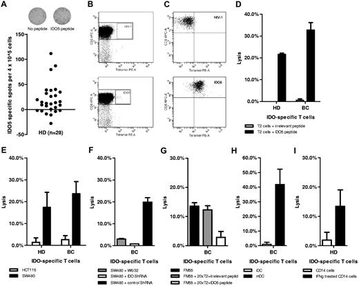
First, we examined PBMCs from healthy donors for the presence of T cells specific for the HLA-A2–restricted IDO-derived epitope IDO5 (IDO199-207; ALLEIASCL). We found that IDO reactivity could readily be detected by IFN-γ enzyme-linked immunospot and HLA-A2/IDO5 tetramer staining (Figure 1A-B). All in all, we examined 28 healthy donors for spontaneous T-cell reactivity against the HLA-A2–restricted IDO epitope and identified specific T cells in 3 persons (Figure 1A). As a control of the HLA-A2/IDO5 tetramer, an IDO5-specific T-cell clone was stained (Figure 1C).
Spontaneous cytotoxic T-cell reactivity against IDO. Spontaneous T-cell reactivity against IDO5 (IDO199-207; ALLEIASCL) in PBMCs, from HLA-A2+ healthy donors (HD), visualized by IFN-γ enzyme-linked immunospot (ELISPOT) assay (A) and flow cytometry (B) after 1 in vitro peptide stimulation. For IFN-γ ELISPOT assay, PBMCs were plated at 4 × 10⁵ PBMCs in duplicates in specialized ELISPOT wells either alone or with added IDO5 peptide. The average number of IDO5-specific spots (after subtraction of spots in wells without added peptide) was calculated per 4 × 10⁵ PBMCs for each donor (black circles; A). For flow cytometry, IDO5-specific T cells were identified with the MHC-tetramer complex HLA-A2/IDO5 and CD8 monoclonal antibody (mAb). For comparison, cells were stained with the MHC-tetramer complex HLA-A2/HIV-1 pol476-484 and CD8 mAb (B). As control, an IDO5-specific T-cell clone was stained with the HLA-A2/HIV-1 pol476-484-PE and HLA-A2/IDO5-PE complexes (C). Lytic capacity of representative IDO5-specific T-cell clones from a healthy donor (HD) or a patient with breast cancer (BC) assayed by 51Cr-release assay. Target cells were TAP-deficient T2 cells pulsed with IDO5 or an irrelevant peptide (HIV-1 pol476-484; D), the HLA-A2+/IDO+ colon cancer cell line SW480 and the HLA-A2+/IDO− colon cancer cell line HCT116 (E), SW480 blocked with the HLA class I–specific mAb W6/32 (F), SW480 transfected with IDO ShRNA for down-regulation of IDO protein expression and SW480 transfected with control ShRNA as a positive control (F), the HLA-A2+/IDO+ melanoma cell line FM55M (G), FM55M added cold T2 cells pulsed with IDO5 peptide or irrelevant peptide (HIV-1 pol476-484) in a inhibitor-to-target ratio of 20:1 (G), autologous in vitro immatured and matured DCs (H), and ex vivo–isolated autologous IDO− CD14+ monocytes as well as IFN-γ–treated IDO+ CD14+ monocytes (I). All 51Cr-release assays were performed in effector-to-target ratio of 5:1, except the experiments regarding ShRNA, which were performed in effector-to-target ratio of 15:1. Data are mean ± SD (n = 3).
Spontaneous cytotoxic T-cell reactivity against IDO. Spontaneous T-cell reactivity against IDO5 (IDO199-207; ALLEIASCL) in PBMCs, from HLA-A2+ healthy donors (HD), visualized by IFN-γ enzyme-linked immunospot (ELISPOT) assay (A) and flow cytometry (B) after 1 in vitro peptide stimulation. For IFN-γ ELISPOT assay, PBMCs were plated at 4 × 10⁵ PBMCs in duplicates in specialized ELISPOT wells either alone or with added IDO5 peptide. The average number of IDO5-specific spots (after subtraction of spots in wells without added peptide) was calculated per 4 × 10⁵ PBMCs for each donor (black circles; A). For flow cytometry, IDO5-specific T cells were identified with the MHC-tetramer complex HLA-A2/IDO5 and CD8 monoclonal antibody (mAb). For comparison, cells were stained with the MHC-tetramer complex HLA-A2/HIV-1 pol476-484 and CD8 mAb (B). As control, an IDO5-specific T-cell clone was stained with the HLA-A2/HIV-1 pol476-484-PE and HLA-A2/IDO5-PE complexes (C). Lytic capacity of representative IDO5-specific T-cell clones from a healthy donor (HD) or a patient with breast cancer (BC) assayed by 51Cr-release assay. Target cells were TAP-deficient T2 cells pulsed with IDO5 or an irrelevant peptide (HIV-1 pol476-484; D), the HLA-A2+/IDO+ colon cancer cell line SW480 and the HLA-A2+/IDO− colon cancer cell line HCT116 (E), SW480 blocked with the HLA class I–specific mAb W6/32 (F), SW480 transfected with IDO ShRNA for down-regulation of IDO protein expression and SW480 transfected with control ShRNA as a positive control (F), the HLA-A2+/IDO+ melanoma cell line FM55M (G), FM55M added cold T2 cells pulsed with IDO5 peptide or irrelevant peptide (HIV-1 pol476-484) in a inhibitor-to-target ratio of 20:1 (G), autologous in vitro immatured and matured DCs (H), and ex vivo–isolated autologous IDO− CD14+ monocytes as well as IFN-γ–treated IDO+ CD14+ monocytes (I). All 51Cr-release assays were performed in effector-to-target ratio of 5:1, except the experiments regarding ShRNA, which were performed in effector-to-target ratio of 15:1. Data are mean ± SD (n = 3).
IDO5-specific T cells are specifically able to kill IDO-expressing cells
IDO-specific T-cell clones were established from bulk cultures by limiting dilution cloning from patients with cancer 16 as well as healthy donors. The lytic capacity of representative clones from a healthy donor and a patient with breast cancer are depicted in Figure 1D-I. The IDO-specific T cells effectively killed IDO5-pulsed TAP-deficient T2 cells, whereas T2 cells pulsed with an irrelevant peptide (HIV-1 pol476-484) were not lysed (Figure 1D). Importantly, IDO-specific T cells also killed the HLA-A2+/IDO+ colon cancer cell line SW480 (Figure 1E). In contrast, IDO-specific T cells did not lyse the HLA-A2+/IDO− colon cancer cell line HCT116 (Figure 1E). HLA restriction was confirmed by blocking HLA class I with the use of the HLA class I–specific monoclonal antibody W6/32, which completely abolished lysis of the SW480 cells (Figure 1F). Using IDO ShRNA we down-regulated IDO protein expression in SW480 and thereby rescued these tumor cells from being killed by IDO-specific T cells, whereas cells transfected with irrelevant control ShRNA were killed (Figure 1F). Cold target inhibition assays with the use of unlabeled T2 cells pulsed with IDO5 peptide confirmed HLA-A2/peptide specificity of the killing. The addition of cold (unlabeled) IDO5-pulsed T2 cells completely abrogated the killing of FM55M melanoma cells, whereas the addition of cold T2 cells pulsed with the irrelevant HIV-1 pol476-484 peptide did not have an effect on the killing of FM55M (Figure 1G). IDO expression is not restricted to tumor cells, but it can also be induced in immune cells. In this regard, IDO-specific T cells specifically killed autologous IDO+ in vitro–generated mature DCs, whereas IDO− immature DCs and IDO− CD14+ monocytes were not killed (Figure 1H-I). IDO is known to be induced by both type I and II IFNs, which are found at sites of immune activation.24,25 Thus, IFN-γ is a well-described inducer of IDO in many cell types, including fibroblasts, endothelial cells, tumor cells, monocyte-derived macrophages, and DCs. We treated CD14+ monocytes with IFN-γ, which indeed induced IDO expression (data not shown). These IDO-expressing CD14+ monocytes were susceptible to killing by autologous IDO-specific T cells (Figure 1I).
IDO-specific T cells boost viral immunity
We used IDO-specific T-cell clones to examine a potential role of such cells in enhancing immune responses. Hence, we added IDO-specific T cells from a healthy donor to autologous PBMC cultures (in ratio of 1:2000) that were stimulated with an HLA-A2–restricted epitope from CMV. The addition of IDO-specific T cells resulted in a vast increase in the number of tetramer-positive, CMV-specific CD8+ T cells in the cultures; on the basis of 4 independent experiments the number of CMV-specific CD8+ T cells increased significantly (P < .05), on average from 18% to 36% (Figure 2A-B). Furthermore, we observed a notable reduction of CD4+CD25highCD127−Foxp3+ Tregs in cultures with added IDO-specific T cells (Figure 2C). Importantly, these changes did not correspond to similar differences in the percentage of CD8+ and CD4+ T cells between the cultures (Figure 2D).
IDO-specific T cells boosted specific immunity toward CMV in PBMCs from a healthy donor. PBMCs from an HLA-A2+ healthy donor cultured with CMV IE1316-324 (VLEETSVML) peptide either alone (top) or added an autologous, IDO5 (IDO199-207; ALLEIASCL)–specific T-cell clone (in a PBMC-to-clone ratio of 2000:1; bottom). The percentage of CMV IE1316-324–specific CD8+ T cells in each culture was identified by flow cytometry with the MHC-tetramer complex HLA-A2/CMV IE1316-324 and CD8 monoclonal antibody (mAb). For comparison, cells were stained with the MHC-tetramer complex HLA-A2/HIV-1 pol476-484 and CD8 mAb. Data from 2 representative experiments are shown (A). Percentage of CMV IE1316-324–specific T cells found in PBMCs cultured alone (□) or added an IDO5-specific T-cell clone (■). Data are mean ± SD (n = 4; P < .05; B). The percentage of CD4+CD25highCD127−Foxp3+ Tregs in each culture was identified by flow cytometry with intracellular staining for Foxp3. For comparison, cells were stained with isotype controls. The data shown are from 1 donor, representative of 4 experiments (C). Distribution of CD4+ and CD8+ T cells in the cultures. Data are mean ± SD (n = 4; D).
IDO-specific T cells boosted specific immunity toward CMV in PBMCs from a healthy donor. PBMCs from an HLA-A2+ healthy donor cultured with CMV IE1316-324 (VLEETSVML) peptide either alone (top) or added an autologous, IDO5 (IDO199-207; ALLEIASCL)–specific T-cell clone (in a PBMC-to-clone ratio of 2000:1; bottom). The percentage of CMV IE1316-324–specific CD8+ T cells in each culture was identified by flow cytometry with the MHC-tetramer complex HLA-A2/CMV IE1316-324 and CD8 monoclonal antibody (mAb). For comparison, cells were stained with the MHC-tetramer complex HLA-A2/HIV-1 pol476-484 and CD8 mAb. Data from 2 representative experiments are shown (A). Percentage of CMV IE1316-324–specific T cells found in PBMCs cultured alone (□) or added an IDO5-specific T-cell clone (■). Data are mean ± SD (n = 4; P < .05; B). The percentage of CD4+CD25highCD127−Foxp3+ Tregs in each culture was identified by flow cytometry with intracellular staining for Foxp3. For comparison, cells were stained with isotype controls. The data shown are from 1 donor, representative of 4 experiments (C). Distribution of CD4+ and CD8+ T cells in the cultures. Data are mean ± SD (n = 4; D).
Likewise, we added IDO-specific T cells from a patient with breast cancer to autologous PBMC cultures (in ratio of 1:2000) stimulated with an HLA-A2–restricted epitope from Flu. Again, the addition of IDO-specific T cells resulted in a vast increase in the number of tetramer-positive, antigen-specific CD8+ T cells in the cultures. This was observed in 3 independent experiments. In one experiment the number of Flu-specific CD8+ T cells almost doubled from 7% to nearly 14% (Figure 3A). The addition of IDO-specific T cells at the same time decreased the amount of CD4+CD25highCD127−Foxp3+ Tregs in the cultures to the half (Figure 3B), while increasing the number of IL-17A–producing CD4+ T cells from 0.2% to 0.6% (Figure 3C). Importantly, these changes did not correspond to similar differences in the percentage of CD8+ and CD4+ T cells between the cultures (Figure 3D). We additionally compared the activity of IDO between the cultures by measuring the amount of tryptophan in the cell culture supernatants. Interestingly, the highest concentration of tryptophan, that is, least IDO activity, was found in cultures with added IDO-specific T cells (Figure 3E). The addition of IDO-specific T cells additionally reduced the concentration of IL-10 in the cell culture supernatants, while increasing concentrations of IL-17A, IL-6, and TNF-α as measured by standard cytokine ELISA (Figure 3F). Notably, the addition of autologous CD8+ T-cell clones of unknown specificities to PBMCs from the same patient did not increase the number of tetramer-positive, Flu-specific CD8+ T cells (data not shown). Likewise, we added a T-cell clone specific for the tumor-associated antigen ML-IAP26 to autologous CMV peptide–stimulated PBMCs (in ratio of 1:2000). The addition of ML-IAP–specific T cells did not change the number of CMV tetramer-positive CD8+ T cells (Figure 3G) or the concentrations of IL-10, IL-17A, IL-6, and TNF-α in the cell culture supernatants (data not shown). The amount of IFN-γ in the cell culture supernatants increased both by the addition of IDO-specific T cells and by the addition of ML-IAP–specific T cells (data not shown).
IDO-specific T cells boosted specific immunity toward Flu in PBMCs from a patient with cancer. PBMCs from a patient with HLA-A2+ breast cancer cultured with Flu matrix p58-66 (GILGFVFTL) peptide either alone (top) or added an autologous, IDO5 (IDO199-207; ALLEIASCL)–specific T-cell clone (in a PBMC-to-clone ratio of 2000:1; bottom). The percentage of Flu matrix p58-66–specific CD8+ T cells in each culture was identified by flow cytometry with the MHC-tetramer complex HLA-A2/Flu matrix p58-66 and CD8 monoclonal antibody (mAb). For comparison, cells were stained with the MHC-tetramer complex HLA-A2/HIV-1 pol476-484 and CD8 mAb (A). The percentage of CD4+CD25highCD127−Foxp3+ Tregs (B) and IL-17A–producing CD4+ T cells (C) in each culture were identified by flow cytometry with intracellular staining for Foxp3 and IL-17A, respectively. For comparison, cells were stained with isotype controls. Distribution of CD4+ and CD8+ T cells in the cultures (D). Tryptophan concentrations in cell culture supernatants before and after the addition of the IDO5-specific T-cell clone measured by competitive ELISA (E). Secreted cytokines (IL-10, IL-17A, IL-6, and TNF-α) in cell culture supernatants quantified by ELISA (F). All data shown are from one patient. Data are mean ± SD (n = 3). White bars indicate Flu matrix p58-66–stimulated PBMCs cultured alone; black bars, Flu matrix p58-66–stimulated PBMCs added an IDO5-specific T-cell clone (A-F). PBMCs from a patient with HLA-A2+ melanoma cancer cultured with CMV pp65495-503 (NLVPMVATV) peptide either alone (top) or added irrelevant autologous, ML-IAP280-289 (QLCPICRAPV)–specific T-cell clone (in a PBMC- to-clone ratio of 2000:1; bottom). The percentage of CMV pp65495-503–specific CD8+ T cells in each culture was identified by flow cytometry with the MHC-tetramer complex HLA-A2/CMV pp65495-503 and CD8 monoclonal antibody. For comparison, cells were stained with the MHC-tetramer complex HLA-A2/HIV-1 pol476-484 and CD8 monoclonal antibody. The data shown are from one patient, representative of 3 experiments (G).
IDO-specific T cells boosted specific immunity toward Flu in PBMCs from a patient with cancer. PBMCs from a patient with HLA-A2+ breast cancer cultured with Flu matrix p58-66 (GILGFVFTL) peptide either alone (top) or added an autologous, IDO5 (IDO199-207; ALLEIASCL)–specific T-cell clone (in a PBMC-to-clone ratio of 2000:1; bottom). The percentage of Flu matrix p58-66–specific CD8+ T cells in each culture was identified by flow cytometry with the MHC-tetramer complex HLA-A2/Flu matrix p58-66 and CD8 monoclonal antibody (mAb). For comparison, cells were stained with the MHC-tetramer complex HLA-A2/HIV-1 pol476-484 and CD8 mAb (A). The percentage of CD4+CD25highCD127−Foxp3+ Tregs (B) and IL-17A–producing CD4+ T cells (C) in each culture were identified by flow cytometry with intracellular staining for Foxp3 and IL-17A, respectively. For comparison, cells were stained with isotype controls. Distribution of CD4+ and CD8+ T cells in the cultures (D). Tryptophan concentrations in cell culture supernatants before and after the addition of the IDO5-specific T-cell clone measured by competitive ELISA (E). Secreted cytokines (IL-10, IL-17A, IL-6, and TNF-α) in cell culture supernatants quantified by ELISA (F). All data shown are from one patient. Data are mean ± SD (n = 3). White bars indicate Flu matrix p58-66–stimulated PBMCs cultured alone; black bars, Flu matrix p58-66–stimulated PBMCs added an IDO5-specific T-cell clone (A-F). PBMCs from a patient with HLA-A2+ melanoma cancer cultured with CMV pp65495-503 (NLVPMVATV) peptide either alone (top) or added irrelevant autologous, ML-IAP280-289 (QLCPICRAPV)–specific T-cell clone (in a PBMC- to-clone ratio of 2000:1; bottom). The percentage of CMV pp65495-503–specific CD8+ T cells in each culture was identified by flow cytometry with the MHC-tetramer complex HLA-A2/CMV pp65495-503 and CD8 monoclonal antibody. For comparison, cells were stained with the MHC-tetramer complex HLA-A2/HIV-1 pol476-484 and CD8 monoclonal antibody. The data shown are from one patient, representative of 3 experiments (G).
Costimulation with IDO peptide boosts T-cell reactivity against viral and tumor-associated antigens
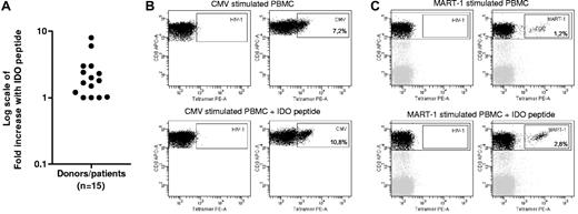
To further analyze the supporting effect of IDO-specific T cells on T-cell responses, PBMCs from 15 HLA-A2+ healthy donors and patients with cancer were stimulated with an HLA-A–restricted CMV or MART-1 epitope either in coculture with IDO5 peptide or an irrelevant HLA-A2–restricted epitope from HIV-1 in the presence of IL-2. After 3-4 in vitro stimulations IDO-specific T cells could be detected in cultures stimulated with IDO5 peptide (0.05%-0.1% IDO-specific CD8+ T cells). In comparison, IDO reactivity could not be detected in any of the HIV-1 peptide–stimulated cultures.
The increase in T-cell reactivity toward CMV or MART-1 was calculated for each donor/patient as fold increase of MHC tetramer–specific CD8+ T cells in coculture with IDO5 peptide (Figure 4A). Notably, we observed from 1.5- to 8-fold increases of tetramer-specific CD8+ T cells in 2 of 3 cultures with IDO reactivity (Figure 4A-C). Importantly, costimulation with IDO5 peptide did not result in a decrease in CMV or MART-1 reactivity in any of the examined persons (Figure 4A). Thus, coactivation of IDO-specific T cells not only boosted T-cell immunity toward viral antigens but also toward the well-known tumor-associated antigen MART-1.
Costimulation with IDO peptide increased frequencies of CMV- and MART-1–specific T cells. PBMCs from HLA-A2+ healthy donors and patients with HLA-A2+ cancer (melanoma and renal cell carcinoma) stimulated in vitro with CMV peptide (CMV pp65495-503 (NLVPMVATV) or CMV IE1316-324 (VLEETSVML)) or MART-126-35 (EAAGIGILTV) peptide either in coculture with IDO5 (IDO199-207; ALLEIASCL) peptide or an irrelevant peptide (HIV-1 pol476-484). The percentage of CMV- or MART-126-35–specific CD8+ T cells in each PBMC culture was identified by flow cytometry with the MHC-tetramer complexes HLA-A2/CMV pp65495-503 (NLVPMVATV), HLA-A2/CMV IE1316-324 (VLEETSVML), or HLA-A2/ MART-126-35 (EAAGIGILTV) and CD8 monoclonal antibody. The differences in tetramer-specific CD8+ T-cell percentages between the cultures are given, for each donor/patient, as fold increase of tetramer-specific CD8+ T cells in coculture with IDO5 peptide. Data are mean differences; n = 15 (A). Example of MHC-tetramer staining of PBMCs from a healthy donor stimulated in vitro with CMV IE1316-324 peptide either in coculture with an irrelevant peptide (HIV-1 pol476-484; top) or IDO5 peptide (bottom). The data shown are from 1 donor, representative of 6 different donors/patients (B). Example of MHC-tetramer staining of PBMCs from a patient with melanoma stimulated in vitro with MART-126-35 peptide either in coculture with an irrelevant peptide (HIV-1 pol476-484; top) or IDO5 peptide (bottom). The data shown are from 1 patient, representative of 4 different patients (C). In all experiments, cells were stained with the MHC-tetramer complex HLA-A2/HIV-1 pol476-484 and CD8 monoclonal antibody for comparison.
Costimulation with IDO peptide increased frequencies of CMV- and MART-1–specific T cells. PBMCs from HLA-A2+ healthy donors and patients with HLA-A2+ cancer (melanoma and renal cell carcinoma) stimulated in vitro with CMV peptide (CMV pp65495-503 (NLVPMVATV) or CMV IE1316-324 (VLEETSVML)) or MART-126-35 (EAAGIGILTV) peptide either in coculture with IDO5 (IDO199-207; ALLEIASCL) peptide or an irrelevant peptide (HIV-1 pol476-484). The percentage of CMV- or MART-126-35–specific CD8+ T cells in each PBMC culture was identified by flow cytometry with the MHC-tetramer complexes HLA-A2/CMV pp65495-503 (NLVPMVATV), HLA-A2/CMV IE1316-324 (VLEETSVML), or HLA-A2/ MART-126-35 (EAAGIGILTV) and CD8 monoclonal antibody. The differences in tetramer-specific CD8+ T-cell percentages between the cultures are given, for each donor/patient, as fold increase of tetramer-specific CD8+ T cells in coculture with IDO5 peptide. Data are mean differences; n = 15 (A). Example of MHC-tetramer staining of PBMCs from a healthy donor stimulated in vitro with CMV IE1316-324 peptide either in coculture with an irrelevant peptide (HIV-1 pol476-484; top) or IDO5 peptide (bottom). The data shown are from 1 donor, representative of 6 different donors/patients (B). Example of MHC-tetramer staining of PBMCs from a patient with melanoma stimulated in vitro with MART-126-35 peptide either in coculture with an irrelevant peptide (HIV-1 pol476-484; top) or IDO5 peptide (bottom). The data shown are from 1 patient, representative of 4 different patients (C). In all experiments, cells were stained with the MHC-tetramer complex HLA-A2/HIV-1 pol476-484 and CD8 monoclonal antibody for comparison.
Stimulation of IDO-specific T cells reduces Tregs numbers while boosting IL-17 production
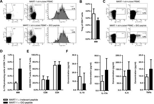
We examined the amount of Tregs and IL-17–producing CD4+ T cells in PBMCs from patients with melanoma stimulated with MART-1 peptide either in coculture with IDO5 peptide or an irrelevant HIV-1 peptide in the presence of IL-2 as described in “Costimulation with IDO peptide boosts T-cell reactivity against viral and tumor-associated antigens.” We observed that activation of IDO-specific T cells with IDO5 peptide not only boosted T-cell immunity toward MART-1 (Figure 4A) but at the same time also decreased the amount of CD4+CD25highCD127−Foxp3+ Tregs (Figure 5A-B) and increased the number of IL-17A–producing CD4+ T cells (Figure 5C-D). This was observed in all 4 examined patients. In cultures in which costimulation with IDO5 peptide did not boost T-cell immunity toward MART-1 (Figure 4A), we could not detect any differences in the number of Tregs (data not shown). Importantly, changes in the amount of MART-1–specific T cells, Treg, and IL-17A–producing CD4+ T cells did not correspond to similar changes in the percentage of CD8+ and CD4+ T cells between the cultures (Figure 5E). Hence, activation of IDO-specific T cells with IDO5 peptide seemed to change the overall composition of regulatory cells. Finally, we compared the secretion of cytokines between cell culture supernatants by cytokine ELISA. PBMC cultures composing IDO-specific T cells, that is, PBMCs cocultured with IDO5 peptide, showed higher concentrations of IL-17A, IL-6, and TNF-α in the cell culture supernatants, whereas a lower amount of IL-10 was detected (Figure 5F).
Costimulation of IDO-specific T cells reduced Treg numbers while boosting IL-17, IL-6, and TNF-α production. PBMCs from patients with HLA-A2+ melanoma cancer stimulated in vitro with MART-126-35 (EAAGIGILTV) peptide either in coculture with IDO5 peptide or an irrelevant peptide (HIV-1 pol476-484). The percentage of CD4+CD25highCD127−Foxp3+ Tregs (A-B) and IL-17A–producing CD4+ T cells (C-D) in each culture was identified by flow cytometry with intracellular staining for Foxp3 and IL-17A, respectively. For comparison, cells were stained with isotype controls. Examples of Treg staining (A) and IL-17A staining (C) of PBMCs stimulated in vitro with MART-126-35 peptide either in coculture with an irrelevant peptide (HIV-1 pol476-484; top) or IDO5 peptide (bottom). Examples shown are from 1 patient, representative of 4 different patients (A,C). Distribution of CD4+ and CD8+ T cells in the cultures (E). Secreted cytokines (IL-10, IL-17A, IL-6, and TNF-α) in cell culture supernatants quantified by ELISA (F). Data are mean ± SD (n = 4 patients). White bars indicate MART-126-35–stimulated PBMCs in coculture with an irrelevant peptide (HIV-1 pol476-484); black bars, MART-126-35–stimulated PBMCs in coculture with IDO5 peptide.
Costimulation of IDO-specific T cells reduced Treg numbers while boosting IL-17, IL-6, and TNF-α production. PBMCs from patients with HLA-A2+ melanoma cancer stimulated in vitro with MART-126-35 (EAAGIGILTV) peptide either in coculture with IDO5 peptide or an irrelevant peptide (HIV-1 pol476-484). The percentage of CD4+CD25highCD127−Foxp3+ Tregs (A-B) and IL-17A–producing CD4+ T cells (C-D) in each culture was identified by flow cytometry with intracellular staining for Foxp3 and IL-17A, respectively. For comparison, cells were stained with isotype controls. Examples of Treg staining (A) and IL-17A staining (C) of PBMCs stimulated in vitro with MART-126-35 peptide either in coculture with an irrelevant peptide (HIV-1 pol476-484; top) or IDO5 peptide (bottom). Examples shown are from 1 patient, representative of 4 different patients (A,C). Distribution of CD4+ and CD8+ T cells in the cultures (E). Secreted cytokines (IL-10, IL-17A, IL-6, and TNF-α) in cell culture supernatants quantified by ELISA (F). Data are mean ± SD (n = 4 patients). White bars indicate MART-126-35–stimulated PBMCs in coculture with an irrelevant peptide (HIV-1 pol476-484); black bars, MART-126-35–stimulated PBMCs in coculture with IDO5 peptide.
IDO-inducing agents stimulate IDO-specific T cells with supporter functions
IDO expression may be either constitutive or secondarily induced by mediators produced as a result of a local immune response. IFN-γ is a potent inducer of IDO,24,25 which could point to a scenario in which IFN-γ indirectly induces or boosts IDO-specific T cells. Therefore, PBMCs from 7 different healthy donors were treated with IFN-γ in the presence of IL-2 and were subsequently examined for IDO-specific T cells. Indeed, we were able to detect HLA-A2/IDO5 tetramer-positive CD8+ T cells (0.05%-0.1%) in 4 of the donors after treatment with IFN-γ (Figure 6A). Triggering of functional IDO requires ligation of B7-1/B7-2 molecules on DCs by CTLA4/CD28 expressed on T cells.27 TLR9 ligation activates DCs to up-regulate surface expression of B7 ligands and thereby increases expression of IDO.28 To determine whether this TLR9 ligand–induced up-regulation of IDO expression in DCs also results in activation of IDO-specific T cells, PBMCs from patients with cancer were treated with the TLR9 ligand CpG ODN in the presence of IL-2 and, subsequently, examined for IDO-specific T cells. TLR9 signaling with CpG ODN induced a measurable number of IDO-specific T cells (0.1%) in 2 of 3 patients (Figure 6B). Likewise, CTLA4 mediated up-regulation of IDO has been shown.27 In this regard, soluble CTLA4 (CTLA4-Ig) has been shown to induce IDO expression in DCs that are competent to express functional IDO and to mediate IDO-dependent T-cell suppression.29 To asses if CTLA4-Ig–induced IDO up-regulation indeed stimulates IDO-specific T cells we cocultured PBMCs with CTLA4-Ig and IL-2 and, subsequently, examined the cultures for the presence of IDO-specific T cells. In 2 of 3 patients HLA-A2/IDO5 tetramer-positive CD8+ T cells (0.1%-0.2%) were detectable after exposure to CTLA4-Ig (Figure 6C).
IDO-inducing agents expanded IDO-specific T cells with supporter functions. Example of reactivity against IDO5 (IDO199-207; ALLEIASCL) in PBMCs from an HLA-A2+ healthy donor, stimulated in vitro with IL-2 and IFN-γ. The percentage of IDO5-specific CD8+ T cells was identified by flow cytometry, ex vivo (top) and after stimulation (bottom), using the MHC-tetramer complex HLA-A2/IDO5 and CD8 monoclonal antibody (mAb). The data shown are from 1 donor, representative of 4 different donors (A). Examples of reactivity against IDO5 in PBMCs from a patient with HLA-A2+ renal cell carcinoma, stimulated in vitro with IL-2 and CTLA4-Ig (B) or CpG ODN (C). The percentage of IDO5-specific CD8+ T cells was identified by flow cytometry, ex vivo (top) and after stimulation (bottom), using the MHC-tetramer complex HLA-A2/IDO5 and CD8 mAb. The data shown are from 1 patient, representative of 2 different patients (B-C). PBMCs from an HLA-A2+ healthy donor stimulated in vitro with CMV pp65495-503 (NLVPMVATV) peptide and cocultured with either autologous, isolated CD8+ T cells (top) or autologous, isolated IFN-γ–induced IDO5-specific T cells (bottom). The percentage of CMV pp65495-503–specific CD8+ T cells in each culture was identified by flow cytometry with the MHC-tetramer complex HLA-A2/CMV pp65495-503 and CD8 mAb (D). PBMCs from an HLA-A2+ healthy donor stimulated with CMV IE1316-324 (VLEETSVML) peptide and cocultured with either autologous, isolated CD8+ T cells (top) or autologous, IDO5-specific T cells isolated after 2 in vitro peptide stimulations (bottom). The percentage of CMV IE1316-324–specific CD8+ T cells in each culture was identified by flow cytometry with the MHC-tetramer complex HLA-A2/ CMV IE1316-324 and CD8 mAb (E). In all experiments, cells were stained with the MHC-tetramer complex HLA-A2/HIV-1 pol476-484 and CD8 mAb for comparison.
IDO-inducing agents expanded IDO-specific T cells with supporter functions. Example of reactivity against IDO5 (IDO199-207; ALLEIASCL) in PBMCs from an HLA-A2+ healthy donor, stimulated in vitro with IL-2 and IFN-γ. The percentage of IDO5-specific CD8+ T cells was identified by flow cytometry, ex vivo (top) and after stimulation (bottom), using the MHC-tetramer complex HLA-A2/IDO5 and CD8 monoclonal antibody (mAb). The data shown are from 1 donor, representative of 4 different donors (A). Examples of reactivity against IDO5 in PBMCs from a patient with HLA-A2+ renal cell carcinoma, stimulated in vitro with IL-2 and CTLA4-Ig (B) or CpG ODN (C). The percentage of IDO5-specific CD8+ T cells was identified by flow cytometry, ex vivo (top) and after stimulation (bottom), using the MHC-tetramer complex HLA-A2/IDO5 and CD8 mAb. The data shown are from 1 patient, representative of 2 different patients (B-C). PBMCs from an HLA-A2+ healthy donor stimulated in vitro with CMV pp65495-503 (NLVPMVATV) peptide and cocultured with either autologous, isolated CD8+ T cells (top) or autologous, isolated IFN-γ–induced IDO5-specific T cells (bottom). The percentage of CMV pp65495-503–specific CD8+ T cells in each culture was identified by flow cytometry with the MHC-tetramer complex HLA-A2/CMV pp65495-503 and CD8 mAb (D). PBMCs from an HLA-A2+ healthy donor stimulated with CMV IE1316-324 (VLEETSVML) peptide and cocultured with either autologous, isolated CD8+ T cells (top) or autologous, IDO5-specific T cells isolated after 2 in vitro peptide stimulations (bottom). The percentage of CMV IE1316-324–specific CD8+ T cells in each culture was identified by flow cytometry with the MHC-tetramer complex HLA-A2/ CMV IE1316-324 and CD8 mAb (E). In all experiments, cells were stained with the MHC-tetramer complex HLA-A2/HIV-1 pol476-484 and CD8 mAb for comparison.
Finally, we isolated IFN-γ–induced, MHC tetramer–positive T cells by fluorescence-activated cell sorting (FACS) and added these to CMV peptide–stimulated PBMCs from the same donor. The addition of the tetramer-sorted IDO-specific T cells boosted T-cell immunity toward CMV compared with cultures with the addition of a similar amount of autologous CD8+ T cells that had undergone FACS (Figure 6D). Next, we examined the effect of coculturing PMBCs from another healthy donor with standard in vitro matured, autologous IDO+ DCs. This indeed induced IDO-specific T cells in the PBMC culture (data not shown). Finally, we sorted HLA-A2/IDO5 tetramer–positive T cells from a healthy donor after 2 in vitro IDO5 peptide stimulations. The tetramer-positive IDO-specific T cells were cocultured with autologous CMV peptide–stimulated PBMCs, which resulted in an increase in the number of CMV-specific T cells compared with cultures with the addition of a similar number of autologous CD8+ T cells that had undergone FACS (Figure 6E). Thus, the addition of HLA-A2/IDO5 tetramer–positive T cells boosted T-cell immunity toward CMV antigens similar to the IDO-specific T-cell clones described in “IDO- specific T cells boost viral immunity.”
Discussion
In the present study, we show that circulating IDO-specific, cytotoxic CD8+ T cells are present in healthy donors, although not as frequent as in patients with cancer. Even more important, we show that coactivation of IDO-specific cytotoxic T cells boosted T-cell immunity toward viral and tumor-associated antigens. We show that IDO-specific T cells were capable of killing IDO-expressing regulatory cells, thereby directly targeting the IDO-dependent counter-regulatory pathway. This “supportive” effect on T-cell immunity by IDO-specific T cells might be mediated in several direct and indirect manners. First of all, IDO is a main inhibitor of the effector phase of the immune response.4,30 IDO expression can suppress effector T cells directly by degradation of the essential amino acid tryptophan or by enhancement of local Treg-mediated immunosuppression. With respect to the former, some of the biologic effects of IDO are mediated through local depletion of tryptophan, whereas others are mediated by immunomodulatory tryptophan metabolites.1,31 Effector T cells starved of tryptophan are unable to proliferate and go into G1 cell cycle arrest.32 These cells are in addition more sensitive to apoptosis.33 We show that the level of tryptophan was elevated after the addition of IDO-specific T cells. This indicates that the addition of IDO-specific T cells directly decreased IDO activity, presumably because of the lysis of IDO-expressing cells. When IDO+ DCs are injected in vivo, they create suppression and anergy in antigen-specific T cells in the lymph nodes draining the injection site.32,34 Constitutive IDO expression in DCs provides T cells with regulatory properties that block T-cell responses to antigenic stimulation.2 The B7 receptors on IDO+ DCs bind to CTLA4 on Tregs, causing them to proliferate and induce antigen-specific anergy.34 Hence, IDO does not only suppress effector T cells directly but also influences Tregs bystander suppressor activity.5,6,35-37 Recently, it has been described that exposure of Tregs to IL-6 and other proinflammatory cytokines induces reprogramming of mature Tregs to acquire a phenotype resembling proinflammatory T helper 17 (Th17) cells.38-40 It has been shown that IDO plays a vital role in this conversion of Foxp3+ Tregs to Th17-like effector cells.6,37 IDO stimulates Treg bystander suppressor activity and simultaneously blocks the IL-6 production that is required to convert Tregs into Th17-like T cells.6,37 Tumor-infiltrating Th17 cells express other cytokines in addition to IL-17, which might be functionally relevant.41,42 A large fraction of Th17 cells produce high levels of effector cytokines such as IL-2, granulocyte-macrophage colony-stimulating factor, IFN-γ, as well as TNF.40 In the study of Sharma et al,37 it was described that the phenotype of reprogrammed Tregs after IDO blocking was similar to that of activated Th17 cells or to “polyfunctional” T helper cells. Hence, such Th17-like cells coexpressed IL-17, IL-22, IL-2, as well as TNF-α.37 These findings suggested that inducible or preexisting IDO activity at local sites of inflammation may dominantly suppresses proinflammatory processes and block effector T-cell responses to antigens encountered. Conversely, when IDO is absent, even strong proinflammatory stimuli do not elicit local Treg suppression, and Tregs are reprogrammed to acquire a proinflammatory Th17-like phenotype. In accordance hereof, we show that the frequency of Tregs decreased, the frequency of IL-17–producing cells increased, while the overall number of CD4+ T cells were constant or decreased slightly when IDO-specific T cells were present, all suggesting an overall decrease in IDO activity. Furthermore, IDO-specific T cells increased the production of both IL-6 as well as the other proinflammatory cytokine TNF-α. In addition, because IDO activation in DCs has been shown to result in IL-10 production and generation of Tregs,43 we examined the effect on IL-10 production. We observed a decrease in IL-10 when IDO-specific T cells were present. Finally, note that the metabolites of tryptophan are directly toxic to CD8+ T cells and CD4+ Th1 cells44 but not Th2 cells. Hence, increased IDO activity seems to tilt helper T-cell polarization toward a Th2 phenotype.45 It is therefore in addition possible that IDO-specific T cells by the killing of IDO-expressing cells were skewing the Th response in a Th1 direction.
Taken together, these different mechanism of actions could explain how IDO-specific T-cells were able to boost CD8+ T-cell immunity in general; the direct killing of IDO-expressing cells diminished the direct IDO-mediated suppression of effector T cells and decreased the local Treg suppression by reprogramming Tregs to acquire a proinflammatory Th17-like phenotype under influence of an increased IL-6 production. It should further be noted that IDO+ cells may be suppressive by other means than IDO. Hence, IDO-specific T cells may not only reduce IDO-mediated suppression but in addition further immune suppression mediated by IDO+ regulatory cells.
All in all, our results suggest that IDO-specific CD8+ T cells are a natural part of the T-cell repertoire in humans. IDO-specific T cells might interact with IDO-expressing cells, hereby eliminating or delaying the local immune suppression and thereby supporting the ongoing immune response. We were able to directly link the up-regulation of IDO with IDO-specific T cells by showing that the addition of the IDO-inducing agents IFN-γ, CTLA4-Ig, or CpG ODN generated measurable numbers of IDO-specific T cells among PBMCs. Thus, it seems apparent that IDO-specific T cells play a supportive role, which might be crucial for the mounting or keeping of an effective immune response during infection. Consequently, we suggest giving such effector T cells with an immune-enhancing function the general term “supporter T cells.”
Interestingly, CD14+ monocytes are major CMV target cells in vivo. Monocytes are responsible for dissemination of the virus throughout the body during acute and late phase of infection.46 CMV has been shown to induce IDO expression in monocytes, which has been suggested to confer an advantage to CMV-infected monocytes to escape T-cell responses.47 CMV is the most immunodominant antigen to be encountered by the human immune system.48 The CD8+ T-cell response to CMV typically constitutes a sizeable percentage of the CD8+ T-cell repertoire in CMV-seropositive persons.49 In light of this, it seems possible that IDO-specific T cells might have evolved to function as supporter T cells for the constitutive anti-CMV CD8+ T-cell response. Notably, we detected the IDO-specific T-cell responses in 3 healthy donors, who in addition all had strong CMV-specific CD8+ T-cells responses. Furthermore, we describe that CMV-specific CD8+ T-cell responses are strongly boosted in the presence of IDO-specific T cells.
In the clinical setting, the targeting of IDO could have synergistic effects in antiviral immune therapy, for example, in HIV vaccines. In this regard, it has been shown that HIV inhibits CD4+ T-cell proliferation by inducing IDO in plasmacytoid DCs and macrophages.50,51 In cancer immune therapy, the boosting of IDO-specific immunity could have both direct and indirect effects. Hence, we describe that IDO-specific T cells are able to recognize and kill IDO+ cancer cells. In fact, it may be possible that the sizable reactivity to this antigen in healthy persons contributes to immune surveillance against cancer. However, the induction of IDO-specific immune responses by therapeutic measures could function highly synergistic with additional anticancer immune therapy not only by eliminating cancer cells but also suppressive DCs. By definition almost any successful anticancer immune therapy strategy aims of inducing immunologic activation and inflammation. Within the limits of acceptable toxicity, as much immune activation as possible is the goal; hence, counter-regulation is not desired. Naturally, one should be cautious of the possible introduction of autoimmunity when targeting a tolerogenic molecule such as IDO. However, the circulation of a measurable number of IDO-specific T cells does not seem to cause autoimmunity. Furthermore, because IDO-specific T cells can be introduced by IDO-inducing agents, this appears to be under tight control. In this regard, an interesting aspect of IDO is that systemic inactivation at the organism level, either pharmacologically or genetically, does not appear to cause autoimmunity.52 IDO may not be involved in tolerance to self but rather in tolerance to nonself antigens where immune nonresponsiveness may be important, for example, fetal antigens.52 Induction of IDO+ tolerogenic DCs occurs during infection of DCs with viruses and intracellular pathogens, such as Listeria monocytogenes. Such IDO+ DCs seem to be involved in protection of the host from granuloma breakdown and pathogen dissemination in advanced human listeriosis. This might have major implications for IDO-based immune therapy because boosting immunity to neoantigens but not normal self antigens by triggering IDO-specific T cells is very attractive. This can naturally only be examined in a clinical setting. Hence, we believe that our data justify and warrant clinical testing to evaluate the efficacy and safety of IDO-based vaccinations. Consequently, a phase 1 vaccination study is ongoing (from June 2010) at the Center for Caner Immune Therapy, Herlev University Hospital, in which patients with non–small cell lung cancer are vaccinated with the IDO5 peptide with Montanide adjuvant (clinical trial NCT01219348).
In conclusion, IDO may serve as an important and widely applicable target for immunotherapeutic strategies in which IDO constitutes a significant counter-regulatory mechanism induced by IFN-γ or other IDO-inducing signals. Until this day Tregs have been defined as suppressor T cells. The data described here add a new dimension to Tregs by illustrating effector T cells acting as Tregs, herein defined as supporter T cells. Our data suggest that antigen-specific T cells play a vital role in immune regulation. We find it realistic that additional proteins are targets for antigen-specific Tregs, depending on the function and expression of these antigens.
An Inside Blood analysis of this article appears at the front of this issue.
The online version of this article contains a data supplement.
The publication costs of this article were defrayed in part by page charge payment. Therefore, and solely to indicate this fact, this article is hereby marked “advertisement” in accordance with 18 USC section 1734.
Acknowledgments
We thank Merete Jonassen and Tina Seremet for excellent technical assistance.
This work was supported by grants from the Novo Nordisk Foundation, the Danish Cancer Society, Danish Medical Research Council, the Lundbeck Foundation, the John and Birthe Meyer Foundation, and Herlev University Hospital.
Authorship
Contribution: R.B.S. designed and performed research, analyzed and interpreted data, and co-wrote manuscript; S.R.H. performed research and contributed vital new reagents and analytical tools; I.M.S. contributed vital new reagents and analytical tools; M.C.H. performed research; P.t.S. interpreted data and gave conceptual advice; and M.H.A. developed the concept and designed the experiments, collected data, analyzed and interpreted data, and wrote the manuscript.
Conflict-of-interest disclosure: M.H.A. and P.t.S. have previously filed a patent application that was based on the use of IDO for vaccination. The rights of the patent application have been transferred to Herlev University Hospital through the Capital Region of Denmark. The remaining authors declare no competing financial interests.
Correspondence: Mads Hald Andersen, Center for Cancer Immune Therapy (CCIT), Department of Hematology, Copenhagen University Hospital, Herlev Ringve 75, Herlev, 2730 Herlev, Denmark; e-mail: mahaan01@heh.regionh.dk.


This feature is available to Subscribers Only
Sign In or Create an Account Close Modal