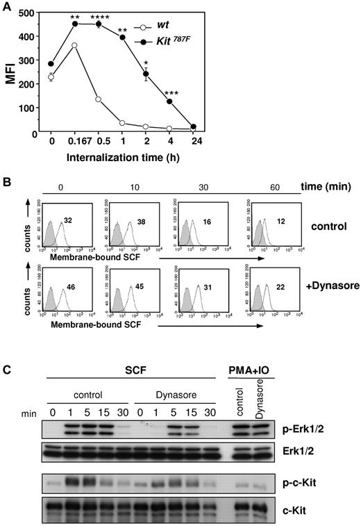
On page 2673 of the 14 October 2010 issue, there is an error in Figure 6. In the FACS data shown in Figure 6B (bottom panel), the histogram for the 30 minutes time point was shown twice. The mean values are correct and correspond with the correct pictures. The corrected Figure 6 is shown.
SCF-mediated c-Kit internalization and downstream signaling. (A) SCF-c-Kit internalization after SCF binding was measured at indicated time points. BMMCs generated from 3 different wild-type and Kit787F mice were compared. One representative experiment from 3 is shown (*P = .009; **P = .001; ***P = 4 × 10⁻⁴; ****P = 2 × 10⁻⁴). (B) SCF-c-Kit internalization was induced after preincubation of cells with Dynasore (100μM) or DMSO (control) for 30 minutes at 37°C and subsequent loading with SCF (50 ng/mL). αSCF staining (white histograms, MFI indicated) and staining of isotype-matched antibodies of irrelevant specificity (gray histograms) were measured at indicated time points by flow cytometry. One representative experiment from 3 is shown. (C) BMMCs from wild-type mice were preincubated with Dynasore (100μM) or DMSO (control) for 30 minutes at 37°C and stimulated for the indicated times with SCF (100 ng/mL) or PMA and ionomycin. Cell lysates were immunoblotted with antibodies specific for (phospho-)Erk1/2, (phospho-)c-Kit, and c-Kit. Immunoblotting with Erk1/2-specific antibodies served as loading control. One representative experiment from 2 is shown.
SCF-mediated c-Kit internalization and downstream signaling. (A) SCF-c-Kit internalization after SCF binding was measured at indicated time points. BMMCs generated from 3 different wild-type and Kit787F mice were compared. One representative experiment from 3 is shown (*P = .009; **P = .001; ***P = 4 × 10⁻⁴; ****P = 2 × 10⁻⁴). (B) SCF-c-Kit internalization was induced after preincubation of cells with Dynasore (100μM) or DMSO (control) for 30 minutes at 37°C and subsequent loading with SCF (50 ng/mL). αSCF staining (white histograms, MFI indicated) and staining of isotype-matched antibodies of irrelevant specificity (gray histograms) were measured at indicated time points by flow cytometry. One representative experiment from 3 is shown. (C) BMMCs from wild-type mice were preincubated with Dynasore (100μM) or DMSO (control) for 30 minutes at 37°C and stimulated for the indicated times with SCF (100 ng/mL) or PMA and ionomycin. Cell lysates were immunoblotted with antibodies specific for (phospho-)Erk1/2, (phospho-)c-Kit, and c-Kit. Immunoblotting with Erk1/2-specific antibodies served as loading control. One representative experiment from 2 is shown.


This feature is available to Subscribers Only
Sign In or Create an Account Close Modal